To the Editor:
Uckun et al1 have reported that bona fide in-frame fusionMLL-AF4 fusion sequences are detectable, using a sensitive nested reverse transcription-polymerase chain reaction (RT-PCR) method, in around 25% of normal fetal bone marrow and liver samples and in 1 of 6 infant bone marrows. It was also reported that similar fusion sequences were present in presumptive normal cells at low levels in 13% of pediatric (noninfant) acute lymphoblastic leukemia (ALL) cases, but were absent in remission bone marrow samples of similar patients. These are very interesting data and require careful consideration. Both these authors1 and editorial articles commenting on the findings2,3 suggested that the MLL-AF4 fusion gene may be necessary but insufficient for the clinical development of infant leukemia. As Hunger and Cleary2 point out in their editorial, it is important that this result is both substantiated by different methods, eg, fluorescence in situ hybridization (FISH), genomic sequencing, and independently confirmed by other investigators.
We have screened a series of unselected normal cord blood samples forMLL-AF4 fusion sequences as part of a program to evaluate the frequency of leukemia fusion genes in normal fetal hematopoiesis. We adapted and standardized a nested RT-PCR assay for maximized sensitivity using primers as previously described for minimal residual disease detection in infant leukemia4 and could, in standard dilution assays with MLL-AF4 positive leukemic cell lines, achieve reproducible detection at 10−5 with variable scores at 10−6. In screening for other translocation products, we have in the past had difficulties with false-positive results due to contamination, and we take appropriate precautions to prevent this. We have now screened a total of 68 samples (60 cord blood samples plus 8 fetal liver samples). All samples were subjected to RT-PCR for an ABL transcript to confirm the presence of intact mRNA. In the first series of 38 cord blood samples and 8 fetal liver samples, 5 bone marrows gave an amplified product. In each case, the product appeared to be slightly different in size from that anticipated from the cDNA prepared from leukemic cells with anMLL-AF4 fusion gene.4 Representative products were cloned and several isolates sequenced, and none were derived from chimeric MLL-AF4 fusion genes. The sequenced products were either unrelated to MLL-AF4 or derived from incompletely spliced AF4 mRNA where the upstream AF4 intron 3 had some fortuitous homology to the 3′ end of the internal MLLprimer. Because our RT-PCR primers4 were somewhat different from those used by Uckun et al, we performed screening on an additional 30 cord blood samples with primers identical to those described,1 but failed to score any positives. We also rescreened the 8 fetal livers and failed to observe any amplified products.
Other groups have similarly failed to identify MLL-AF4 fusion sequences in newborn samples5 (J. Trka, personal communication, September 1998). Intragenic fusions or partial tandem duplications of AF4 or MLL have been recorded in normal blood samples,4-7 the significance of which remains uncertain. Therefore, we are unable to confirm the report of Uckun et al1 and cannot at present suggest a likely explanation for the discrepancy. Although there are precedents for the finding of fusion genes in normal tissue samples,8-10 there are difficulties in accepting that infant leukemia is “primed” by widespread or common MLL-AF4 fusions in utero. First, even if such fusion genes were reproducibly detectable, this would not necessarily indicate the presence of preleukemic cells awaiting a second strike. The cellular context is likely to be critical. Such genes (as with other mutant oncogenes in normal individuals) could reside in cells that are irrelevant to leukemogenesis. TheMLL-AF4 fusion genes of infant leukemia do arise during fetal hematopoiesis11,12 and most probably in a stem cell with B/monocytic lineage potential. The presence of an MLL-AF4 gene product, albeit in frame, in, say, a T-cell or granulocyte progenitor, would be of no immediate significance with respect to leukaemogenesis. Second, MLL-AF4 fusions differ significantly in one particular respect from other aberrant leukemia- or cancer-associated genes detectable in normal individuals. Infant ALL/acute myeloblastic leukemia and secondary acute leukemia13withMLL fusion genes are associated with uniquely brief latent periods measurable in months as opposed to the norm of years or decades. Furthermore, the concordance rate of infant ALL in identical twins derived from an MLL fusion gene in one fetus is very high—somewhere around 50% to 100% of those with a monochorionic placenta with vascular connections facilitating spread of the preleukemic clone from one fetus to the other.10 14 One plausible interpretation of these data is that once an in-frameMLL fusion gene occurs in an appropriate cell type, evolution of the leukemic clone to a clinical diagnosis is both highly likely and rapid. This could occur either if MLL fusion genes are in themselves sufficient for leukemogenesis or if they very efficiently provoke other necessary secondary genetic events. Either way, if this interpretation is valid, it sits ill at ease with the presence of MLL-AF4 fusion-positive cells primed for leukemia in 25% of normal newborns. Back to the drawing board?
Response to Trka et al and Greaves et al
MLL-AF4 Fusion Transcripts in Normal and Leukemic Hematopoietic Cells
In a recent study,1-1 we used nested polymerase chain reaction (PCR) assays to determine the expression frequency of MLL-AF4 in infant and childhood acute lymphoblastic leukemia (ALL). In infants, nested PCR assays were in agreement with the standard cytogenetics data. Of 17 infants, 9 were MLL-AF4 positive by nested PCR and 8 of these 9 infants had a cytogenetically detectable t(4;11).1-2 In contrast, nested PCR assays showed MLL-AF4 positivity in 17 of 127 children with ALL, including children without a cytogenetically detectable t(4;11), although standard PCR showed MLL-AF4 positivity only in one case.1-1 Surprisingly, nested PCR assays (but not standard PCR) detected MLL-AF4 fusion transcript expression in some normal fetal liver or bone marrow cells as well. We1-1 and others1-3 commented that our results suggest that the t(4;11) translocation may be a common occurrence in utero, and may be a critical but not sufficient step for leukemogenesis.
In their Letter to Editor,1-4 Trka et al provide several suggestions and criticisms regarding our study. Their statements require further clarification and comment. The authors state that they have no data to support our hypothesis that MLL-AF4 expression is not sufficient for leukemogenesis, which was based upon detection of MLL-AF4 fusion transcripts in fetal hematopoietic tissues. This should not be surprising when one considers the fact that the authors have not examined MLL-AF4 expression in any fetal tissue. Furthermore, we studied only a very limited number of fetal tissue specimens and therefore cannot comment on the frequency of MLL-AF4 expression. Fresh samples need to be analyzed since MLL-AF4 transcripts in normal cells may be unstable. We were unable to detect MLL-AF4 transcripts by nested RT-PCR in any of 21 cryopreserved fetal liver specimens. The authors based their statement on their failure to detect MLL-AF4 positivity in cord blood samples from 103 full-term healthy newborns. Unfortunately, the authors did not include any positive controls in their study to show that their failure to detect MLL-AF4 fusion transcripts was not a false-negative result due to technical difficulties. Specifically, it is prudent that the authors show the presence of MLL-AF4 positive leukemic cells added to their cord blood samples at a 0.01% level. Furthermore, the authors do not comment on the cellular composition of the cord blood samples. The nucleated cell content as well as freshness of cord blood samples will undoubtedly affect the ability of the authors to detect MLL-AF4 positivity, which required even for highly hematopoietic cell–enriched mononuclear cell preparations from fetal tissues the use of a highly sensitive nested PCR assay.1-1 It should also be noted that the authors used a different PCR assay with different primers for nested PCR. Specifically, the authors report that they used primers in MLL exon 5 for both standard and nested PCR even though our sequencing results provide conclusive evidence that exon 6 of the MLL gene is frequently fused to AF4a in normal hematopoietic cells as well as non-t(4;11) ALL cells.1-1 Another interesting aspect of the letter by Trka et al is their statement that the authors usually find up to six bands in their gel analysis of their nested PCR products and therefore find the results shown in Fig 3 of our report1-1 surprising. While most investigators, including our group, frequently find multiple bands in the post-PCR gels of the MLL-AF4 standard PCR product of a given case, this is not to be expected after a nested PCR reaction. Therefore, the question arises of how the nested PCR reactions were performed and how the band profiles differed from those of standard PCR products. The post-PCR gels reported in our paper are indeed very similar to those reported by Downing et al,1-2 who used the same nested PCR methodology. Nevertheless, the results reported by Trka et al,1-4 if confirmed by others and validated after inclusion of appropriate positive controls, would certainly expand our knowledge of MLL-AF4 expression in hematopoietic tissues. I certainly agree with the authors that MLL-AF4 positive cord blood specimens cannot be considered free of leukemic cells and find nothing in our report to suggest this rather unusual interpretation. To the contrary, the detection of in utero rearrangements reported to date1-5-1-7 indicates that extreme caution is needed when autologous or syngeneic cord blood stem cells are to be used for transplantation, as recently discussed by Rowley.1-3 For example, Greaves reported the development of a T-cell leukemia with identical T-cell receptor rearrangements at the ages of 9 and 10 years in twins, indicating that the transforming events occurring in utero may lead to leukemia after a very long latency period.1-3
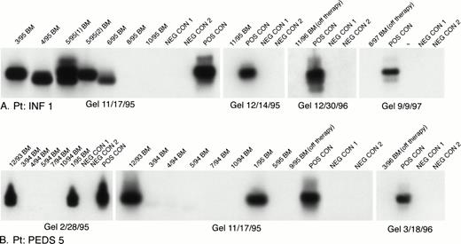
Regarding the failure of the authors to detect MLL-AF4 positive cells in remission bone marrow samples, I would like to point out that this is consistent with our own experience as well.1-1Specifically, in Table 5 of our paper, we reported that none of the 44 remission bone marrow samples expressed MLL-AF4 by standard or nested PCR.1-1 We were interested to know if patients with t(4;11) ALL would show persistent MLL-AF4 fusion transcript expression after chemotherapy. As shown in Fig 1-1A, nested PCR analysis of sequential bone marrow specimens from a t(4;11)+ infant ALL case (INF 1 from Table 2 of ref 1-2) showed disappearance of MLL-AF4 positivity 5 months after initiation of chemotherapy. More than 2 years after the first disappearance of MLL-AF4 positivity and in complete remission off therapy for more than a year, nested PCR continues to show no evidence of MLL-AF4 expression in this patient. Therefore, we postulate that all MLL-AF4 positive cells in this case were involved in the original leukemic transformation. Similarly, we were interested to know if the MLL-AF4 positivity detectable only by nested PCR in a non-t(4;11) ALL case represents low-level fusion transcript expression in leukemic cells or detection of MLL-AF4 fusion transcript expression in nonleukemic cells. As shown in Fig 1-1B, the MLL-AF4 positivity of bone marrow samples from a normal diploid ALL patient (PEDS 5) disappeared after remission induction, was transiently detectable again towards the end of the maintenance chemotherapy (ie, 1/95 bone marrow sample), and has been undetectable by nested PCR for more than 2 years as the patient continues to be in complete remission off chemotherapy. These results suggest that the MLL-AF4 positivity in this case was likely caused by low-level fusion transcript expression in leukemic cells. In view of the disappearance of MLL-AF4 positivity in nested PCR positive cases after remission induction and the nested PCR negativity of remission bone marrow specimens from 44 children with standard-risk ALL, I do not believe that bone marrow specimens from healthy children (excluding infants) would be nested PCR positive for MLL-AF4 expression.
Chemotherapy-induced elimination of MLL-AF4 positive cells in bone marrow specimens from ALL patients. (A) INF1 is an infant t(4;11) ALL case diagnosed in March 1995. (B). PEDS5 is a normal diploid ALL case diagnosed in December 1993. Sequential bone marrow specimens were analyzed for the presence of MLL-AF4 fusion transcripts using nested RT-PCR, as described. Amplified mRNA from the RS4;11 cell line was used as a positive control (POS CON). Negative controls (NEG CON) were PCR products from RNA-free and DNA-polymerase-free reaction mixtures.1-2
Chemotherapy-induced elimination of MLL-AF4 positive cells in bone marrow specimens from ALL patients. (A) INF1 is an infant t(4;11) ALL case diagnosed in March 1995. (B). PEDS5 is a normal diploid ALL case diagnosed in December 1993. Sequential bone marrow specimens were analyzed for the presence of MLL-AF4 fusion transcripts using nested RT-PCR, as described. Amplified mRNA from the RS4;11 cell line was used as a positive control (POS CON). Negative controls (NEG CON) were PCR products from RNA-free and DNA-polymerase-free reaction mixtures.1-2
We have not performed Southern blot analyses on a routine basis to detect MLL gene rearrangements. Since the nested PCR assay did not appear to be a useful diagnostic test because it failed to distinguish patients with t(4;11) ALL and those who might have MLL-AF4 in a minor population of cells, we used Southern blotting in only 9 patients, 2 fetal livers, and 1 infant bone marrow to highlight these distinctions. Interestingly, cells from all t(4;11) ALL patients, including a 4.7-year-old child (PEDS87) whose leukemic cells had MLL-AF4 positivity detectable by nested PCR only, showed 11q23 gene rearrangements by Southern blot analysis. Thus, in some cases with 11q23 gene rearrangements and MLL-AF4 positivity, the level of expression for MLL-AF4 may be so low that a nested PCR is required for detection. This conclusion is also supported by findings of Hilden et al.1-8 Do patients with no 11q23 gene rearrangement detectable by Southern blotting and MLL-AF4 positivity detectable only by nested PCR have a small subpopulation of MLL-AF4 positive cells or a cryptic MLL gene rearrangement in all leukemic cells with a very low-level fusion transcript expression detectable only by nested PCR? This question will be addressed using in situ nested PCR assays in our future studies. The authors refer to two patients (INF-6, PEDS-5) with normal diploid ALL and MLL-AF4 positivity with nested PCR. The detection of MLL gene rearrangements by Southern blot analysis has been extensively reported by others who examined normal diploid ALL cases.1-8-1-10 We do not believe that the nested PCR positivity in INF-6 or PEDS-5 has much to do with the Southern blot evidence of MLL gene rearrangement.
Finally, I would like to mention that recent results from our laboratory indicate the presence of additional molecular abnormalities in t(4;11) infant leukemia cells, which further supports our hypothesis that the expression of MLL-AF4 may be critical but not sufficient for leukemic transformation.1-11 However, our published findings1-1 that were obtained using nested RT-PCR still need to be validated using genomic sequencing, which we have not yet performed. I would like to thank the authors for their interest in our work and this opportunity to clarify a number of issues regarding MLL-AF4 fusion transcript expression in human lymphocyte ontogeny.


This feature is available to Subscribers Only
Sign In or Create an Account Close Modal