Abstract
To evaluate our diagnostic and therapeutic guidelines, clinical and long-term follow-up data of 219 patients with primary or secondary cutaneous CD30+ lymphoproliferative disorders were evaluated. The study group included 118 patients with lymphomatoid papulosis (LyP; group 1), 79 patients with primary cutaneous CD30+ large T-cell lymphoma (LTCL; group 2), 11 patients with CD30+ LTCL and skin and regional lymph node involvement (group 3), and 11 patients with secondary cutaneous CD30+ LTCL (group 4). Patients with LyP often did not receive any specific treatment, whereas most patients with primary cutaneous CD30+ LTCL were treated with radiotherapy or excision. All patients with skin-limited disease from groups 1 and 2 who were treated with multiagent chemotherapy had 1 or more skin relapses. The calculated risk for systemic disease within 10 years of diagnosis was 4% for group 1, 16% for group 2, and 20% for group 3 (after initial therapy). Disease-related 5-year-survival rates were 100% (group 1), 96% (group 2), 91% (group 3), and 24% (group 4), respectively. The results confirm the favorable prognoses of these primary cutaneous CD30+ lymphoproliferative disorders and underscore that LyP and primary cutaneous CD30+ lymphomas are closely related conditions. They also indicate that CD30+ LTCL on the skin and in 1 draining lymph node station has a good prognosis similar to that for primary cutaneous CD30+ LTCL without concurrent lymph node involvement. Multiagent chemotherapy is only indicated for patients with full-blown or developing extracutaneous disease; it is never or rarely indicated for patients with skin-limited CD30+ lymphomas.
Primary cutaneous T-cell lymphomas (CTCL) represent a heterogeneous group of neoplasms derived from skin-homing T cells. Apart from mycosis fungoides (MF), primary cutaneous CD30+lymphoproliferative disorders are the most common group, accounting for approximately 25% of all CTCL. This group includes primary cutaneous CD30+ (anaplastic) large T-cell lymphomas (LTCL) and lymphomatoid papulosis (LyP), a chronic recurrent, self-healing papulonodular skin eruption with histologic features of a (CD30+) CTCL.1-3 Because of overlapping clinical, histologic, and immunophenotypical characteristics, these conditions are considered to represent a spectrum of primary cutaneous CD30+lymphoproliferations.4,5 It is well established that these primary cutaneous CD30+ lymphoproliferations have favorable prognoses in most patients.6-12 Previous studies by our group demonstrated significant differences between primary cutaneous CD30+ (anaplastic) LTCL and morphologically similar systemic CD30+ anaplastic large-cell lymphomas.13 In contrast to these systemic lymphomas, primary cutaneous CD30+ LTCL are rare in children; they generally express the cutaneous lymphocyte antigen (CLA) characteristic of skin-homing T cells but usually not the epithelial membrane antigen (EMA). They are not associated with Epstein–Barr virus or, according to most studies,14-17 with the t(2;5) translocation (ALK/p80-negative), and they have a significantly better prognosis. For these reasons, primary CD30+ CTCL is included as a separate group in the revised European–American classification for non-Hodgkin lymphomas (REAL classification)18 and in the proposed World Health Organization classification.19 Notwithstanding the many publications describing the characteristic features of these primary cutaneous CD30+ lymphomas and emphasizing their favorable prognosis, referral centers for cutaneous lymphomas are confronted regularly with patients who have been misdiagnosed or who have been treated with unnecessarily aggressive treatment regimens.20-22 In particular, the diagnosis LyP is often overlooked. Based on the histologic diagnosis of a CD30+(anaplastic) LTCL and the presence of multifocal skin lesions, patients with LyP are routinely treated with multiagent chemotherapy by physicians unaware of the spectrum of LyP. Unfortunately, skin relapses during or shortly after systemic chemotherapy are almost the rule and give the false impression of a highly aggressive T-cell lymphoma requiring even more aggressive therapy.
The current report describes the results of a recent evaluation of 219 patients with cutaneous CD30+ lymphoproliferation who were included in the Dutch registry for cutaneous lymphomas between 1983 and 1998. This study was conducted to evaluate our current diagnostic and therapeutic approaches for these patients and to define potential risk factors for tumor progression. Detailed clinical and long-term follow-up data of well-defined groups of primary and secondary cutaneous CD30+ lymphoproliferations are presented. Based on these data, practical guidelines for proper diagnosis, management, and treatment are presented.
Patients and methods
Patients
Between 1983 and 1998, 247 patients with primary or secondary cutaneous CD30+ lymphoproliferation had been included in the database of the Dutch Cutaneous Lymphoma group. Patients with a follow-up less than 12 months unless they died of lymphoma (n = 14), patients with a CD30+ LTCL originating from MF (n = 8), and patients with HIV-associated (n = 4) or posttransplant (n = 2) CD30+ LTCL were excluded. The final study group contained 219 patients, including 118 patients with LyP, 79 patients with primary cutaneous CD30+LTCL, and 22 patients with cutaneous CD30+ (anaplastic) LTCL with concurrent or prior extracutaneous disease at the time of presentation. Of this latter group, 11 of 22 patients with skin lesions and regional lymph node involvement restricted to 1 site were evaluated as a separate group. All diagnoses were based on combinations of clinical, histologic, and immunohistochemical criteria23and were made by an expert panel of dermatologists and hematopathologists at the time of diagnosis, before entry in the database. Because correct categorization (ie, diagnosis) of these lymphomas was crucial, these diagnostic criteria have been summarized in Table 1. It should be emphasized that, because of the histologic similarities between LyP and other types of CTCL (MF or CD30+ ALCL), the presence of a recurrent, self-healing papular or papulonodular eruption is used as a decisive criterion for the diagnosis of LyP. For all patients in groups 2, 3, and 4 and for all patients with LyP type C (Table 1), adequate staging procedures including physical examination, routine examination of blood morphology and blood chemistry, radiography of the chest, and computerized tomography of the thorax (most patients) and abdomen were conducted, and bone marrow biopsies were taken. No extracutaneous disease was found in groups 1 and 2. In most other patients with LyP, staging procedures other than physical examination and routine blood examination were not performed.
Diagnostic criteria and histologic subgroups
| Category . | Diagnostic criteria . | Histologic subtype . |
|---|---|---|
| Group 1 Lymphomatoid papulosis (n = 118) | ▸ Recurrent, self-healing papulonodular eruption. ▸ Note: spontaneous complete remission of every individual skin lesion, irrespective of the appearance of new lesions. ▸ Histologic features suggesting (CD30+) CTCL. | LyP, type A: (n = 93) LyP, type B: (n = 6) LyP, type C: (n = 8) LyP, mixed (A, B, or both): (n = 11) |
| Group 2 Primary cutaneous CD30+ large T-cell lymphoma (n = 79) | ▸ Most (>75%) or large clusters of neoplastic cells express the CD30 antigen and have a T- or null cell phenotype ▸ No clinical evidence of LyP ▸ No prior or concurrent LyP, MF, or other type of (cutaneous) lymphoma ▸ No extracutaneous localizations at the time of diagnosis, as assessed with adequate staging. | Anaplastic ▸ Diffuse (n = 55) ▸ LyP-like (n = 10) Nonanaplastic ▸ Pleomorphic (n = 12) ▸ Immunoblastic (n = 2) |
| Group 3 CD30+ LTCL presenting with skin lesions and 1 involved regional lymph node station (n = 11) | ▸ Selection criteria of group 2 ▸ Histologically confirmed involvement of 1 regional lymph node station. | Anaplastic ▸ Diffuse (n = 8) ▸ LyP-like (n = 1) Nonanaplastic ▸ Pleomorphic (n = 2) |
| Group 4 Secondary cutaneous CD30+ large T-cell lymphoma (n = 11) | ▸ CD30+(anaplastic) LTCL ▸ Patients with concurrent cutaneous and extracutaneous disease (other than 1 regional lymph node) (n = 7) or ▸ Systemic CD30+(anaplastic) LTCL developing specific skin lesions during follow-up (n = 4) | Anaplastic ▸ Diffuse (n = 8) Nonanaplastic ▸ Pleomorphic (n = 3) |
LyP, type A: scattered CD30+ blast cells in an extensive inflammatory infiltrate.
LyP, type B: mycosis fungoides-like histology with atypical CD30− T cells with cerebriform nuclei.
LyP, type C: large clusters of CD30+ cells with few inflammatory cells, histologically suggesting a CD30+(anaplastic) large cell lymphoma.
LyP-like: anaplastic large T-cell lymphomas with relatively few CD30+ tumor cells in an extensive inflammatory infiltrate, histologically suggesting LyP.
Follow-up data had been collected yearly for each patient and could therefore easily be retrieved from the database. If necessary, additional information was obtained from the referring physician or from the patient.
Risk factors for tumor progression
As endpoints of tumor progression, extracutaneous disease in patients who initially only had cutaneous disease (groups 1 and 2) and lymphoma-related death were selected. As potential risk factors, the following parameters were scored for each patient: age at diagnosis, sex, morphology, localization and extent of skin lesions (solitary or localized vs multifocal skin lesions), type and result of initial treatment, occurrence and site of relapse (cutaneous or extracutaneous), disease-free survival, and histologic subtype (type A vs type B vs type C in LyP; anaplastic vs nonanaplastic morphology in the other 3 groups). Definitions of the different histologic subgroups have been published previously5 23 and are summarized in Table 1.
Statistical analysis
As indicators of survival, both disease-specific survival (including only death related to lymphoma as event) and overall survival (including death related to any cause as event) were investigated. Actuarial survival curves were calculated from the date of diagnosis to the date of death or last contact using the Kaplan–Meier technique. Univariate analysis of possible risk factors for tumor progression was performed using log-rank test and Cox proportional hazards regression analysis. Multivariate analysis was performed using significant univariate variables from Cox proportional hazards regression analysis. All analyses were performed with the SPSS statistic software (SPSS, Chicago, IL).
Results
The main clinical characteristics for the 4 groups studied are summarized in Table 2. Additional information for these groups is given separately below.
Main clinical findings and follow-up data
| . | LyP . | Primary cutaneous CD30+ LTCL . | CD30+ LTCL in skin and regional lymph nodes . | Secondary cutaneous CD30+ LTCL . |
|---|---|---|---|---|
| Number | 118 | 79 | 11 | 11 |
| Sex (M:F) | 69:49 | 59:20 | 4:7 | 6:5 |
| Age (y) | ||||
| Median | 45.5 | 60 | 64 | 55 |
| Range | 4-88 | 16-89 | 27-74 | 7-87 |
| Extent of skin lesions | ||||
| Solitary | 0 | 42 (53%) | 7 (64%) | 1 (9%) |
| Regional | 15 (13%) | 20 (25%) | 3 (27%) | 1 (9%) |
| Generalized | 103 (87%) | 17 (22%) | 1 (9%) | 8 (82%) |
| Type of skin lesions | ||||
| Papule | 80 (68%) | 6 (7%) | 1 (9%) | 3 (27%) |
| Nodule | 4 (3%) | 23 (29%) | 0 | 5 (46%) |
| Papules and nodules | 26 (22%) | 0 | 0 | 0 |
| Tumor | 5 (4%) | 47 (60%) | 10 (91%) | 3 (27%) |
| Plaques | 3 (3%) | 3 (4%) | 0 | 0 |
| Spontaneous remission | ||||
| Absent | 0 | 46 (58%) | 8 (73%) | 6 (55%) |
| Partial | 0 | 17 (22%) | 1 (9%) | 5 (45%) |
| Complete | 118 (100%) | 16 (20%) | 2 (18%) | 0 |
| B symptoms at diagnosis | ||||
| Absent | 118 (100%) | 79 (100%) | 11 (100%) | 5 (45%) |
| Present | 0 | 0 | 0 | 6 (55%) |
| Initial therapy | ||||
| Radiotherapy | 5 (4%) | 38 (48%) | 0 | 2 (18%) |
| Multiagent chemotherapy | 0 | 6 (8%) | 9 (82%) | 9 (82%) |
| Excision | 0 | 15 (19%) | 1 (9%) | 0 |
| PUVA/UV-B | 41 (35%) | 3 (4%) | 0 | 0 |
| Other | 6 (5%) | 2 (2%) | 0 | 0 |
| None/topical steroids | 66 (56%) | 15 (19%) | 1 (9%) | 0 |
| Result of initial therapy | ||||
| Complete remission | 16 (14%) | 78 (99%) | 10 (91%) | 3 (27%) |
| Partial remission | 31 (26%) | 1 (1%) | 0 | 5 (55%) |
| No response | 61 (60%) | 0 | 1 (9%) | 3 (27%) |
| Disease-free survival (mo) | ||||
| Median | NR | 23 | 38 | 0 |
| Range | NR | 1-192 | 0-84 | 0-115 |
| Relapse | ||||
| Skin only | 111 (94%) | 32 (41%) | 4 (36%) | 2 (18%) |
| Regional lymph node | 5 (4%) | 5 (6%) | 1 (9%) | 1 (9%) |
| Systemic | 2 (2%) | 3 (4%) | 1 (9%) | 55% |
| Follow-up (mo) | ||||
| Median | 77 | 61 | 58 | 22 |
| Range | 12-350 | 13-288 | 15-284 | 6-133 |
| Current status | ||||
| No evidence of disease | 38 (32%) | 51 (65%) | 6 (55%) | 2 (18%) |
| Alive with disease | 73 (62%) | 13 (16%) | 3 (27%) | 1 (9%) |
| Died of lymphoma | 2 (2%) | 4 (5%) | 1 (9%) | 8 (73%) |
| Died of other cause | 5 (4%) | 11 (14%) | 1 (9%) | 0 |
| Disease-related survival | ||||
| 5 years | 100% | 96% | 91% | 24%* (44%) |
| 10 years | 100% | 96% | 91% | 24%* (24%) |
| Overall survival | ||||
| 5 years | 98% | 83% | 76% | 24%* (44%) |
| 10 years | 95% | 78% | 76% | 24%* (24%) |
| Calculated risk for extracutaneous disease | ||||
| 5 years | 2% | 9% | 20%† | 80%† |
| 10 years | 4% | 16% | 20%† | 80%† |
LTCL, large T-cell lymphoma; NR, not relevant.
Denotes survival after the development of specific skin lesions; percentages between brackets indicate survival after initial diagnosis.
Denotes risk of new extracutaneous disease (either progression or relapse) after initial therapy.
Lymphomatoid papulosis
Group 1 consisted of 69 males and 49 females with a median age of 45.5 years at diagnosis (range, 4 to 88 years). Patients initially sought treatment for papular, papulonecrotic, or nodular skin lesions, and 8 patients (7%) also sought it for additional plaques or tumors. Characteristically, skin lesions in different stages of evolution were found next to each another (Figure 1). There was no preferential anatomic site of involvement. The group included 12 patients (10%) younger than 20 years of age at the time of diagnosis (median, 12 years; range, 4 to 19 years). Interestingly, 3 of these children (6, 13, and 16 years of age) had papules and rapidly growing ulcerating nodules or tumors. Despite this alarming clinical presentation, the lesions slowly subsided within 6 to 10 weeks without any specific treatment. None of the 12 children had extracutaneous disease or died of lymphoma after a median follow-up of 52 months (range, 25 to 131 months).
Lymphomatoid papulosis.
(A) Characteristic papular and papulonecrotic lesions at different stages of evolution. (B) Mixed inflammatory infiltrate with scattered CD30+ blast cells.
Lymphomatoid papulosis.
(A) Characteristic papular and papulonecrotic lesions at different stages of evolution. (B) Mixed inflammatory infiltrate with scattered CD30+ blast cells.
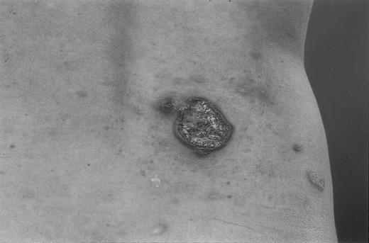
Throughout the course of their disease, 52 of 118 patients received no therapy other than topical steroids, which was not always recorded. Therapies most commonly applied in the other patients included psoralen–UV-A (PUVA) or UV-B phototherapy or chemotherapy (46 patients), topical nitrogen mustard (8 patients), and low doses of methotrexate (8 patients). Although partial or even complete remission was common, none of these therapies resulted in sustained complete remission. Similarly, remission after systemic chemotherapy or total skin electron beam irradiation, given for associated malignancies, was short-lived and did not affect the natural course of LyP. Associated lymphomas, either before, after, or concurrently with the development of LyP, were observed in 23 of 118 (19%) patients (Table3). One of these 23 patients had systemic CD30+ ALCL, which went into complete remission after multiagent chemotherapy, 6 years before LyP developed. Eleven of these 23 patients had concurrent LyP and MF. The diseases ran indolent clinical courses in all these patients. Large, persistent tumors developed in 4 patients with LyP; they were treated with radiotherapy (2 patients) or disappeared spontaneously (2 patients) (Figure2). Extracutaneous disease developed in only 7 of 118 patients. Skin tumors with involved regional lymph nodes developed in 4 of these 7 patients. Although 2 of these 4 patients were treated with cyclophosphamide-doxorubicin-vincristine-prednisone (CHOP) courses, skin and lymph node localizations disappeared spontaneously in the other 2 patients, and planned chemotherapy was not given. Systemic lymphoma developed in the other 3 of those 7 patients (CD30+ LTCL with lung localization in 1 patient and Hodgkin disease in 2 patients). All 3 patients were treated with multiagent chemotherapy. After a median follow-up of 77 months (range, 12 to 350 months), 111 patients (94%) are alive, 5 patients (4%) died of nonrelated disease, and only 2 patients (2%) died of systemic CD30+ LTCL or Hodgkin disease 14 and 19 years, respectively, after the diagnosis of LyP. Both patients have been described in more detail.24 25
Associated lymphomas in patients with lymphomatoid papulosis
| Patient . | Sex/age (y) . | Associated lymphoma . | Time interval3-150 (y) . | Localization . | Therapy . | Current status (follow-up in mo) . | ||
|---|---|---|---|---|---|---|---|---|
| Skin . | ec . | Lymphoma . | LyP . | |||||
| 1 | F/48 | CD30+ LTCL | B (5) | − | + | RT | A0 (120) | A+ (57) |
| 2 | M/46 | CD30+ LTCL | A (1) | + | − | RT | A0 (81) | A+ (94) |
| 3 | M/28 | CD30+ LTCL | A (25, 27) | + | − | RT/None3-151 | A0 (50, 20) | A+ (350) |
| 4 | F/24 | CD30+ LTCL | A (2, 3) | + | − | None3-151 | A0 (57, 43) | A+ (77) |
| 5 | M/12 | CD30+ LTCL | A (1) | + | − | None3-151 | A0 (4) | A+ (21) |
| 6 | M/23 | CD30+ LTCL | A (1) | + | +3-152 | CHOP | A0 (144) | A+ (156) |
| 7 | M/31 | CD30+ LTCL | A (5) | + | + | CHOP | A0 (39) | A+ (99) |
| 8 | F/53 | CD30+ LTCL | A (1) | + | +3-152 | None3-151 | A0 (105) | A+ (115) |
| 9 | M/43 | CD30+ LTCL | A (1) | + | +3-152 | None3-151 | A0 (14) | A+ (27) |
| 10 | M/52 | CD30+ LTCL | A (18) | + | + | CHOP | D+ (12) | D+ (228) |
| 11 | F/64 | Hodgkin disease | A (6) | − | + | MOPP | D0 (77) | D0 (142) |
| 12 | M/33 | Hodgkin disease | A (13) | − | + | MOPP | D+ (6) | D+ (168) |
| 13 | M/38 | Mycosis fungoides (Ib) | A (4) | + | − | PUVA | A+ (98) | A+ (142) |
| 14 | F/72 | Mycosis fungoides (Ib) | B (4) | + | − | RT | A+ (143) | A0 (97) |
| 15 | F/56 | Mycosis fungoides (Ib) | A (10) | + | − | HN2 | A0 (244) | A+ (118) |
| 16 | M/45 | Mycosis fungoides (Ia) | A (4) | + | − | HN2 | A+ (131) | A+ (177) |
| 17 | F/88 | Mycosis fungoides (Ib) | C | + | − | Retinoids + PUVA | D0 (23) | D0 (23) |
| 18 | M/73 | Mycosis Fungoides (Ib) | A (8) | + | − | UVB | A0 (102) | A0 (199) |
| 19 | F/24 | Mycosis Fungoides (Ib) | C | + | − | PUVA | A+ (94) | A+ (94) |
| 20 | M/59 | Mycosis Fungoides (Ib) | A (12) | + | − | TSEB | A0 (218) | A+ (77) |
| 21 | M/64 | Mycosis Fungoides (Ia) | A (2) | + | − | UVB | A+ (34) | A+ (55) |
| 22 | M/51 | Mycosis Fungoides (Ib) | C | + | − | PUVA | A+ (20) | A+ (20) |
| 23 | M/73 | Mycosis Fungoides (Ib) | B (8) | + | − | PUVA | D0 (160) | D0 (78) |
Associated lymphoma before (B), after (A), or concurrent (C) with the development of lymphomatoid papulosis.
Complete spontaneous resolution before initiation therapy.
Involved draining lymph node only.
ec, extracutaneous; LyP, lymphomatoid papulosis; CD30+LTCL, CD30+ large T-cell lymphoma; RT, radiotherapy; A0, alive no evidence of disease; A+, alive with disease; D0, died of unrelated disease; D+, died of lymphoma; CHOP, cyclophosphamide, doxorubicin, vincristine, prednisone; MOPP, mechlorethamine, vincristine, procarbazine, prednisone; HN2, topical nitrogen mustard; TSEB, total skin electron beam irradiation; mycosis fungoides, in all patients patch/plaque stage MF affecting <10% (Ia) or >10% (Ib) of the skin surface.
A large ulcerating tumor (diameter, 5 cm) developed in a 12-year-old boy with lymphomatoid papulosis.
The tumor disappeared spontaneously within 2 months.
A large ulcerating tumor (diameter, 5 cm) developed in a 12-year-old boy with lymphomatoid papulosis.
The tumor disappeared spontaneously within 2 months.
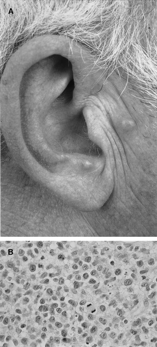
Evaluation of risk factors for tumor progression by univariate analysis did not reveal statistically significant results (P > 0.1), which may be explained by the small proportion (4%) of patients with LyP in whom extracutaneous disease developed. Notably, none of the 8 patients with LyP whose skin biopsies revealed cohesive sheets of CD30+ cells (LyP type C) died of lymphoma. In addition, after a follow-up of 23 to 156 months (median, 43 months), extracutaneous disease developed in only 1 (Figure3).
Lymphomatoid papulosis (type C).
(A) Approximately 10 firm papules on the face and the chest. (B) Detail of dermal infiltrate showing a monotonous population of large anaplastic (CD30+) cells. During follow-up there was complete spontaneous resolution of all skin lesions, whereas new lesions continued to develop.
Primary cutaneous CD30+ large cell lymphoma
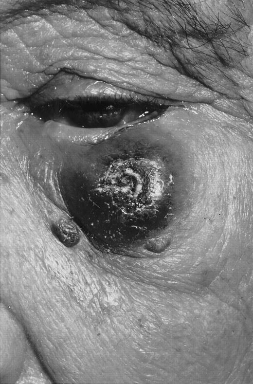
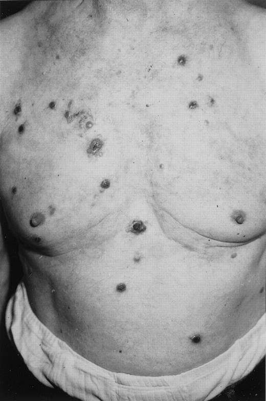
Group 2 consisted of 59 males and 20 females with a median age of 60 years at diagnosis (range, 16 to 89 years). Only 1 patient was younger than 20 years of age, illustrating that there is no bimodal age distribution in this group. Patients usually sought treatment for a solitary (ulcerating) tumor (42 patients) or several grouped nodules or papules restricted to 1 skin area (20 patients) (Figure4). Only 17 patients (22%) had multifocal skin disease, ie 2 (n = 9) or more (n = 8) lesions at multiple anatomic sites (Figure 5). Partial or even complete spontaneous remission of skin lesions, either at diagnosis or at relapse, were observed in 33 (42%) patients.
Primary cutaneous CD30+ large T-cell lymphoma with 1 large, partly ulcerating tumor.
Primary cutaneous CD30+ large T-cell lymphoma with 1 large, partly ulcerating tumor.
Primary cutaneous CD30+ large T-cell lymphoma with multifocal persistent skin lesions.
There was no spontaneous remission of any of these lesions.
Primary cutaneous CD30+ large T-cell lymphoma with multifocal persistent skin lesions.
There was no spontaneous remission of any of these lesions.
Initial treatment consisted of radiotherapy (47%) or surgical excision (22%). Only 6 patients (8%) received doxorubicin-based multiagent chemotherapy, including 4 of 17 patients who sought treatment for multifocal skin lesions. Twelve patients (15%) received no treatment because of spontaneous, complete remission of the skin lesions. Although 33 of 79 patients (42%) had 1 or moreskin relapses, extracutaneous disease developed in only 8 of 79 patients (10%). Treatment consisted of CHOP courses (6 patients) or autologous bone marrow transplantation. After a median follow-up of 61 months (range, 13 to 288 months), only 4 patients died of lymphoma.
Patients with multifocal skin lesions more often acquired extracutaneous disease (17% vs 8%) and more often died of lymphoma (12% vs 3%) than patients with solitary or localized skin lesions. However, univariate analysis demonstrated that neither extent of skin lesions, age at diagnosis, presence of spontaneous remission, histologic subtype, or any other variable tested was significantly related to disease progression or survival. Multivariate analysis, therefore, was not performed.
CD30+ LTCL with skin and draining lymph node involvement
Group 3 consisted of 4 men and 7 women with a median age of 64 years (range, 27 to 74 years). Ten of 11 patients had 1 (8 patients) or several (2 patients) ulcerating tumors in 1 anatomic area and enlarged, histologically involved draining lymph nodes at that site. Initial therapy consisted of CHOP-like multiagent chemotherapy in 9 of 11 patients (82%) and resulted in complete remission in 8 of them. However, 5 of these 8 patients had skin relapses during follow-up. In 2 other patients not treated with systemic chemotherapy, skin and nodal lesions disappeared spontaneously after diagnostic excision of the skin tumor, and no further treatment was instituted. Neither of them had a relapse, and both are still in complete remission 24 and 79 months after diagnosis, respectively. After a median follow-up of 58 months (range, 15 to 248 months), 9 patients are alive, 1 died of unrelated disease, and 1 died of lymphoma. The disease-related 5-year survival rate in this group was 91% (Figure 6).
Survival curve of different groups of primary and secondary cutaneous CD30+ lymphoproliferations.
Highly significant differences in survival were found between patients with secondary cutaneous CD30+ LTCL on the one hand and patients with LyP (P < .0001), primary cutaneous CD30+ LTCL (P < .0001), and cutaneous CD30+ LTCL with concurrent involvement of a regional lymph node (P < .003) on the other hand. There were no significant differences in survival between these last 3 groups.
Secondary cutaneous CD30+ large T-cell lymphoma
Group 4 consisted of 6 males and 5 females with a median age of 55 years (range, 7 to 87 years). Seven patients had extensive cutaneous and extracutaneous disease. The age distribution was typically bimodal: 3 patients were younger than 22 years and 4 were older than 55 years. Treatment with multiagent chemotherapy resulted in complete remission in only 2 of 7 patients; 6 of these 7 patients died after a median period of only 13 months (range, 6 to 46 months). The remaining patient is alive and well more than 10 years after autologous bone marrow transplantation.
Skin lesions developed in the other 4 patients 5 to 108 months (median, 53 months) after the diagnosis of systemic CD30+ ALCL was made. The ages of these patients (15, 24, 60, and 70 years) also suggested a bimodal age distribution. Treatment consisted of multiagent chemotherapy (3 patients) or radiotherapy at multiple sites (1 patient). Only 1 of 4 patients achieved complete remission. Two patients died 10 and 26 months after the development of skin lesions (63 and 132 months after initial diagnosis), the other 2 are alive 43 and 131 months after the development of skin lesions (48 and 185 months after initial diagnosis).
Discussion
In the current study, clinical and histologic variables of a major group of 219 cutaneous CD30+ lymphoproliferations were evaluated. The primary goals of this analysis were a critical evaluation of our current diagnostic and therapeutic approaches and a definition of potential risk factors for tumor progression. Relevant features of the different groups of cutaneous CD30+ lymphomas are discussed, and guidelines for correct diagnosis and treatment are presented.
Primary cutaneous CD30+ large T-cell lymphoma
Primary cutaneous CD30+ LTCL predominantly affected adults but rarely affected children (see below). They generally appeared as solitary or localized skin lesions (78%), they had the tendency to regress spontaneously, they frequently relapsed on the skin (41%) but uncommonly disseminated to extracutaneous sites (10%), and their prognosis was excellent with 5- and 10-year disease-related survival rates exceeding 95%. These findings confirm and extend the results of previous studies on smaller groups of patients.5-11
Risk factors predicting tumor progression or dissemination could not be found. This may be related to the small number of patients in whom extracutaneous disease developed (8 of 79 patients) and the small number of tumor-related deaths (4 of 79 patients). No differences in survival rates for patients with lymphoma with or without typical anaplastic morphology were found. However, CD30+ LTCL with an LyP-like histology (few CD30+ cells) more often showed complete spontaneous remission (50%) than CD30+ LTCL with large clusters of CD30+ anaplastic (29%) or nonanaplastic cells (28%). Neither extent of skin lesions (solitary or localized vs multifocal skin lesions) nor age was significantly related to survival.
Interestingly, the group of patients with CD30+ LTCL with skin lesions and only 1 involved draining lymph node station (group 3) had the same clinical features (presentation with solitary or localized skin lesions, no bimodal age distribution, good response to therapy) and a similarly good prognosis as the group with primary cutaneous CD30+ LTCL. Recent studies demonstrated that systemic CD30+ anaplastic large-cell lymphomas expressing ALK have a much better prognosis than ALK-negative lymphomas (5-year survival rates, 93% vs 37%).26 Immunohistochemical studies of 5 patients in group 3 demonstrated an ALK−, EMA−, CLA+ immunophenotype in each of them. As a comparison, the results of ALK staining in 28 other patients showed a positive reaction in 3 of 4 patients with secondary cutaneous CD30+ LTCL but not in 15 patients with primary cutaneous CD30+ LTCL, and they showed neither in 9 patients with LyP.17 The favorable prognosis for our group 3 patients cannot simply be attributed to the fact that 9 of 11 patients were treated with systemic chemotherapy. If this were representative of skin localizations of an ALK-negative systemic lymphoma, a less favorable outcome would have been expected for this group. Our clinical and immunohistochemical observations suggest rather that these patients are in fact patients with primary cutaneous CD30+ LTCL with (early) dissemination to the regional lymph nodes.
Major differences were found between the above-mentioned groups and patients with systemic CD30+ anaplastic large T-cell lymphomas with concurrent or secondary skin involvement. These patients more often sought treatment for generalized skin lesions. They had a bimodal age distribution, poor responses to often intensive treatment, and poor prognoses, with 5-year-survival rates of 44% after diagnosis and 23% after the development of skin lesions—suggesting that the appearance of skin lesions in CD30+ ALCL is a poor prognostic sign.11
Therapy.
In patients with a solitary or few localized nodules or tumors, local radiotherapy is the first choice of treatment. However, follow-up data indicate that if the lesion has been excised completely or has disappeared spontaneously, no further therapy is required. If a skin lesion relapses, spontaneous resolution can be awaited for some weeks or the patient can undergo radiotherapy or surgical excision. According to our existing guidelines formulated in 1991, patients with multifocal skin lesions should be treated with doxorubicin-based multiagent chemotherapy. However, only 7 of 17 patients with multifocal skin lesions in this study had ever been treated with systemic chemotherapy. More important, all patients who were treated with CHOP courses because of multifocal skin lesions (6 of 7 patients) had 1 or several skin relapses afterward. Moreover, in 3 of these 17 patients, all initial skin lesions disappeared spontaneously. Unlike LyP lesions, however, relapse did not occur during the 32- to 72-month follow-up. These observations suggest that multifocal skin-restricted CD30+LTCL should not be treated routinely with multiagent chemotherapy. If spontaneous regression does not occur, these patients can best be treated with radiotherapy for a few lesions or with low-dose methotrexate as for LyP.20 27 In patients with full-blown or developing regional lymph node involvement, multiagent chemotherapy is still considered the safest option. However, skin relapses after chemotherapy are common, but they are not associated with an aggressive clinical behavior and therefore do not require an aggressive approach. Moreover, complete spontaneous resolution of skin and lymph node localizations may occur. In such patients an expectative approach seems justified.
Lymphomatoid papulosis
In this very large group of patients with LyP with a median follow-up of 77 months and of 29 patients with a follow-up of more than 10 years, 23 of 118 patients (19%) had associated malignant lymphoma either before, after, or concurrent with LyP. However, only 7 of 118 had extracutaneous disease, and only 2 of 118 patients died of systemic lymphoma. The calculated risk for systemic lymphoma within the first 10 or 15 years after diagnosis was 4% and 12%, respectively, in contrast with the 80% at 15 years in a study of Cabanillas et al.21However, in that study, only patients with LyP who had initially been misdiagnosed or had another type of malignant lymphoma were included, suggesting that the striking difference between both studies results from differences in patient selection. As in earlier studies on smaller groups of patients,25,28 29 risk factors for disease progression could not be found.
Therapy.
Because a curative therapy is unavailable and none of the available treatment modalities affects the natural course of the disease, the short-term benefits of active treatment should be balanced carefully against the potential side effects.5,20 We believe that in patients with relatively few and nonscarring lesions, active treatment is not necessary. In patients with cosmetically disturbing lesions (eg, scarring lesions or many lesions), low-dose oral methotrexate (5 to 20 mg/wk) or PUVA therapy can be administered to reduce skin lesions.20,27 30 When larger skin tumors develop in the course of LyP, spontaneous remission can be awaited for 4 to 6 weeks. If spontaneous resolution does not occur, such lesions can be excised or treated with radiotherapy. Whether such isolated skin tumors developing in the course of LyP should be considered progression to CD30+ LTCL is debatable.
Primary cutaneous CD30+lymphoproliferations in children.
Of the 12 children with LyP in this study, 3 had large, rapidly growing ulcerating lesions in addition to papular lesions. Because of this distressing clinical presentation suggesting aggressive lymphoma, multiagent chemotherapy was first considered in all 3 children. However, because the histologic appearance was highly suggestive of LyP and staging procedures did not show any evidence of extracutaneous disease, it was decided to wait for several weeks. In all 3 patients the larger skin lesions disappeared completely within 2 to 3 months, whereas the papular lesions continued to develop. This illustrates that the physician should be cautious before using aggressive chemotherapy to treat children with skin-limited CD30+ lymphoproliferations because they often simply have LyP (12 of 13 patients younger than 20 in this study). A short period of watchful waiting is warranted and may avert unnecessary treatment. Previous studies also report an indolent course for children with LyP.28,31 32
Guidelines for diagnosis, management, and treatment
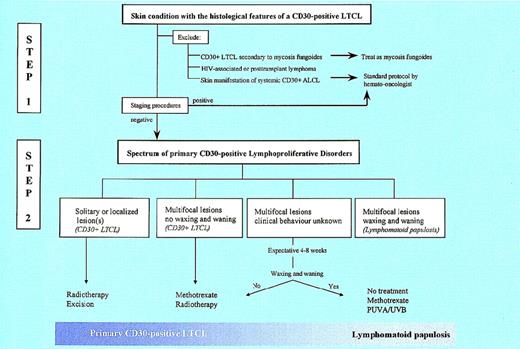
The central problem in the correct diagnosis and classification of this group of diseases is that there are no reliable histologic criteria to differentiate between the different types of primary and secondary cutaneous CD30+ lymphoproliferations. Patients with cutaneous CD30+ LTCL may show an LyP-like histology, whereas patients with the characteristic clinical presentation and clinical behavior of LyP may show large sheets of CD30+cells with only few infiltrating reactive cells (LyP, type C). Consequently, the histologic diagnosis should always be considered as a differential diagnosis and should never serve as a basis for therapeutic decisions. The definite diagnosis should always be based on a combination of histological and clinical criteria. This diagnostic process involves 2 steps (Figure7). The first and most important question is whether it is a primary or a secondary cutaneous CD30+lymphoproliferation. The second question is whether it is LyP or primary cutaneous CD30+ LTCL.
Algorithm for the diagnosis and treatment of cutaneous CD30+ lymphoproliferations.
Algorithm for the diagnosis and treatment of cutaneous CD30+ lymphoproliferations.
Step 1: primary or secondary cutaneous CD30+ lymphoma?
Confronted with a patient with the histologic diagnosis of a cutaneous CD30+ lymphoproliferation, distinctions should first be made among primary cutaneous CD30+lymphoproliferation, cutaneous CD30+ lymphoma secondary to MF (or another type of CTCL), and skin localizations of a systemic CD30+ (anaplastic) LTCL. Cutaneous CD30+lymphomas secondary to MF can generally be recognized easily because of the presence of prior or concurrent patches or plaques with the typical histology of MF. If no extracutaneous disease can be demonstrated, patients can be treated routinely as for tumor-stage MF, primarily with skin-directed therapies such as local radiotherapy or total skin electron beam irradiation. Patients with skin and extracutaneous localizations require multiagent chemotherapy and generally have an unfavorable prognosis. MF with transformation into a CD30+LTCL should not be confused with MF with concurrent LyP, a combination observed in 9% of patients with LyP (current study) and in approximately 3% of patients with MF33 and carrying, almost without exception, a very good prognosis.25,33 34
To exclude secondary cutaneous CD30+ (anaplastic) LTCL, we suggest adequate staging in all patients with cutaneous CD30+ lymphoproliferation. An exception is made for patients with clinical and histologic features characteristic of LyP in whom meticulous physical examination suffices.20
Step 2: management of primary cutaneous CD30+lymphoproliferations.
Previous studies emphasize the many similarities between primary cutaneous CD30+ LTCL and LyP, suggesting that these are closely related conditions within a continuous spectrum.4,5Distinction between these 2 ends of the spectrum was considered important because patients with CD30+ LTCL were thought to be at much higher risk for extracutaneous disease than patients with LyP and, unlike patients with LyP, always required adequate staging and even systemic chemotherapy if they had multifocal skin lesions.5,11 However, the results of the current study suggest that the differences in disease progression and survival between both groups are smaller than anticipated previously (Table 2). In addition, skin relapses after systemic chemotherapy were not only observed in patients with LyP but also in all patients with skin-limited CD30+ LTCL,22 which argues against the use of systemic chemotherapy for patients with only skin lesions. Taken together, our observations indicate that the choice of treatment should be based above all on the size, the extent, and the clinical behavior of the skin lesions and not as much on the diagnosis of CD30+ LTCL or LyP (Figure 6).
Following this approach, distinction is first made between patients with a solitary or few localized nodules or tumors and patients with multifocal skin lesions. The term localized refers to a few clustered lesions restricted to 1 anatomic area generally not exceeding 15 × 15 cm. These primary cutaneous CD30+ LTCL may show spontaneous resolution, either partial or complete, but they do not wax and wane as do LyP lesions. As discussed before, local radiotherapy is the first choice of treatment. Most patients with multifocal skin lesions have LyP. Characteristically, skin lesions wax and wane, and skin lesions coexist in different stages of evolution. These include early small red papules, fully developed papules or nodules with or without central ulceration, brown–red disappearing lesions, residual lesions, and sometimes residual scars. Complete disappearance of every single lesion, irrespective of the appearance of new lesions, is a prerequisite. Most patients do not require any specific treatment. If a patient has cosmetically disturbing lesions, low-dose oral methotrexate (5 to 20 mg/wk) or PUVA therapy may be considered. Multifocal skin lesions that do not wax and wane as in LyP are less common. Nevertheless, skin lesions in these multifocal primary cutaneous CD30+ LTCL may also show partial, and sometimes even complete, spontaneous remission, which can often be recognized clinically by the skin lesions turning from red to brown. As discussed before, patients with such lesions should no longer be treated routinely with doxorubicin-based multiagent chemotherapy because skin relapses after chemotherapy are likely to occur. If spontaneous remission does not occur, radiotherapy or low-dose methrotrexate is the best treatment if there are only a few lesions.
In some patients with a brief history of multifocal skin lesions, the clinical behavior may still be unknown, and a definite diagnosis cannot yet be made. It is our experience in particular that such patients—not uncommonly colleagues or close relatives—are often treated with systemic chemotherapy. However, because extracutaneous disease has already been excluded, and because not only LyP but also primary cutaneous CD30+ LTCL has a low tendency to progress to extracutaneous disease, it is better to wait for several weeks while monitoring closely the natural evolution of the skin lesions. After a period of 4 to 8 weeks, a definite diagnosis can normally be made, and the appropriate treatment can then be selected.
In conclusion, the results of this study confirm the indolent clinical behavior and excellent prognosis of a group of primary cutaneous CD30+ lymphoproliferations. Close collaboration between dermatologists, pathologists, and hematologists/oncologists is the best guarantee for a correct diagnosis and proper treatment. If a definite diagnosis cannot yet be made, an expectative approach seems justified. Multiagent chemotherapy is only indicated for patients with existent or developing extracutaneous disease. It is indicated rarely or not at all for patients with only skin lesions. For all patients, however, long-term follow-up is required.
Reprints:Rein Willemze, Leiden University Medical Center,Department of Dermatology, B1-Q-93, P.O. Box 9600, 2300 RC Leiden, The Netherlands; e-mail: willemze.dermatology@lumc.nl orrein.willemze@planet.nl.
The publication costs of this article were defrayed in part by page charge payment. Therefore, and solely to indicate this fact, this article is hereby marked “advertisement” in accordance with 18 U.S.C. section 1734.



This feature is available to Subscribers Only
Sign In or Create an Account Close Modal