Abstract
To investigate the tropism of the T-lymphotropic human herpesvirus 7 (HHV-7) for hematopoietic progenitors, cord blood CD34+ cells were inoculated in vitro with HHV-7 and then induced to differentiate along the granulocytic and erythroid lineages by the addition of appropriate cytokine cocktails. In semisolid assays, HHV-7 modestly affected the growth of committed (granulocytic/macrophagic and erythroid) progenitors, whereas it significantly decreased the number of pluripotent (granulocytic/erythroid/ monocytic/megakaryocytic) progenitors. Such inhibitory effect was completely abrogated by incubating HHV-7 inoculum with anti–HHV-7 neutralizing serum. In liquid cultures, HHV-7 hastened maturation along the myeloid but not the erythroid lineage, as demonstrated by the up-regulation of CD33 early myeloid antigen at day 7 of culture, and of CD15 and CD14 antigens at day 15. Moreover, HHV-7 messenger RNA was detected by reverse transcriptase–polymerase chain reaction (RT-PCR) in cells maturating along both the myeloid and the erythroid lineages. To evaluate the relevance of these in vitro findings, the presence of HHV-7 was investigated in bone marrow (BM) unfractionated mononuclear cells (MCs) as well as in purified CD34+ and CD34− cell subsets, obtained from 14 normal adult donors. HHV-7 DNA was detected by DNA-PCR in 4 of 7 BMMC samples, and it was found to be associated with both the CD34− (2 of 7) and the CD34+ (1 of 7) fractions. These data indicate that HHV-7 infects BM cells in vivo and shows the ability to affect the survival/differentiation of CD34+ hematopoietic progenitors in vitro by inhibiting more ancestral progenitors and perturbing the maturation of myeloid cells.
Human herpesvirus-7 (HHV-7) is a CD4+T-lymphotropic herpesvirus1 closely related to human herpesvirus-6 (HHV-6) and human cytomegalovirus (HCMV).2-4HHV-7 was first isolated from the peripheral blood lymphocytes of healthy individuals,1,5,6 and it turned out to be a prevalent virus, with more than 90% of the population seropositive by adulthood.7-9 Although the only clinical manifestation clearly associated with primary HHV-7 infection is exanthem subitum,10-13 the high seroprevalence of HHV-7 in the general population7-9 suggests that this virus is a potential opportunistic agent in immunocompromised individuals.14 In fact, owing to its selective tropism for CD4+ T lymphocytes,1 15 once reactivated, acute HHV-7 infection might worsen the state of immunodeficiency.
In vitro studies have demonstrated that the CD4 antigen acts as a critical component of the receptor for HHV-7.15,16Moreover, we have previously shown that HHV-7 induces several cytopathic effects on cultured mature CD4+ T lymphocytes, such as induction of cell death,17 perturbation of the cell cycle,18 and formation of giant balloon-like cells.1,2,6,17,18 Of note, the CD4 antigen is expressed at high density on the surface of the helper/inducer subset of peripheral blood T lymphocytes19 and at low density on the surface of other cell types, including hematopoietic progenitors.20-22This suggests that CD34+ hematopoietic progenitors are also a potential target of HHV-7.
Previous studies have shown that HHV-6 can infect in vitro hematopoietic progenitor cell lines as well as primary CD34+ hematopoietic progenitors.23 Moreover, bone marrow (BM) myeloid and erythroid progenitors may also be infected with type B HHV-6 in vivo.24 Apparently, HHV-6 can infect clonogenic progenitors without eliminating their ability to proliferate and differentiate, thus creating a pool of infected BM progenitors that can serve as a reservoir of latent virus.24 HCMV, which causes myelosuppression in immunodeficient patients, is another human herpesvirus capable of infecting myeloid progenitors in vitro.25-31
The aim of our study was to investigate the effects of HHV-7 on CD34-derived cells differentiating along the granulocytic and erythroid lineages in liquid culture as well as on clonogenic progenitors in semisolid cultures. In parallel, we sought to evaluate the possibility of direct infection of hematopoietic cells by HHV-7, both in vitro and in vivo. To investigate these issues, CD34+ hematopoietic progenitor cells were purified from cord blood (CB), infected in vitro with HHV-7, and seeded in both semisolid and liquid cultures. In parallel, unfractionated BM mononuclear cells (BMMCs) as well as BM CD34− and CD34+ cell subsets, obtained from adult donors, were analyzed ex vivo for the presence of HHV-7 DNA.
Materials and methods
Isolation of human CB and BM CD34+ hematopoietic progenitors
CB specimens, collected according to institutional guidelines, were obtained during 12 normal full-term deliveries. BM samples were obtained from 14 normal adult donors. CB and BMMCs were separated on Ficoll-hypaque gradient (density 1.077 g/mL) (Pharmacia, Uppsala, Sweden). CD34+ cells were then isolated with the use of a magnetic cell-sorting program (Mini-MACS) and the CD34 isolation kit (both from Miltenyi Biotec, Auburn, CA) in accordance with the manufacturer's recommendations and as previously described.32 Purity of CD34-selected cells was analyzed for each isolation by FACScan (Becton Dickinson, San José, CA) by double-staining with a monoclonal antibody (mAb), which recognizes a separate epitope of the CD34 molecule (HPCA-2, Becton Dickinson), directly conjugated to fluorescein isothiocyanate (FITC) plus an anti-CD3 mAb, directly conjugated to phycoerythrin (PE) (Becton Dickinson). Flow cytometry analysis, performed as described below, showed that CB and BM CD34+ cell purity ranged between 85% and 98%, while the presence of contaminating CD3+ mature T lymphocytes was constantly less than 3%.
In vitro HHV-7 infection of CB CD34+ cells
The procedures for HHV-7 propagation in SupT1 lymphoblastoid T-cell line and the preparation of viral stocks have been previously described.16-18 HHV-7 infection of human CB CD34+ cells was carried out by incubation with 0.45 μm filtered infectious supernatant obtained from HHV-7–infected SupT1 cells. Supernatant from uninfected SupT1 cells was used for mock infections. After overnight incubation with the HHV-7 or mock inoculum (MOI = 0.1-0.02) in X-VIVO-20 (BioWhittaker, Walkersville, MD) medium supplemented with stem cell factor (SCF, 50 ng/mL) plus interleukin (IL)–3 (10 ng/mL), cells were seeded in either liquid or semisolid cultures, as described below.
For virus-neutralization assays, viral and mock inocula were preincubated for 30 minutes at room temperature with an anti–HHV-7 neutralizing human serum (Advanced Biotechnologies, Columbia, MD). The optimal dilution for the serum was determined in a neutralizing assay carried out on SupT1 cells. Briefly, viral inoculum was pretreated for 30 minutes at room temperature with serial 2-fold (from 1:25 to 1:400) dilutions of the human serum and then used to inoculate 1.5 × 10⁵ SupT1 cells in 24-well plates. The appearance of giant cells17 18 was scored starting at 72 hours after infection. The optimal neutralizing antibody titer was determined as the reciprocal of the highest dilution of serum (1:100) that completely prevented giant cell formation.
The supernatants of both uninfected and HHV-7–infected SupT1 cells, used as inocula, were tested for the presence of IL-1β, IL-6, IL-15, IL-3, granulocyte/macrophage-colony stimulating factor (GM-CSF), and granulocyte-colony stimulating factor (G-CSF). These cytokines were measured on specific commercial Elisa plates (R&D Systems, Minneapolis, MN), in accordance with the manufacturer's recommendations. Intra-assay and interassay coefficients of variation did not exceed 10%.
Semisolid and liquid cultures
Pluripotent granulocytic/erythroid/monocytic/megakaryocytic (CFU-GEMM) colonies and committed granulocytic/macrophagic (CFU-GM) and erythroid (BFU-E) colonies were assayed in plasma clot cultures, as previously described.20 Briefly, 5 × 10³ purified CB CD34+ cells were cultured in 1 mL of Iscove's Modified Dulbecco's Medium (GIBCO, Grand Island, NY), containing 10% detoxified bovine serum albumin (BSA, fraction V Chon) (Sigma Chemical, St Louis, MO), 10% heat-inactivated pooled human AB sera, 10% citrated bovine plasma (GIBCO), 20 μg of L-asparagine (Sigma), 3.4 mg/mL CaCl2. The standard sources of growth factors were SCF (50 ng/mL) + IL-3 (10 ng/mL) + G-CSF (10 ng/mL) for the growth of CFU-GM; SCF (50 ng/mL) + IL-3 (10 ng/mL) + erythropoietin (EPO, 4U/mL) for the growth of CFU-GEMM and BFU-E. All cytokines were purchased from Genzyme (Cambridge, MA).
At day 14 of culture, for CFU-GM and BFU-E identification, plasma clots were fixed in situ with acetone for 20 minutes and stained with 3,3′ dimethoxybenzidine and hematoxylin. Colonies of more than 50 hemoglobinized cells were scored as BFU-E; colonies of more than 50 nonhemoglobinized cells were scored as CFU-GM; and colonies of more than 200 mixed hemoglobinized and nonhemoglobinized cells were scored as CFU-GEMM.
For liquid suspension cultures, purified CB CD34+ cells were seeded in X-VIVO-20 medium. Cells were adjusted to an optimal cell density (5 × 10⁴/mL) and seeded in culture in the presence of the same cytokine cocktails used for semisolid cultures. Granulocytic and erythroid differentiation was induced by adding SCF (50 ng/mL) + IL-3 (10 ng/mL) + G-CSF (10 ng/mL) or SCF (50 ng/mL) + IL-3 (10 ng/mL) + EPO (4 U/mL), respectively. Every 4 days, cells were counted, and an aliquot of cells was stained and analyzed by flow cytometry as described below. Cell density was readjusted to 5 × 10⁴ cells/mL by adding fresh medium and the appropriate cocktail of cytokines.
Phenotypic analysis of cell surface molecules
At various days of liquid culture, the expression of CD34, CD3, CD33, CD14, CD15, and glycophorin A surface markers was evaluated by direct staining with PE- or FITC-conjugated anti-CD34, anti-CD3, anti-CD33, anti-CD14, anti-CD15, and anti-glycophorin A mAbs (all from Becton Dickinson). Briefly, aliquots of 1.5 × 10⁵cells were stained with 5 μL of each mAb in 200 μL of phosphate buffered saline (PBS) containing 2% fetal bovine serum (FBS), at 4°C for 30 minutes. Isotype-matched controls were used to assess nonspecific fluorescence. After staining procedures, samples were analyzed by FACScan. Data collected from 10 000 cells are presented as the percentage of positive cells.
Analysis of HHV-7 DNA and messenger RNA
The nested polymerase chain reaction (PCR) assay, used to detect HHV-7 genomic sequence in DNA from BM cells, was performed as previously described.33 The HV7 and HV8 primers were used for the first round of amplification (PCR product: 186 base pairs [bp]), whereas the HV9 and HV10 primers were used for the second round of amplification (PCR product: 142 bp). Briefly, unfractionated BMMCs (from 7 donors) and CD34+ and CD34−cell populations (from 7 different donors) were counted and resuspended in lysis buffer (50 mmol/L Tris-HCl [pH 8.3], 50 mmol/L KCl, 1.5 mmol/L MgCl2, 0.5% Tween 20, 0.5% NP-40) supplemented with proteinase K (100 μg/mL). After incubation at 56°C for 2 hours, the proteinase K was inactivated by heating the tube at 98°C for 10 minutes, and 10 μL of this extract was used for the PCR reaction. The strictest precautions were taken to avoid false positive results from PCR contamination. In particular, purification of BM CD34+ cells was carried out in a cell biology laboratory separated from the one in which in vitro experiments of HHV-7 infection on CB CD34+ cells were performed. The samples that could be amplified by β-globin PCR, were next tested for HHV-7 PCR.
In CB CD34+-derived erythroid and granulocytic liquid cultures, the presence of HHV-7 messenger RNA (mRNA) transcripts was examined by reverse transcriptase (RT)–PCR at day 7. RNA purification from uninfected and HHV-7–infected cells was performed with the use of the SV total RNA isolation system (Promega, Madison, WI) following the manufacturer's protocol and with the use of the HV7 and HV8 primers. Synthesis of first-strand complementary DNA (cDNA) and amplification were performed with the use of the Access RT-PCR system (Promega) following the manufacturer's protocol. As a control for DNA contamination, equal amounts of RNA were used for PCR without template retrotranscription.
Statistical analysis
Results are expressed as means ± SD of 3 or more experiments performed in duplicate. Statistical analysis was performed with the 2-tailed Student t test.
Results
Selective decline of CFU-GEMM progenitors induced by HHV-7
Initially, we sought to establish whether HHV-7 could affect the clonogenic ability of hematopoietic progenitors. For this purpose, highly purified CD34+ cells were inoculated with HHV-7 and then dispersed in plasma clot immediately after virus adsorption. To avoid the potential interference of endogenous HHV-7 infection, we carried out the in vitro infection of hematopoietic progenitor cells with HHV-7 on CB-derived CD34+ cells, taking into account that primary HHV-7 infection usually occurs during the first years of postnatal life.7-9
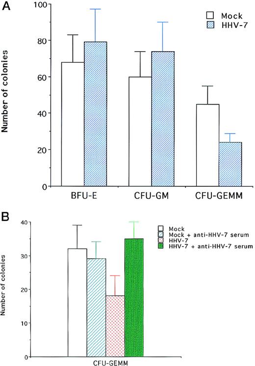
The number of colonies was scored 14 days following infection (Figure 1). Although no significant differences (P > .1) were observed in lineage-committed progenitors (CFU-GM and BFU-E) between HHV-7–infected and mock-infected cultures, pluripotent CFU-GEMM progenitors were significantly (P < .01) reduced by HHV-7 (Figure 1A). The specificity of HHV-7–mediated inhibition of CFU-GEMM was demonstrated by the fact that the number of BFU-E, obtained and scored in the same plates of CFU-GEMM, was comparable in HHV-7–infected and mock-infected cultures.
Effect of HHV-7 on CB CD34–derived clonogenic progenitors.
Data are expressed as means ± SD of 3 to 7 separate experiments performed in triplicate. (A) The number of BFU-E, CFU-GM, and CFU-GEMM, scored after 14 days of semisolid cultures in HHV-7–infected and mock-infected cultures. (B) The effect of pretreatment of HHV-7 inoculum with anti–HHV-7 neutralizing serum on the number of CFU-GEMM.
We have considered the possibility that the reduction of CFU-GEMM colonies observed in HHV-7–inoculated cultures could be due to the presence of cytokines in the viral inoculum. However, both mock and HHV-7 inocula showed undetectable levels of a series of cytokines (IL-1β, IL-6, IL-15, IL-3, GM-CSF, G-CSF, tumor necrosis factor α) analyzed by enzyme-linked immunosorbent assay. Moreover, in further experiments performed by incubating the HHV-7 and mock inocula with anti–HHV-7 human neutralizing serum, the ability of HHV-7 to inhibit the formation of CFU-GEMM was completely reverted (P < .01) (Figure 1B). On the other hand, anti–HHV-7 serum did not show any significant effect on BFU-E progenitors in both HHV-7–infected and mock-infected cultures (data not shown). Taken together, these results indicated that the decrease of CFU-GEMM was directly due to HHV-7 virions rather than to soluble cellular components contaminating the viral inoculum.
Perturbation of the myeloid maturation of CD34+ cells in liquid cultures induced by HHV-7
The experiments illustrated above indicated that only the most immature hematopoietic progenitors were inhibited by HHV-7 in semisolid assays. However, it was possible that under culture conditions that promote virus–cell interaction(s), HHV-7 might affect hematopoietic maturation. Thus, in the following experiments, CB CD34+cells were inoculated with HHV-7 and then placed in liquid culture, an approach allowing secondary infection.
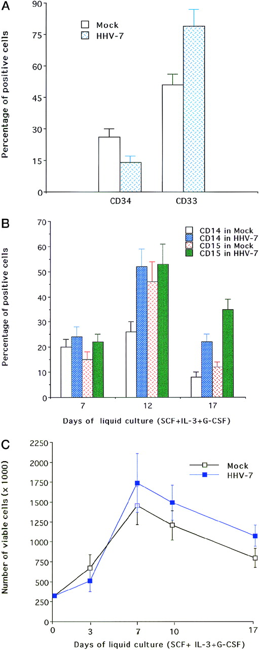
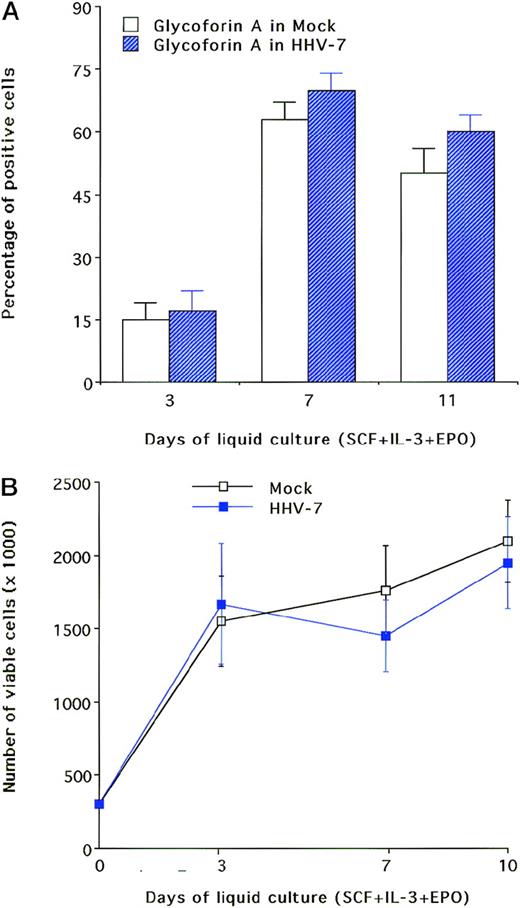
At different days of liquid cultures in the presence of SCF + IL-3 ± G-CSF, cells were harvested, counted, labeled with lineage-specific mAb, and analyzed by flow cytometry. Under these culture conditions, a progressive differentiation along the myeloid lineage was observed, as shown by the loss of CD34 antigen and by the sequential expression of CD33, CD14, and CD15 (Figure 2A, B). Flow cytometry analysis of infected versus mock-infected cultures indicated that at day 7 of liquid culture, HHV-7 induced a significant up-regulation (P < .05) of CD33 myeloid antigen coupled with a hastened down-regulation (P < .05) of CD34 (Figure2A). At later culture days, HHV-7 induced a significant (P < .05) up-regulation of late (CD14, CD15) myeloid markers compared with mock-treated cultures (Figures 2B). These phenotypic changes were not accompanied by significant (P > .1) modifications in the total number of viable cells between HHV-7–infected and mock-infected cultures (Figure 2C). CB CD34+ cells grown in the presence of SCF + IL-3 + EPO displayed a progressive erythroid differentiation, as demonstrated by monitoring of the erythroid-specific glycophorin A marker (Figure3A). The addition of HHV-7 to erythroid cultures only modestly (P > .1) affected the glycophorin A expression in these cells (Figure 3A). Also in this case, the total number of viable cells in HHV-7–infected cultures did not significantly (P > .1) differ from that in mock-infected ones (Figure 3B).
Effect of HHV-7 on CB CD34–derived granulocytic cultures.
Data are expressed as means ± SD of 4 to 6 independent experiments performed in duplicate. (A) Phenotypic analysis of surface CD34 and CD33, performed after 7 days of liquid culture supplemented with SCF + IL-3 ± G-CSF in HHV-7–infected and mock-infected cultures. The surface expression of CD34 and CD33 was analyzed by FACS and reported as the percentage of positive cells in cultures analyzed at the time indicated. (B) Phenotypic analysis of surface CD14 and CD15 performed after 7 to 14 days of liquid culture supplemented with SCF + IL-3 + G-CSF in HHV-7–infected and mock-infected cultures. The surface expression of CD14 and CD15 was analyzed by FACS and reported as the percentage of positive cells in cultures analyzed at the time indicated. (C) Viable cell counts performed at the time indicated by trypan blue dye exclusion.
Effect of HHV-7 on CB CD34–derived erythroid cultures.
Data are expressed as means ± SD of 4 to 7 independent experiments performed in duplicate. (A) Phenotypic analysis of surface glycophorin A was performed at various time points of liquid culture on erythroid cells infected or mock-infected with HHV-7. The surface expression of glycophorin A was analyzed by FACS and reported as the percentage of positive cells. (B) Viable cell counts performed at the time indicated by trypan blue dye exclusion.
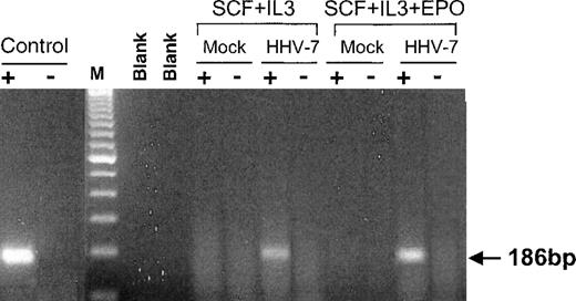
The presence of HHV-7 infection/expression in liquid cultures was examined by RT-PCR performed on 200 ng of total RNA extracted from cells at day 7 postinfection (Figure 4). Remarkably, in CD34-derived cells differentiating along the myeloid and erythroid lineages, the expected 186-bp specific HHV-7 RNA signal was found. End-point dilution experiments still showed a detectable signal, corresponding to 10 to 100 HHV-7 cDNA copies after amplification of 40 ng of total RNA (not shown).
HHV-7 expression determined by specific RT-PCR in liquid cultures.
RNA was extracted from uninfected (mock) and HHV-7–infected cultures at day 7 of culture. For RT-PCR, equivalent amounts (200 ng) of RNA samples before (−) and after (+) RT were used as template for the amplification reaction with the HV7 and HV8 primers (U10 ORF). Control indicates positive control (RNA from HHV-7–infected SupT1 cells) showing the expected amplification products of 186 bp; lane M, 100-bp ladder of molecular weight marker; blank lanes are samples without RNA. The data are representative of 3 experiments from separate infections.
Presence of HHV-7 DNA in adult BM cells
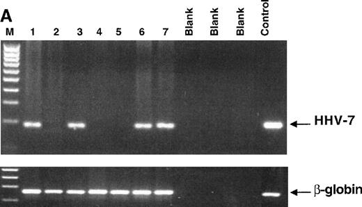
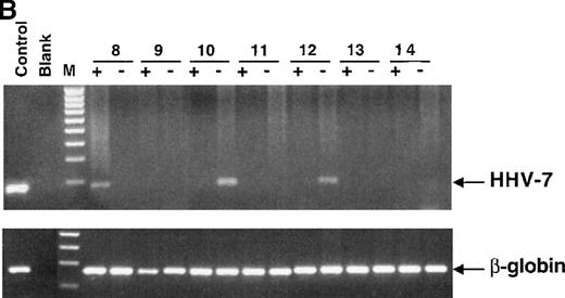
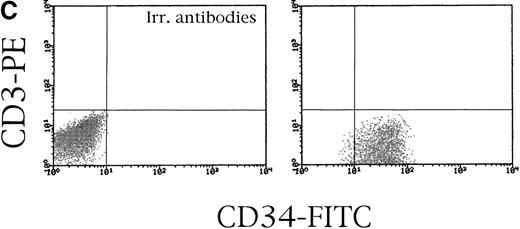
In the last group of experiments, we sought to establish whether HHV-7 is able to infect BM accessory and/or hematopoietic progenitors in vivo. DNA extracted from 14 BM specimens, which could be amplified by β-globin PCR, were analyzed for the presence of HHV-7 DNA by nested PCR. The expected 142-bp nested PCR product was amplified in 4 of 7 unfractionated BMMCs (Figure 5A). In addition, DNA was extracted from both the CD34+ and the CD34− cell fractions of 7 additional BM specimens. In 2 of 7 cases, HHV-7 DNA could be amplified from the CD34− cell population but not from the corresponding CD34+ cell fraction (Figure 5B). On the other hand, in 1 of 7 BM samples, HHV-7 was detected only in the CD34+ cell subset (Figure 5B), showing a purity of more than 97% (Figure 5C). The CD34− cell population obtained from the same donor did not show the presence of HHV-7 DNA, clearly indicating that HHV-7 DNA was present in CD34+ cells and not in potentially contaminating mature leukocytes of the CD34− fraction in this particular donor.
Nested PCR amplification of DNA extracted from different BM samples to detect HHV-7 genomic sequences.
(A) The results obtained from 7 unfractionated BMMC samples (lanes 1-7). (B) Both CD34+ and CD34− subsets (indicated with + and −, respectively) separated from each BM specimen (samples 8 to 14) have been tested. In panels A and B, the products of either the HHV-7 nested PCR (top gel) or β-globin PCR (bottom gel) are visualized by ethidium bromide staining. Blank lane indicates samples without DNA; lane M, 100-bp ladder of molecular weight marker, lane C, positive control (DNA from HHV-7–infected SupT1 cells), showing the expected amplification products of 142 bp. (C) The degree of CD34 purification (more than 97%) is shown for BM 8. CD34+ cells were double-stained with anti-CD34–FITC and anti-CD3–PE or irrelevant isotype-matched mAb.
Discussion
Although there is a consensus that β-herpesviruses, such as HCMV and HHV-6, may exert clinically important effects on the hematopoietic system,25-31,34-37 no data are available on the ability of HHV-7 to affect hematopoiesis. Mechanisms postulated to explain how HCMV and HHV-6 may suppress hematopoiesis include direct infection of progenitor cells and inhibition of colony formation,25-31decreased cytokine production associated with the infection of stromal cells,34 and diminished responsiveness of CD34+hematopoietic progenitors to growth factors.25
We have here demonstrated for the first time that HHV-7 can directly infect normal hematopoietic progenitor cells. The presence of HHV-7 DNA in 1 of 7 samples of purified CD34+ cells obtained from adult normal BM specimens is remarkable because it indicates that infection of hematopoietic progenitor cells occurs in vivo. In turn, the presence of HHV-7 DNA in the CD34− cell fraction of 2 additional donors suggests that HHV-7 can also infect either more mature hematopoietic progenitor cells, which have already lost the CD34 marker, or accessory cells composing the bone marrow microenvironment.
The potential detrimental effects of HHV-7 on hematopoiesis were 2-fold, as depicted by the experiments of in vitro infection. The first and more relevant inhibitory effect was underscored with the use of a primary colony-forming unit assay, which revealed a selective suppression of the more ancestral (CFU-GEMM) progenitors. The ability of HHV-7 to suppress CFU-GEMM is similar to the previously reported ability of type B strains of HHV-6.35 However, at variance with HHV-6, which also suppressed the growth of BFU-E and CFU-GM,35-37 HHV-7 was completely ineffective on lineage-committed progenitors. Although it has been previously shown that both human T-lymphotropic viruses, HHV-6 and HHV-7, are able to induce the production of several cytokines,35,36 38-40 we were unable to find detectable levels of IL-1β, IL-6, IL-15, IL-3, GM-CSF, or G-CSF in the inocula derived from HHV-7–infected and mock-infected SupT1 cells. Moreover, the specificity of the virus-induced effect was unequivocally demonstrated by the ability of anti–HHV-7 neutralizing serum to completely revert the HHV-7–mediated suppression of colony formation.
We have not addressed the mechanism by which HHV-7 impairs the growth of pluripotent hematopoietic progenitors (direct infection and lysis or induction of cell death). However, previous studies have demonstrated that a subset of CD34+ cells, including more ancestral progenitors, coexpresses CD420-22 and CXC–chemokine receptor 4 (CXCR4)41-44 surface antigens. In this respect, there is evidence that both CD416,17 and CXCR445 play a role in the entry of HHV-7 in mature CD4+ T-lymphoid cells. These findings suggest that HHV-7 has the potential to infect early hematopoietic progenitors and impairs their survival/proliferation.
The second detrimental effect of HHV-7 on hematopoiesis was disclosed in liquid suspension cultures in which CB CD34+ cells were induced to proliferate and differentiate along the granulocytic and erythroid lineages in the presence of an appropriate cytokine cocktail. In liquid cultures, HHV-7 did not show any gross cytopathic effect (ie, increase of cell death), but perturbed the maturation along the granulocytic lineage. This effect was reminiscent of a previously observed alteration of the cell cycle progression in HHV-7–infected mature T cells involving an unscheduled expression of the M-phase promoting factor, composed by the association of cyclin B with cdc2.18 Moreover, the RT-PCR analysis indicated that, once HHV-7 has infected hematopoietic progenitors, some of these cells are likely to survive the initial infection, and viral mRNA can be expressed in their progeny.
We believe that these findings may have important implications for groups at high risk of HHV-7 disease, including BM tranplant patients, recipients of solid organ transplants, developing fetuses, and patients with AIDS.15 In fact, it is possible that when the pressure exerted by the immune system declines, HHV-7 released from latently infected hematopoietic cells in vivo may be a source of HHV-7 dissemination and possibly of clinical disease.
Supported by the AIDS project of the Italian Ministry of Health, Telethon Foundation (grant 279/bi of P.S.), G. D'Annunzio University of Chieti local funds, and University of Ferrara local funds.
Reprints:Paola Secchiero, Department of Morphology and Embryology, Human Anatomy Section, University of Ferrara, Via Fossato di Mortara 66, 44100 Ferrara, Italy; e-mail: secchier@umbi.umd.edu.
The publication costs of this article were defrayed in part by page charge payment. Therefore, and solely to indicate this fact, this article is hereby marked “advertisement” in accordance with 18 U.S.C. section 1734.

This feature is available to Subscribers Only
Sign In or Create an Account Close Modal