Abstract
Recent studies have suggested that variations in levels of caspases, a family of intracellular cysteine proteases, can profoundly affect the ability of cells to undergo apoptosis. In this study, immunoblotting was used to examine levels of apoptotic protease activating factor-1 (Apaf-1) and procaspases-2, -3, -7, -8, and -9 in bone marrow samples (at least 80% leukemia) harvested before chemotherapy from adults with newly diagnosed acute myelogenous leukemia (AML, 42 patients) and acute lymphocytic leukemia (ALL, 18 patients). Levels of each of these polypeptides varied over a more than 10-fold range between specimens. In AML samples, expression of procaspase-2 correlated with levels of Apaf-1 (Rs = 0.52, P < .02), procaspase-3 (Rs = 0.56,P < .006) and procaspase-8 (Rs = 0.64, P < .002). In ALL samples, expression of procaspases-7 and -9 was highly correlated (Rs = 0.90,P < .003). Levels of these polypeptides did not correlate with prognostic factors or response to induction chemotherapy. In further studies, 16 paired samples (13 AML, 3 ALL), the first harvested before induction therapy and the second harvested at the time of leukemia regrowth, were also examined. There were no systematic alterations in levels of Apaf-1 or procaspases at relapse compared with diagnosis. These results indicate that levels of initiator caspases vary widely among different leukemia specimens but cast doubt on the hypothesis that this variation is a major determinant of drug sensitivity for acute leukemia in the clinical setting.
Introduction
Studies performed over the last decade indicate that chemotherapeutic agents induce apoptosis in human leukemia cell lines in vitro1-4 and in clinical leukemia in vivo.5,6 Additional experiments have suggested that failure to activate the apoptotic machinery can result in resistance to the cytotoxic effects of multiple chemotherapeutic agents.7-10 These observations raise the possibility that factors regulating the apoptotic process might play a role in drug resistance in the clinical setting.
Recent experiments have pointed to caspases, members of a unique family of intracellular cysteine proteases, as critical participants in the apoptotic process.11-15 According to current models,13-15 different caspases play 2 distinct roles during apoptosis. Effector or downstream caspases selectively cleave a small subset of cellular polypeptides.1,11,14 These cleavages destabilize certain structural components of the cell; inactivate polypeptides involved in DNA repair, replication, and transcription; and activate a limited set of intracellular enzymes, including the caspase-activated deoxyribonuclease CAD16and certain kinases,14,17 18 thereby setting into motion many of the biochemical and morphologic changes that constitute the apoptotic process. The effector caspases are in turn activated by initiator or upstream caspases, molecules that are uniquely capable of transducing various signals into proteolytic activity.
Procaspases-8 and -9, the most widely studied initiator caspases, are activated by different cellular processes.14,15Interactions between cell surface death receptors and their ligands19-21 trigger the apoptotic process by activating procaspase-8. For example, binding of CD95 ligand to CD95 results in recruitment of the adaptor molecule FADD, binding of procaspase-8 to the FADD-containing complex, and cleavage of procaspase-8 to an active form that can proteolytically activate downstream caspases.19-22 In contrast, apoptosis-inducing stimuli that cause release of cytochrome c from mitochondria23-26initiate the apoptotic process by activating caspase-9. Once released to the cytosol, cytochrome c interacts with the docking protein Apaf-1,27 causing a dATP-dependent conformational change that enables Apaf-1 to bind and activate procaspase-9,28-30 which in turn proteolytically activates procaspases-3 and -7.28 31
A growing body of evidence has suggested that levels of Apaf-1 and caspase precursors can affect sensitivity to apoptotic stimuli. Overexpression of the Drosophila caspase drICE sensitizes S2 dipteran tissue culture cells to etoposide-induced apoptosis.32Similarly, overexpression of Apaf-1 sensitizes HL-60 human leukemia cells to paclitaxel and etoposide.33 Conversely, deletion of the procaspase-9 gene slows34 or abolishes35 the triggering of apoptosis in mouse embryonic stem cells, fibroblasts, or thymocytes treated with doxorubicin, etoposide, and/or glucocorticoids. Likewise, deletion of the Apaf-1 gene renders thymocytes resistant to killing by etoposide, staurosporine, dexamethasone, and γ-irradiation.36Collectively, these observations suggest that levels of caspase-9 and/or Apaf-1 might play a critical role in determining sensitivity to various anticancer drugs.
The roles of other caspases have proven unexpectedly controversial. Some experiments have suggested that chemotherapeutic agents might also trigger apoptosis by activating the CD95 pathway.37,38Consistent with this possibility, Jurkat cells that lack procaspase-8 are less sensitive to etoposide-induced apoptosis than parental cells.39 In contrast, however, fibroblasts from mice containing a targeted deletion of the procaspase-8 gene remain fully sensitive to etoposide-induced apoptosis.40 Likewise, it has been reported that oocytes lacking procaspase-2 are resistant to doxorubicin-induced apoptosis.41 Other studies, however, have indicated that procaspase-2 is activated downstream of the principal effector caspases42 or is not even activated during chemotherapy-induced cell death.43 Finally, targeted disruption of the caspase-3 gene has been reported to diminish doxorubicin-induced apoptosis in oncogenically transformed fibroblasts44 but was not observed to have any effect on drug-induced apoptosis in thymocytes.45 Collectively, these observations raise the possibility that altered levels of some procaspases might affect drug sensitivity in a cell type-specific manner.
Evaluation of procaspase expression in clinical neoplasms has been limited. Immunohistochemical examination revealed an association between procaspase-1 expression in marrow blasts from 14 patients with AML and response to induction chemotherapy.46 Subsequent studies, however, failed to reveal a role for caspase-1 in chemotherapy-induced apoptosis.43,47 Estrov et al48 reported that high levels of procaspase-2 and procaspase-3 in peripheral blood mononuclear cells from patients with AML portended poor survival, whereas the presence of cleaved caspase-3 correlated with a favorable prognosis. In contrast, Campos et al49 observed no relationship between procaspase-2 or procaspase-3 and response of patients with AML to therapy. Levels of Apaf-1 and the initiator caspases were not previously measured in human leukemia.
In view of these limited previous studies, we have utilized immunoblotting to examine the relative expression of Apaf-1 and procaspases-2, -3, -7, -8, and -9 in bone marrow aspirates from patients with AML and ALL. These studies were designed to assess the degree of variability of these polypeptides among individual leukemia specimens, determine whether expression correlates with leukemia subtype, examine the relationship between expression of these polypeptides and response to therapy, and explore the possibility that levels of these polypeptides might change during the course of therapy.
Materials and methods
Antibodies
Monoclonal antibodies to procaspases-2, -3, and -7 were purchased from Transduction Laboratories (Lexington, KY). Monoclonal antibodies to procaspase-9 and Apaf-1,29 as well as rabbit sera that recognize procaspase-3, procaspase-8, and Bcl-2,50 51 were generated as described. Monoclonal antibodies to histone H1 and poly(ADP-ribose) polymerase (PARP) were provided by Drs James Sorace (Veteran's Affairs Medical Center, Baltimore, MD) and Guy Poirier (Laval University, Ste-Foy, Quebec, Canada), respectively. Peroxidase-coupled antibodies were from KPL (Gaithersburg, MD).
Tissue culture
HL-60 cells were cultured in RPMI 1640 medium containing 10% heat-inactivated fetal bovine serum, 100 U/mL penicillin G, 100 μg/mL streptomycin, and 2 mmol/L glutamine. To induce granulocytic maturation, cells were diluted to a density of 3 × 10⁵per milliliter, treated for the indicated length of time with 1.3% dimethylsulfoxide (DMSO),52 53 and harvested on Ficoll-Hypaque step gradients (density = 1.119 gm/cm3).
Buffers
Buffer A contained RPMI 1640 medium with 10 mmol/L HEPES (pH 7.4 at 21°C). Alkylation buffer consisted of 6 mol/L guanidine hydrochloride, 250 mmol/L Tris-HCl (pH 8.5 at 21°C), and 10 mmol/L EDTA, with 1% (vol/vol) β-mercaptoethanol and 1 mmol/L α-phenylmethylsulfonyl fluoride added to each aliquot immediately before use. Sodium dodecylsulfate (SDS) sample buffer contained 4 mol/L deionized urea, 2% (wt/vol) SDS, 62.5 mmol/L Tris-HCl (pH 6.8), and 1 mmol/L EDTA. Blocking solution contained 10% (wt/vol) powdered milk, 150 mmol/L NaCl, 10 mmol/L Tris-HCl (pH 7.4 at 21°C), 100 U/mL penicillin G, 100 μg/mL streptomycin, and 1 mmol/L sodium azide.
Sample preparation
From September 1987 through December 1992, samples were prepared from all patients with newly diagnosed acute leukemia admitted to the Adult Leukemia Service of the Johns Hopkins Hospital, except patients treated emergently at night or on weekends. In conjunction with institutional review board–approved treatment protocols, heparinized bone marrow aspirates obtained from the posterior iliac crests of these patients before the initiation of chemotherapy were prospectively prepared for SDS-polyacrylamide gel electrophoresis. Within 2 hours of aspiration, marrows were sedimented on Ficoll-Hypaque step gradients (density = 1.077 and 1.119 gm/cm3). Cells collected from the upper interface were diluted with buffer A, sedimented at 200g for 10 minutes, and resuspended in buffer A. Aliquots were removed for counting and to prepare Wright's stained cytospins for morphologic examination. Samples were then sedimented at 200g for 10 minutes and immediately solubilized in alkylation buffer.
Immunoblotting and quantitation
After sonication, samples were treated with iodoacetamide and dialyzed at 4°C into 0.1% (wt/vol) SDS as described.54Multiple separate aliquots were lyophilized to dryness and stored at −20°C. Immediately before electrophoresis, aliquots were solubilized in SDS sample buffer at a final concentration of 5 × 10⁷cell equivalents per milliliter and heated to 65°C for 20 minutes. Aliquots containing 5 × 10⁵ cells were applied to gels containing linear 5% to 15% acrylamide gradients. To provide a standard curve, aliquots containing 0.5 × 10⁵, 1.25 × 10⁵, 2.5 × 10⁵, and 5.0 × 10⁵ HL-60 cells were also applied to each gel. To insure that this standard was reproducible from gel to gel, a large aliquot of HL-60 cells was prepared for electrophoresis as described previously and lyophilized in multiple single-use vials. After samples were transferred to nitrocellulose, blots were stained with 0.1% (wt/vol) Fast Green FCF in 50% (vol/vol) methanol-5% (vol/vol) acetic acid; treated with blocking solution for at least 6 hours at 21°C; incubated overnight with primary antibodies diluted in fresh blocking solution, washed, and reacted with peroxidase-coupled affinity-purified secondary antibodies using techniques previously described in detail.55 Bound secondary antibody was detected using enhanced chemiluminescence reagents from Amersham (Arlington Heights, IL). Signals on the resulting x-ray film were scanned on a Kodak UMax Supervista S-12 scanner, quantified (area × intensity) using NIH Image version 1.61 software, and compared with signals resulting from the serial dilution of HL-60 cells on the same blot. When the signal in the clinical sample was higher than the signal in the undiluted HL-60 cells, the relative quantity of the polypeptide in question was estimated by linear extrapolation from the highest 2 points on the HL-60 standard curve. To correct for loading differences, blots were reprobed with antibody to histone H1, a polypeptide present in constant amounts in diploid cells. The relative caspase expression values in the text represent the caspase:histone H1 ratio in the clinical sample divided by the caspase:histone H1 ratio in HL-60 cells. A value of 1.0 indicates that the average leukemia cell contains as much caspase as an average HL-60 cell; a value of 0.1 indicates that the average leukemia cell contains one tenth as much caspase as an average HL-60 cell.
Patient treatment
The vast majority of patients with newly diagnosed AML were treated on protocol 8410,56 which involved induction therapy with cytarabine (667 mg/m2 per day by continuous infusion, days 1-3), daunorubicin (DNR, 45 mg/m2 per day, days 1-3), and amsacrine (200 mg/m2 per day, days 8-10). Etoposide (400 mg/m2 per day, days 8-10) was substituted for amsacrine if there was a contraindication to amsacrine therapy or if patients received infusions of granulocyte-macrophage colony-stimulating factor (GM-CSF)56 or interleukin-3 (IL-3)57 before cytotoxic therapy. Patients who achieved a complete response (CR) received consolidation therapy on day 60 ± 7, as previously described56,57 and were then followed without further therapy until relapse. Patients with newly diagnosed ALL received induction therapy on protocol 8802, which consisted of prednisone (60 mg/m2 per day by mouth, days 1-21), vincristine (1.4 mg/m2 intravenously [IV], days 1, 8, and 15), etoposide (400 mg/m2 per day IV, days 1-3), andl-asparaginase (10 000 U/m2 per day, days 10-14), followed by cytarabine (2 gm/m2 per day continuous infusion, days 22-24) and DNR (45 mg/m2 per day, days 22-24 and 30-32).58 For purposes of this study, response of patients with ALL was assessed at recovery from cytarabine and DNR.
Patients who died before day 10 with leukemia or before day 45 with no evidence of leukemia were considered unevaluable (UE). Patients who had regrowth of leukemia or who died beyond day 45 with persistent aplasia were considered to have no response (NR). Patients who had less than 5% marrow blasts and reconstitution of normal hematopoiesis that was sustained for at least 30 days beyond discharge without intervening cytotoxic therapy were considered to have a CR.
Statistics
Correlation coefficients were calculated using Instat2 software (Graphpad Software, San Diego, CA) with a Bonferroni correction ofP values for multiple comparisons. The relative frequencies of various parameters in the analyzed subsets and the overall AML or ALL populations were compared using 2-tailed Fisher exact tests. Sample means were compared using 2-tailed unpaired ttests.
Results
Changes in expression of Apaf-1 and procaspases during granulocytic maturation of HL-60 cells
This study was undertaken to evaluate expression of Apaf-1 and procaspases in AML and ALL specimens from cohorts of patients who received relatively uniform chemotherapy at a single institution. Before studying the clinical specimens, we investigated the possibility that levels of these polypeptides might vary during granulocytic maturation. In initial experiments, HL-60 cells were treated with DMSO under conditions previously shown to induce granulocytic maturation.52,53 and harvested for immunoblotting. As indicated in Figure 1, the reagents used for this study recognized single polypeptide species of Apaf-1 and procaspases-2, -3, -7, and -9 as well as 2 previously described59 splice variants of procaspase-8. Within 24 hours of adding DMSO, procaspases-3, -7, -8, and -9 began to increase. Levels of these procaspases peaked at various times during the course of granulocytic differentiation and then declined toward (procaspases-3, -7, and -9) or even below (procaspase-8) baseline. Levels of Apaf-1 also rose 2 to 4 days after addition of DMSO and then declined. Additional analysis using the same blots revealed that Bcl-2 decreased by 50% within one day of the addition of DMSO and declined further during the course of granulocytic maturation. The DNA repair-associated protein PARP also decreased after the addition of DMSO, albeit with a different time course. In contrast, levels of 2 housekeeping proteins, histone H1 and lamin B1 did not change, providing a loading control. The patterns observed with these other polypeptides argue against the possibility that the changes observed in Apaf-1 and procaspases-3, -7, -8, and -9 are an artifact of aberrant loading or transfer. Instead, these observations raised the possibility that Apaf-1 and certain procaspases might change as cells undergo granulocytic maturation. As a result of this concern, subsequent studies were limited to leukemia samples that contained at least 80% blasts.
Alterations in Apaf-1 and procaspases during granulocytic maturation of HL-60 cells.
After cultures were treated with 1.3% DMSO for 0 to 7 days as indicated, aliquots containing 3 × 10⁵, 1.5 × 10⁵, 0.75 × 10⁵, and 0.3 × 10⁵ untreated cells (lanes 1-4, respectively) or 3 × 10⁵ treated cells (lanes 5-11) were subjected to SDS-PAGE, followed by transfer to nitrocellulose and blotting with reagents that recognize the indicated polypeptide. Note that histone H1 and lamin B1 served as loading controls.
Expression of Apaf-1 and procaspases in AML and ALL samples at diagnosis
Expression of Apaf-1 and various procaspases was examined in bone marrow aspirates from patients with acute leukemia using the same reagents. As previously described,56 58 samples were harvested from 140 patients with AML and 29 patients with ALL before initial chemotherapy. Of these 169 specimens, 60 (42 AML and 18 ALL) contained at least 80% blasts and yielded sufficient cells to allow a new set of gels to be run. These 60 specimens are compared with the original 169 specimens in Table 1. For the AML specimens, the distribution of FAB subtypes, patient age, and white blood cell count (WBC) at diagnosis were roughly equivalent. Likewise, the frequency of poor risk karyotypes, CD34 expression, and antecedent hematologic malignancies did not differ significantly. For the ALL specimens, the patient age, WBC, and frequency of T-cell phenotype were similar. Although the ALL samples analyzed in this study came from a subset of patients with a lower incidence of t(9;22) translocations and a slightly higher response to therapy, these differences did not reach statistical significance.
Comparison of clinical leukemia specimens obtained and analyzed
| . | Samples obtained . | Samples analyzed in present study . |
|---|---|---|
| AML | ||
| Number of patients | 140 | 42 |
| FAB classification | ||
| M0 | 3 | 1 |
| M1 | 17 | 9 |
| M2 | 34 | 10 |
| M3 | 11 | 1 |
| M4 | 33 | 7 |
| M5 | 17 | 8 |
| M6 | 16 | 4 |
| M7 | 9 | 2 |
| Treatment | ||
| Timed sequential Ac-D-X* | 129 | 38 |
| Individual therapy | 8 | 2 |
| No treatment | 3 | 2 |
| Age, median (range) | 52 (18-79) | 56 (18-79) |
| WBC, median (range) | 4800 (400-313 000) | 9400 (800-258 000) |
| AHD†, no. (%) | 32 (22%) | 8 (19%) |
| Secondary leukemia‡, no. (%) | 16 (11%) | 2 (5%) |
| CD34-positive, no. (%) | 85/135 (63%) | 30/42 (71%) |
| Poor risk karyotype1-153, no. (%) | 58/131 (44%) | 21/38 (55%) |
| % blasts in analyzed sample, median | 67 | 88 |
| CR with first cycle, no. (%)1-155 | 80/116 (69%) | 18/34 (53%) |
| ALL | ||
| Number of patients | 29 | 18 |
| T-cell phenotype, no. (%) | 10 (34) | 5 (28) |
| Age, median (range) | 34 (18-74) | 29 (18-74) |
| WBC × 10−3, median (range) | 26 (0.8-600) | 46 (3-312) |
| Ph1chromosome, no. (%) | 7/28 (25) | 2/17 (12) |
| % blasts, median | 85 | 88 |
| CR by day 60, no. (%)1-155 | 11/21 (52%) | 9/12 (75%) |
AML = acute myelogenous leukemia; ALL = acute lymphocytic leukemia; WBC = white blood cell count; CR = complete response.
Includes patients treated with cytarabine, daunorubicin, and amsacrine56 as well as a small number of patients treated with cytarabine, daunorubicin, and etoposide with or without priming doses of GM-CSF56 or interleukin-3.57 In cases where cytokine was administered, samples harvested before cytokine were analyzed.
AHD, antecedent hematologic disorder, ie, myelodysplastic or myeloproliferative syndrome.
Leukemia occurring after prior cytotoxic chemotherapy for a nonhematologic malignancy.
Deletions involving chromosomes 5 or 7, trisomy 8, and complex karyotypes.
Number of CR ÷ (number of CR + NR).
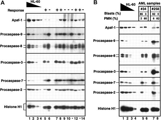
The 60 specimens were analyzed by immunoblotting as illustrated in Figure 2A. Examination of the results revealed a wide variety of expression patterns. Many specimens contained abundant levels of each of these polypeptides (eg, lanes 8, 9, 11, and 13). Some specimens, however, expressed low levels of Apaf-1 (eg, lanes 6 and 7), procaspase-9 (eg, lanes 12 and 14), or both of these polypeptides (eg, lane 5). Other specimens (eg, lane 10) expressed low levels of procaspase-3, as well as procaspase-9 and Apaf-1. Initial examination did not reveal an obvious correlation between expression of any of the polypeptides and response to induction chemotherapy (indicated by + and − signs above each lane in Figure 2A).
Variations in Apaf-1 and procaspases in various pretreatment bone marrow samples.
(A) Aliquots containing 5 × 10⁵ marrow mononuclear cells (at least 80% blasts) from patients with newly diagnosed AML (lanes 5-14) were subjected to SDS-PAGE, followed by blotting with reagents that recognize the indicated polypeptide. To provide a standard curve, aliquots containing 5 × 10⁵, 2.5 × 10⁵, 1.25 × 10⁵, and 0.5 × 10⁵ untreated HL-60 cells (lanes 1-4, respectively) were loaded onto each blot. The response of each patient to the first cycle of induction therapy is denoted above each lane as follows: − indicates NR; + indicates CR; 0 indicates UE. (B) Aliquots containing 5 × 10⁵ mononuclear cells (lanes 5 and 7) or granulocytes (lanes 6 and 8) from the same AML marrows were analyzed as in panel A. Differential counts in each sample are indicated above each lane.
Of the 42 AML specimens, 2 contained mononuclear cell fractions with more than 80% blasts and granulocyte fractions with more than 80% neutrophils. Immunoblotting (Figure 2B) demonstrated increased Apaf-1 and decreased procaspases-2 and -7 in both granulocyte samples. Procaspases-3, -8, and -9 also decreased in one granulocyte fraction. These differences between blasts and neutrophils further reinforced the decision to limit the present analysis to samples containing at least 80% blasts.
In subsequent analyses, the blots were scanned, the signal (area × intensity) was quantitated for each sample, and the results were compared with the serial dilution of HL-60 cells that was included on each blot as a positive control. To correct for variations in sample loading, the blots were probed with antibodies to histone H1, a polypeptide that is present in equal amounts in all diploid cells. Data were recorded as the polypeptide:histone H1 ratio of the sample divided by the polypeptide:histone H1 ratio of HL-60 cells. A value of 1.0 indicates that a sample contained as much polypeptide per unit histone as HL-60 cells. For purposes of comparison, we also determined that HL-60 cells contain approximately 2 × 10⁵ copies of Apaf-1, 3 × 10⁴ copies of procaspase-9, 4 × 10⁵ copies of procaspase-8, and 2 × 10⁵ copies of procaspase-3 per cell (P.A.S., Y.A.L., and S.H.K., unpublished observations).
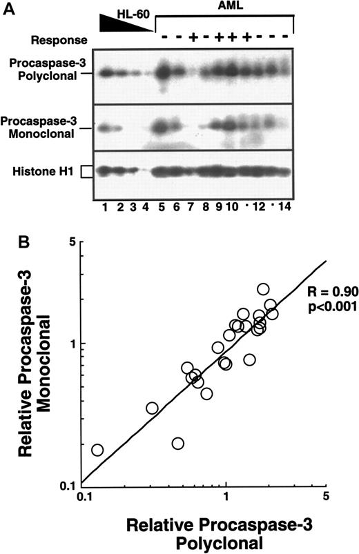
The reproducibility of this quantitation was assessed in 2 ways. First, blots containing 24 AML specimens were probed with mouse monoclonal and rabbit polyclonal antiprocaspase-3 (Figure3A). Results of this analysis revealed that both antibodies yielded qualitatively (Figure 3A) and quantitatively (Figure 3B) similar results. Second, a subset of 15 AML samples and an HL-60 cell dilution were applied to another gel 2 years after the initial analysis. After the resulting blot was probed with 4 irrelevant antibodies and erased twice, it was probed with yet another anticaspase-3 antiserum. Quantitation of the signal on this highly manipulated blot revealed correlation coefficients of ≥0.83 compared with either of the blots shown in Figure 3A. These results illustrate both the reproducibility and limitations of attempting to quantitate immunoblots.
Evaluation of the reproducibility of caspase quantitation by immunoblotting.
Duplicate blots containing AML samples and a serial dilution of HL-60 cells were prepared as described in the legend to Figure 2. (A) Results obtained when one pair of blots was probed with affinity-purified polyclonal rabbit anticaspase-3 antiserum (top) or monoclonal anticaspase-3 antibody (middle). Histone H1 (bottom) served as a loading control. Responses to therapy are indicated as described in the legend to Figure 2. (B) The blots shown in panel A, along with an additional pair of blots, were probed with both caspase-3 reagents, scanned, and quantitated as described in the “Materials and methods.” A value of 1.0 indicates that a particular leukemia sample contained as much procaspase-3 as an equal number of HL-60 cells after correction for loading by normalization of histone H1 contents.
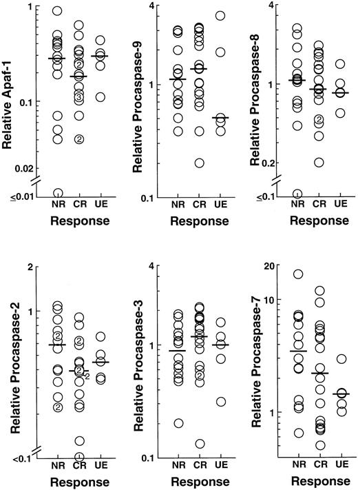
Analysis of the results obtained in newly diagnosed AML patients revealed that levels of Apaf-1 varied over 20-fold at the time of diagnosis, from 0.04 to 0.9 times the levels observed in HL-60 cells (Figure 4). There was a weak correlation (Rs = 0.52, P < .02) between levels of Apaf-1 and procaspase-2 (Table 2 and Figure5A) but no significant correlation between Apaf-1 and other caspases, patient age, WBC, FAB classification, or response to therapy (Table 2, Figure 4, and data not shown). A similar degree of variation was observed in relative expression of the 2 initiator caspases, procaspases-8 and -9 (Figure4). Although relative levels of procaspase-8 correlated with expression of procaspase-2 (Rs = 0.64,P < .002, Figure 5B), levels of the 2 initiator caspases did not correlate with prognostic factors, including age (Table2), WBC (Table 2), FAB classification (not shown), or presence of an antecedent hematologic disorder (not shown), nor did they correlate with response to therapy (Figure 4). Likewise, there was no relationship between relative levels of procaspases-2, -3, or -7 and various prognostic factors (Table 2 and data not shown) or response to therapy. Results of this analysis also failed to correlate with response to therapy when levels of Apaf-1 or various procaspases were examined without correction for histone H1 content (eg, Figure 2).
Relationship between expression of Apaf-1 or procaspase-2, -3, -7, -8, or -9 and response of previously untreated patients with AML to induction chemotherapy.
From the blots shown in Figure 2, as well as 3 additional sets of blots containing 29 more AML samples, relative levels of Apaf-1 and the indicated procaspases were quantitated. The relationship between relative polypeptide content and response of patients to the first cycle of induction chemotherapy is indicated. Bars represent median peptide levels.
Correlations between levels of Apaf-1 and procaspases-2, -3, -7, -8, and -9
| . | Apaf-1 . | Procaspase-2 . | Procaspase-3 . | Procaspase-7 . | Procaspase-8 . | Procaspase-9 . |
|---|---|---|---|---|---|---|
| AML | ||||||
| Apaf-1 | ||||||
| Caspase-2 | .52* | |||||
| Caspase-3 | .39 | .56† | ||||
| Caspase-7 | .18 | .09 | .30 | |||
| Caspase-8 | .44 | .64‡ | .39 | .25 | ||
| Caspase-9 | .43 | .41 | .42 | −.04 | .40 | |
| WBC | .22 | .02 | .09 | .01 | .24 | .18 |
| Age | −.30 | −.19 | −.26 | −.02 | −.31 | −.42 |
| ALL | ||||||
| Apaf-1 | ||||||
| Caspase-2 | .23 | |||||
| Caspase-3 | .41 | .24 | ||||
| Caspase-7 | .44 | .52 | .33 | |||
| Caspase-8 | .73 | .48 | .24 | .35 | ||
| Caspase-9 | .28 | .50 | .43 | .902-153 | .28 | |
| WBC | −.21 | −.26 | .17 | .13 | −.23 | .37 |
| Age | −.58 | .01 | −.39 | .01 | −.26 | .04 |
AML indicates acute myelogenous leukemia; ALL, acute lymphocytic leukemia; WBC, white blood cell count.
,
, and
: P < .02, .006, and .002, respectively, after correction for multiple comparisons.
P < .003 after correction for multiple comparisons.
Correlations between levels of various polypeptides in pretreatment specimens of newly diagnosed leukemia.
(A) Relationship between Apaf-1 and procaspase-2 in AML. (B) Relationship between procaspase-8 and procaspase-2 in AML. (C) Relationship between procaspase-7 and procaspase-9 in ALL.
Correlations between levels of various polypeptides in pretreatment specimens of newly diagnosed leukemia.
(A) Relationship between Apaf-1 and procaspase-2 in AML. (B) Relationship between procaspase-8 and procaspase-2 in AML. (C) Relationship between procaspase-7 and procaspase-9 in ALL.
Similar variations in expression of Apaf-1 and procaspases-2, -3, -7, -8, and -9 were observed in marrow specimens obtained from patients with newly diagnosed ALL (Figure 6). Because of limitations in sample volume, Apaf-1 and procaspases-7 and -9 could not be analyzed in a few of these samples. Nonetheless, this analysis revealed that Apaf-1, procaspase-7, and procaspase-9 varied over a 2-log range. There was a strong correlation (Rs = 0.9, P < .003) between the expression of procaspases-7 and -9 (Table 2 and Figure 5C). Levels of procaspases-2, -3, and -8 also varied over a greater than 10-fold range. Nonetheless, there was no discernible relationship between relative polypeptide levels and prognostic factors such as white count, age, or presence of the Philadelphia chromosome (Table 2 and data not shown). The overlap in expression between responders and nonresponders (Figure 6) suggested that expression patterns in the 2 groups would not be distinct, but the number of nonresponders was too small to directly assess the relationship between polypeptide expression and response to therapy.
Relationship between expression of Apaf-1 or procaspase-2, -3, -7, -8, or -9 and response of previously untreated patients with ALL to induction chemotherapy.
From blots like the one shown in Figure 2, relative levels of Apaf-1 and the indicated procaspases were quantitated. The relationship between relative polypeptide content and response of patients to induction chemotherapy is indicated.
Relative expression of Apaf-1 and procaspases at the time of leukemic relapse
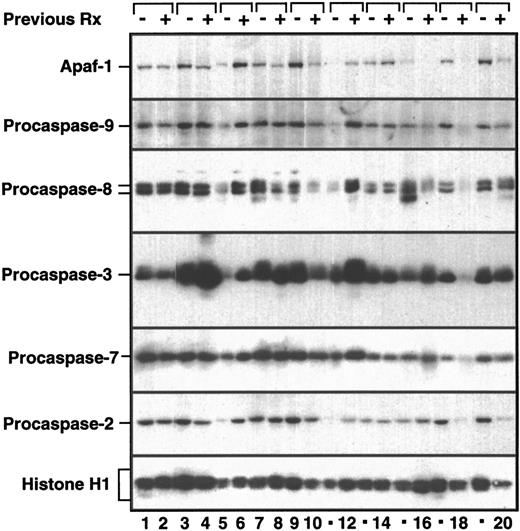
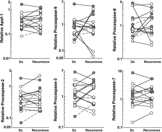
An alternative approach to determining the significance of polypeptide expression levels would be the examination of serial samples that came from individual patients before treatment and at the time of relapse. If diminished levels of a particular polypeptide (eg, Apaf-1 or procaspase-9) were responsible for resistance of leukemia cells to chemotherapy, then diminished expression of that polypeptide might be observed in samples obtained at the time of relapse. To assess this possibility, we examined the expression of Apaf-1 and procaspases-2, -3, -7, -8, and -9 in paired samples, the first harvested before induction chemotherapy and second at the time of leukemic regrowth, whether separated by a remission or not. Samples from 16 adult patients (13 AML and 3 ALL) were available for this analysis. Seven of these patients achieved a CR with their initial therapy, whereas 9 patients did not. The paired specimens were obtained a median of 196 days apart (range 22 to 380 days) and were run in adjacent wells of polyacrylamide gels. Representative immunoblots are presented in Figure 7, and the results of this analysis are summarized in Figure 8.
Comparison of Apaf-1 and procaspases in paired leukemia samples.
Aliquots containing 5 × 10⁵ marrow mononuclear cells harvested before initial chemotherapy (−, odd lanes) and again from the same patient at the time of leukemia regrowth (+, even lanes) were subjected to SDS-PAGE, followed by blotting with reagents that recognize the indicated polypeptide. To provide a standard curve, aliquots containing 5 × 10⁵, 2.5 × 10⁵, 1.25 × 10⁵, and 0.5 × 10⁵ untreated HL-60 cells were loaded onto each blot as shown in Figures 1 through 3. Blots were scanned and quantitated as described in “Materials and methods.”
Graphical representation of Apaf-1 and procaspase levels from paired samples after corrections for loading based on content of histone H1 as described in “Materials and methods.”
Analysis using this same methodology demonstrated increased levels of the apoptotic regulator Mcl-1 in over half of the same paired samples.54 Shaded circles indicate patients who achieved a CR; open circles, patients who did not achieve a CR; solid lines, AML; dotted lines, ALL.
Relative levels of Apaf-1 increased at least 2-fold in 2 of 16 paired specimens (eg, Figure 7, lanes 5 and 6) and decreased at least 2-fold in 2 specimens (Figure 7, lanes 15, 16, 19, 20). In contrast, changes were less than 2-fold in either direction in 12 of 16 specimens. Likewise, relative levels of procaspase-9 increased at least 2-fold in 3 of 16 specimens at relapse and decreased 2-fold in 3 of 16 specimens at relapse. Similarly, procaspase-8 levels increased at least 2-fold in 2 of 16 samples and decreased at least 2-fold in one sample. Collectively, these results failed to provide any evidence for systematic changes in Apaf-1 or initiator procaspase at the time of relapse. Procaspase-7 levels decreased 2-fold in one of 16 samples and did not increase 2-fold in any sample. In contrast, relative procaspase-3 levels increased at least 2-fold (eg, Figure 7, lanes 3, 4, 11, 12) in 6 of 16 samples, whereas it decreased 2-fold in only 2 of 16 pairs (Figure 8).
Discussion
Studies performed in tissue culture cell lines and in cells derived from animals after targeted gene deletion have suggested that levels of Apaf-1 or procaspases-2, -3, -8, and -9 might affect sensitivity to various agents, including anthracyclines and etoposide (see “Introduction”). On the basis of these data, we have examined the potential prognostic significance of levels of expression of these polypeptides in blasts from patients with newly diagnosed AML and ALL. To our knowledge, this study represents the first examination of Apaf-1 and procaspases-7, -8, and -9 in clinical leukemia specimens. Our study demonstrated that expression of Apaf-1 and procaspases-2, -3, -7, -8, and -9 varied widely. However, no relationship was found between the expression of any of these polypeptides and the response to induction chemotherapy. These results have important implications for current models that stress variations in components of the apoptotic machinery as potential determinants of drug sensitivity in the clinical setting.
Previous studies of caspase levels in acute leukemia cells have focused on caspases-1, -2, and -3.46,48,49,60 Results of one previous analysis suggested that elevated levels of procaspase-2 and procaspase-3 were associated with a poor prognosis in AML.48 That study examined peripheral blood mononuclear cells containing a median of 39% blasts. Subsequent investigations have suggested that procaspase-2 is activated downstream of procaspases-3 and-642 and has little effect on chemotherapy-induced apoptosis.51 Likewise, deletion of procaspase-3 causes only a mild transient delay in the induction of apoptosis by anthracycline44 and has no effect on sensitivity to other agents (T.J. Kottke, A.L. Blajeski, and S.H.K., unpublished observations).45 In this study, we did not observe any correlation between pretreatment levels of procaspase-2 or procaspase-3 and response of patients with newly diagnosed AML to induction chemotherapy (Figures 2 and 4). It should be noted, however, that the present study differs from that of Estrov et al48in several respects: (i) we examined bone marrow aspirates rather than peripheral blood mononuclear cells; (ii) we focused on samples containing at least 80% leukemia cells rather than examining samples that in some cases lacked blasts; and (iii) we examined response to induction chemotherapy rather than survival as an endpoint. One or more of these differences might account for the different conclusions of the 2 studies. While the present study was in progress, Campos et al49 reported that expression of procaspase-2 and procaspase-3 in AML blasts as assessed by flow cytometry also failed to correlate with response to induction chemotherapy.
In view of these negative results, we investigated the possibility that expression of procaspase-8, procaspase-9, or Apaf-1, molecules involved in the initiation of apoptosis, might vary among different leukemia specimens. Procaspase-8 clearly plays a role in initiating apoptosis after ligation of various death receptors.14,15,20,22,40The role of this procaspase in chemotherapy-induced apoptosis is less well defined.38 Several studies have demonstrated that procaspase-8 is activated in leukemia cell lines treated with various anticancer drugs61,62; but it appears that this might reflect cleavage of procaspase-8 downstream from other caspases rather than a caspase-8–initiated cascade.42 61 Although the present experiments revealed a 10-fold variation in procaspase-8–levels among various leukemia specimens, there was no apparent relationship between procaspase-8 expression and response to antileukemia therapy.
A number of studies have indicated that cells lacking procaspase-9 or Apaf-1 exhibit prolonged survival after exposure to chemotherapy.34-36 These effects have been observed in a variety of cell types, including embryonic stem cells, embryonic fibroblasts, and thymocytes, after exposure to a number of different agents, including glucocorticoids, epipodophyllotoxins, and anthracyclines. Because the chemotherapy administered to our patients relied heavily on the same classes of agents, we predicted that blasts from patients who failed to respond to this chemotherapy might have lower levels of procaspase-9 and/or Apaf-1 than blasts from patients who responded. Despite the strong preclinical data supporting this hypothesis, a clear-cut relationship between expression of Apaf-1 or procaspase-9 and therapeutic response was not discernible (Figures 2and 4). Further analysis revealed that relative caspase-9 levels were 24% lower in samples from patients with AML who did not respond compared with those who achieved a CR (1.30 ± 0.95 vs 1.70 ± 0.99, P > .1). Given the current sample size, a 34% decrease (to 1.13 ± 0.95) would have reached statistical significance. Even if statistical significance had been reached, however, the wide variation within each group makes it difficult to evaluate the biologic importance of a 34% difference in mean caspase-9 levels in the 2 patient populations. Moreover, the same analysis revealed that mean Apaf-1 levels were 32% higher (rather than lower) in samples from patients with AML who did not respond compared with those who did (0.29 ± 0.23 vs 0.22 ± 0.15, P > .1), suggesting that the population differences probably reflect chance variation.
As an alternative approach, Apaf-1 and procaspase-9 were examined in paired samples harvested at the time of diagnosis and recurrence (Figures 7 and 8). Although the number of paired samples with sufficient blast percentages was small, this approach previously demonstrated that the antiapoptotic Bcl-2 homolog Mcl-1 was more than 2-fold higher at the time of relapse in over half of the same pairs.54 In contrast, the present analysis indicated that Apaf-1 and procaspase-9 changed by a factor of 2 in only a small number of samples, with equal numbers showing increases or decreases (Figure8).
In evaluating the present negative study, several potential limitations must be kept in mind. First, because many of the immunologic reagents utilized in this study gave good signals by immunoblotting but not immunolocalization or flow cytometry, this study was limited to a subset of leukemia samples with a high percentage of blasts and a large enough number of cells to permit running of a new set of gels. This practical requirement might have resulted in a slightly skewed subset of leukemia specimens that underrepresents certain leukemia subgroups. Second, the levels of Apaf-1 and procaspases were measured before drug exposure. In view of recent reports that procaspase-3 expression in tissue culture cells can change after drug exposure,63 it is conceivable that a relationship between induced levels of these molecules and response was missed because of the design of the present study. Third, it is possible that an underlying relationship between caspase expression and drug sensitivity of the clonogenic leukemia cells has been obscured by analyzing the bulk leukemia population. Until a means of rapidly separating large numbers of leukemia stem cells for biochemical analysis becomes widely available, this potential limitation will apply to all studies that attempt to correlate biochemical parameters with clinical response. Fourth, the samples analyzed in the present study came from patients enrolled in phase II trials of regimens that are not widely used. It should be noted, however, that these regimens induced CRs in 65% of evaluable AML patients and 77% of evaluable ALL patients,56,58 results that are similar to those reported using other regimens in the same era. Moreover, these regimens utilized anthracyclines and epipodophyllotoxins, which are among the classes of drugs that have been shown to be affected when Apaf-1 and procaspase-9 are deleted.34-36 Although we cannot rule out the possibility that levels of Apaf-1 or procaspase-9 would have prognostic significance if a broader group of leukemia patients treated on different regimens were examined, our results raise the possibility that levels of these polypeptides might not have prognostic significance at all.
How can this tentative conclusion be reconciled with the results observed after gene deletion in mice? Two potential explanations are possible. First, it is possible that the mouse knockout data failed to adequately predict the prognostic significance of molecules such as Apaf-1 and caspase-9. In this context, it is important to note that some expression of Apaf-1 and caspase-9 was detectable in virtually every leukemia specimen (Figures 2, 4, and 6), whereas tissues from knockout mice have no expression of these molecules. It is possible that low-level expression of Apaf-1 or caspase-9 will permit cells to undergo apoptosis after treatment with chemotherapy. Second, experiments using cells from knockout animals examined cell survival over a very limited time frame.34-36 It is possible that cells deficient in Apaf-1 or caspase-9 undergo apoptosis, albeit at later time points after drug treatment.34 Indeed, given the fact that activation of caspase-9 is preceded by release of cytochrome c from mitochondria,14,15 it is possible that cells treated with chemotherapeutic agents will ultimately die even if procaspase-9 is not present or cannot be activated.64-66
On the other hand, the observation that the rate of growth factor withdrawal-induced apoptosis in AML samples in vitro correlates with response to antileukemic therapy67 suggests that inhibition of apoptosis might play an important role in the clinical setting. It is possible that other factors affecting the ability of cells to trigger apoptosis might be more important in AML and ALL than relative levels of Apaf-1 and the caspases. The present study did not assess posttranslational modifications of caspases,14especially phosphorylation,68,69 which has been reported to affect caspase activity. Likewise, variations in expression or posttranslational modification of Bcl-2 family members70might contribute to resistance of AML in the clinical setting. Finally, it is possible that variations in expression of IAPs, which are polypeptide regulators of caspase activation and activity,71 might contribute to drug resistance. Testing of these alternative hypotheses appears to be warranted.
Acknowledgments
We gratefully acknowledge the assistance of Sharon McLaughlin, Sandra Kiesewetter, Tim Soos, and Lisa Prichard in processing these specimens, as well as the assistance of Deb Strauss in preparing the manuscript.
This article is dedicated to the memory of Ken Hall, whose skillful care made possible the clinical and translational research on the Adult Leukemia Service of the Johns Hopkins Oncology Center.
Supported in part by grants CA69008 (S.H.K.), CA55164 (J.C.R.), and CA13106 (Y.A.L) from the NIH, and grants from the Seraph Foundation (Y.A.L).
The publication costs of this article were defrayed in part by page charge payment. Therefore, and solely to indicate this fact, this article is hereby marked “advertisement” in accordance with 18 U.S.C. section 1734.
References
Author notes
Scott H. Kaufmann, Division of Oncology Research, Guggenheim 1342C, Mayo Clinic, 200 First St, SW, Rochester, MN 55905; e-mail: kaufmann.scott@mayo.edu.




This feature is available to Subscribers Only
Sign In or Create an Account Close Modal