Abstract
In a Hungarian family with severe decrease in triosephosphate isomerase (TPI) activity, 2 germ line–identical but phenotypically differing compound heterozygote brothers inherited 2 independent (Phe240Leu and Glu145stop codon) mutations. The kinetic, thermodynamic, and associative properties of the recombinant human wild-type and Phe240Leu mutant enzymes were compared with those of TPIs in normal and deficient erythrocyte hemolysates. The specific activity of the recombinant mutant enzyme relative to the wild type was much higher (30%) than expected from the activity (3%) measured in hemolysates. Enhanced attachment of mutant TPI to erythrocyte inside-out vesicles and to microtubules of brain cells was found when the binding was measured with TPIs in hemolysate. In contrast, there was no difference between the binding of the recombinant wild-type and Phe240Leu mutant enzymes. These findings suggest that the missense mutation by itself is not enough to explain the low catalytic activity and “stickiness” of mutant TPI observed in hemolysate. The activity of the mutant TPI is further reduced by its attachment to inside-out vesicles or microtubules. Comparative studies of the hemolysate from a British patient with Glu104Asp homozygosity and with the platelet lysates from the Hungarian family suggest that the microcompartmentation of TPI is not unique for the hemolysates from the Hungarian TPI-deficient brothers. The possible role of cellular components, other than the mutant enzymes, in the distinct behavior of TPI in isolated form versus in hemolysates from the compound heterozygotes and the simple heterozygote family members is discussed.
Introduction
Triosephosphate isomerase (TPI) is a glycolytic enzyme that catalyzes the interconversion of D-glyceraldehyde-3-phosphate and dihydroxyacetone phosphate (DHAP) with an equilibrium of 1:20. The enzyme is a dimer, consisting of 2 identical subunits of 248 amino acids in humans.1 The x-ray crystal structures of TPIs from different sources have been determined.2 The 3-dimensional structure of recombinant human TPI complexed with the transition-state analog 2-phosphoglycerate (2-PG) was determined at a resolution of 0.28 nm (2.8 Å),2 which showed an asymmetric unit containing 1 complete dimer of 53 kd, with only 1 of the subunits binding the inhibitor. Three residues, Lys13, His95, and Glu165, are involved in the active site formation.
TPI deficiency is characterized by chronic hemolytic anemia, neurologic disturbances, and early death of homozygotes and compound heterozygotes.3,4 The metabolic block in glycolysis results in very high level of DHAP as determined in red blood cells (RBCs).4 Two compound heterozygote Hungarian brothers (both beyond 20 years) with TPI activity less than 5%5 inherited 2 separate mutations: Phe240Leu6 and Glu145stop codon7 from their mother and from their father, respectively. The propositus, the younger brother, has chronic hemolytic anemia and extrapyramidal neurodegeneration.5 The mother and father are symptom-free heterozygotes. Recently, we reported8 enhanced association of TPI from the hemolysate of the propositus to inside-out vesicles (IOVs) of erythrocytes and to brain microtubules (MTs) as compared with the normal control. We have suggested that abnormal TPI interactions may play a crucial role in the development of the neurodegeneration.8
In the present work, human wild-type (wTPI) and mutant (Phe240Leu) (mTPI) from normal and TPI-deficient RBC hemolysates were compared with recombinant wTPI and mTPI. Blood samples of a British patient with hemolytic anemia and neurologic symptoms, who was homozygous for the most common Glu104Asp mutation,9 10were also used for comparison. Our data show that the missense mutation (Phe240Leu) is not exclusively responsible either for the low TPI activity or the enhanced “stickiness” of TPI to RBC membrane or MTs.
Patients, materials, and methods
Patients
The clinical and biochemical characteristics of the TPI-deficient Hungarian family5 and the British child who died recently at the age of 59 10 were published earlier. Informed consents were obtained from the family members and the age-matched controls. Blood samples of the affected child were obtained with informed parental consent.
Materials and methods
TPI and glycerol-3-phosphate dehydrogenase from rabbit muscle, glyceraldehyde-3-phosphate, β-nicotinamide adenine dinucleotide phosphate, reduced form (NADH), paclitaxel (Taxol), 2-[N-morpholino]ethanesulfonic acid (MES), ethylene glycol-bis(β-aminoethyl ether)N, N, N′, N′-tetraacetic acid, dithioerythritol, and guanosine 5-triphosphate (GTP) were purchased from Sigma Chemical (St Louis, MO). Polyethylene glycol 6000 (PEG) was bought from Fluka AG (Buchs, Germany). All other chemicals were reagent-grade commercial preparations. Milli-Q (Millipore, Bedford, MA) ultrapure water was used for preparing the solutions.
Recombinant TPIs.
Phe240Leu mutant was obtained by site-directed mutagenesis using the QuickChange kit from Stratagene (La Jolla, CA), according to the manufacturer's instructions, using the following oligonucleotides: 5′CCTCAAGCCCGAACTCGTGGACATCATC3′ and 5′GATGATGTCCACGAGTTCGGGCTTGAG3′. The wTPI and mTPI recombinant proteins were expressed and purified as previously described.2
Tubulin and MT preparation.
MT-associated protein–free tubulin was purified from bovine brain by the method of Na and Timasheff.11 Tubulin was dialyzed in 50 mM MES buffer, pH 6.8, at 4°C for 3 hours and then centrifuged at 4°C at 100 000g for 20 minutes. The tubulin in the supernatant was polymerized by adding 20 μM paclitaxel to 10 mg/mL tubulin, followed by incubation at 37°C for 30 minutes.
IOV preparation.
Hemolysate.
Packed RBCs from control and from the propositus were prepared from washed isotonic RBC preparations by centrifugation at 1000gat 4°C for 20 minutes, and the separated cells were then lysed by diluting 3-fold into 10 mM Tris-HCl buffer, pH 7.0, containing 0.1 mM ethylenediamine tetraacetic acid and 5 mM mercaptoethanol (buffer A), followed by 3 cycles of freezing in liquid N2 and thawing.14 These lysed cells were used as hemolysate after centrifugation at 24 000g at 4°C for 25 minutes.
Lysate of platelet.
Blood was drawn into a Vacutainer collection system with sodium citrate anticoagulant solution (Becton Dickinson, Franklin Lake, NJ) and centrifuged at 300g at 25°C for 10 minutes to prepare platelet-rich plasma. The pellet of the original sample was centrifuged at 2000g for 20 minutes, and the supernatant cell-free plasma was used for the dilution of platelet-rich plasma to the final concentration of 2 × 10⁸ cell/mL. The separated cells were lysed by 3 cycles of freezing in liquid N2 and thawing and were then centrifuged at 24 000g at 4°C for 25 minutes. The supernatant was used as lysate of platelet.
Protein determination.
Routine measurements of protein concentration were done by the Bradford method.15 Hemoglobin concentration was determined spectrophotometrically using an absorption coefficient (414 nm, 0.1%) of 8.77.
DHAP level.
Enzyme activity assays.
TPI activity was measured according to Beutler17 using glyceraldehyde-3-phosphate as substrate.
Thermal inactivation measurements.
Hemolysates or purified TPIs were diluted in buffer A and were incubated at 52°C. Aliquots were withdrawn at different time intervals, and the remaining enzyme activities were assayed.
Circular dichroism.
Circular dichroism (CD) spectra were recorded with a Jasco J-720 spectropolarimeter (Tokyo, Japan) in the 190- to 250-nm (1-mm cell) and 250- to 320-nm (1-cm cell) wavelength ranges in thermostatted cuvets, diluting the proteins to 7.5 μg/mL with 10 mM phosphate buffer, pH 7.4. Spectra were corrected by subtracting spectrum of the buffer from that of the sample. The α-helical content of TPIs was calculated as described by Chen et al.18
Binding to IOVs.
A total of 100 μg IOVs was washed twice by suspension in buffer A and centrifuged at 24 000g at 4°C for 25 minutes and resuspended with 300 μL diluted hemolysates or purified TPIs using 100 mM MES buffer, pH 7.0, containing 5 mM MgCl2. The suspensions were incubated for 30 minutes at 25°C and centrifuged again as above. The pellets were resuspended in 150 μL 100 mM Tris, pH 8.0, containing 100 mM KCl, and aliquots of both the pellets and supernatants were assayed for TPI activity. The SE of the determinations was ± 15% (n = 3-4).
Binding to MTs.
Paclitaxel-stabilized MTs at a concentration of 2 mg/mL (except if otherwise stated) were incubated with 300 μL hemolysates of normal and deficient RBCs or with TPIs (mTPI, wTPI) in 100 mM MES buffer, pH 7.0, containing 5 mM MgCl2 and 20 μM paclitaxel at 37°C for 15 minutes. The samples were centrifuged (50 000g, 30 minutes, 37°C), and the pellets were resuspended in 150 μL 100 mM Tris, pH 8.0, containing 100 mM KCl. Both the pellet and supernatant fractions were analyzed by sodium dodecyl sulfate–polyacrylamide gel electrophoresis and TPI activity assay. The SE of the determinations was ± 15% (n = 3-6).
Turbidity measurements.
A total of 10 μM tubulin was assembled to MTs at 37°C in 50 mM MES buffer, pH 6.6, containing 2 mM dithioerythritol, 1 mm ethylene glycol-bis(β-aminoethyl ether)N, N, N′, N′-tetraacetic acid, and 5 mM MgCl2, adding 20 μM paclitaxel. TPIs were mixed with tubulin before starting the polymerization. Absorbance was monitored at 350 nm by a Cary 50 (Varian) spectrophotometer (Mulgrave, Australia).
Molecular dynamics.
Molecular dynamics (MD) simulations were based on the crystal structure of human recombinant TPI dimer complexed with 2-phosphoglycerate (Protein Data Bank identifier: 1hti).2 MD calculations on the truncated molecules were carried out with the Sybyl program package, version 6.6 (Tripos, St Louis, MO), with 500 ps length and 1.5 fs steps. Snapshots were taken every 0.5 ps and analyzed after 50 ps of equilibration time. Temperature was kept constant at 25°C. Calculations were performed using the all-atom implementation of the Amber force field19available in the Sybyl package. Electrostatic treatment of the systems involved a distance-dependent dielectric constant (ε = 4r).20 Substrate atoms carried their ESP charges calculated at HF6-31G* level, and Amber all-atom charges were used for amino acid residues. Because crystal structures even refined at high resolution might contain several unfavorable interactions, the crystal structure of TPI was first relaxed using 20 steps of steepest descent minimization.21 Relaxation was followed by conjugate gradient optimization utilizing the Polack-Ribiere algorithm and the convergence criteria of 0.41868 J × mol−1 × nm−1 to obtain the starting geometry for MD simulations.
Results
Enzyme activity measurements
As reported previously, the TPI activity detected in the hemolysates of the propositus and his brother is very low (< 5%) in comparison with normal controls.5,11 To explore the role of mutations in the decrease of the isomerase activity, the activity of purified recombinant wild-type and mutant enzymes were compared. The wTPIs and mTPIs were overexpressed in Escherichia coli and purified until homogeneity. Table 1 shows that the relative specific activity of the recombinant mTPI is much higher (about 30% of wTPI) than expected on the basis of the activities measured in the hemolysates (Table2), which suggests that the mutations alone cannot be responsible for the drastic reduction of the mTPI activity in the hemolysate of the brothers. The missense mutation caused slight (if any) increase in theK value of the recombinant mTPI, similarly to the data obtained with hemolysate.5
Kinetic properties of recombinant TPIs
| Enzymes . | Specific activity, U/mg . | kcat/Km, min/μM . | TPI bound to MT,*U . | Activity, % . | |||
|---|---|---|---|---|---|---|---|
| TPI added 1.8 U . | 18 U . | MT added . | |||||
| −PEG . | +PEG . | −PEG . | 0.6 mg . | 3 mg . | |||
| wTPI | 8000 | 18.6 | 0.10 | 0.23 | 0.28 | 87 | 70 |
| mTPI | 2400 | 6.3 | 0.10 | 0.27 | 0.29 | 59 | 66 |
Different amounts of recombinant TPIs were incubated with MT (2 mg/mL) in 300 μL of 100 mM MES buffer, pH 7.0, containing 5 mM MgCl2 and 20 μM paclitaxel, in the presence or absence of PEG (10%), at 37°C for 15 minutes. The samples were centrifuged (50 000 g, 30 minutes, 37°C), and the bound and free TPI was assayed enzymatically as described in “Materials and methods.”
Kinetic and binding properties of TPIs from blood cells
| . | TPI activity expected, % . | TPI activity measured in hemolysates U/g hemoglobin . | TPI activity measured in lysates of platelets U/1010 cells . | DHAP level in RBC μM RBCs . | DHAP level in platelets nmol/1010 cells . | Binding from hemolysates . | Binding from platelet lysates . | |||
|---|---|---|---|---|---|---|---|---|---|---|
| to IOV . | to MT . | to MT . | ||||||||
| −PEG, % . | +PEG, % . | −PEG, % . | +PEG, % . | −PEG, % . | ||||||
| Control | 100 | 1398 ± 361 | 181 | 11.4 ± 0.7 | 136 | 0.53 | 1.1 | 1.5 | 3.1 | 1.4 |
| Mother | 65 | 553 ± 84 | 120.7 | 19.8 | 188 | 1.3 | 2.7 | 5.9 | 7.4 | 1.9 |
| Father | 50 | 772 ± 123 | 86.8 | 23.5 | 183 | n.m. | n.m. | 2.2 | 10.8 | 1.3 |
| Brother | 15 | 40.9 ± 17.9 | 39.8 | 547 ± 53 | 163 | 6.5 | 10.2 | 5.0 | 12.0 | 6.9 |
| Propositus | 15 | 33.0 ± 16.4 | 39.7 | 701 ± 98 | 267 | 4.2 | 5.3 | 11.0 | 11.0 | 4.3 |
| British | — | 29.8 | n.m. | 935 | n.m. | 4.9 | 5.9 | 18.8 | 18.7 | n.m. |
The data are the mean of 3 to 10 measurements. SEs are given only if at least 5 different sets of experiments were carried out.
n.m. indicates not measured.
The propositus and his brother are compound heterozygotes having the Phe240Leu mutation inherited from their mother and a stop codon mutation from their father. While the missense mutation does not alter the messenger RNA level, the nonsense mutation, as in several other cases,22 results in about 10-fold reduced messenger RNA level.6 The truncated fragment (roughly half of the intact TPI monomer), expressed probably in reduced amount, is catalytically inactive because it does not have the construct of the active site residues. The residual activity of the compound heterozygotes due to the mutations can be predicted as shown in Table 2. At least 15% activity in the hemolysate of the propositus ought to be detected. The discrepancy between the enzyme activity of the recombinant mTPI and that in hemolysate suggests the presence of additional interacting factors in the hemolysates of the 2 compound heterozygote brothers.
The analysis of the hemolysates of the parents rendered it possible to search the origin of the factor(s). The measured and expected data are also shown in Table 2. The overall activity of the mother's TPI is about 40% instead of 65%, which ought to be due to the presence of completely active native plus the partially active mutant enzyme. This value in the case of the father roughly correspond to the predicted one or even higher. The isomerase activities measured in the hemolysates of the heterozygote parents and the compound heterozygote brothers are inversely proportional with the DHAP levels of their cells (Table 2). In fact, the lowest TPI activity and highest DHAP level was measured for the propositus, and these data similarly are extremes as obtained for a British homozygote patient who carried Glu104Asp mutation (Table2). The very low TPI activity of the British homozygote patient was also coupled with very high level of DHAP.
Comparative data are presented in Table 2 for platelets. The TPI activities in the platelets of the 2 compound heterozygotes are also low as compared with their parents. Their values are, however, in accordance with those expected, and DHAP levels appear to be only slightly higher than the normal ones.
Binding studies
We have recently reported that the TPI in the hemolysates from both the normal control and the propositus associate with IOVs and to MTs from brain. The binding affinity of the isomerase from the deficient cell was higher.8 To investigate whether the difference in the binding of TPI is due exclusively to the Phe240Leu mutation or additional factors contributing to the enhanced “stickiness” of mTPI, binding experiments were carried out. In one set of experiments the recombinant enzymes were preincubated with MTs, and the MTs were then pelleted to separate the free and bound enzymes as described previously for other glycolytic enzymes.23 24TPI was assayed both in the supernatants and pellets for the calculation of its partition. As shown in Table 1, the purified enzymes are bound with comparable affinities to MTs at both lower and higher enzyme concentrations, indicating that the missense mutation does not alter significantly the association of TPI with MTs.
Table 2 shows the binding data of TPI in the hemolysates from the heterozygote parents and the compound heterozygote brothers to IOVs and MTs. The most extensive binding of the TPI was found in the hemolysates from the brothers. Similar extensive association of TPI was observed in the hemolysate from a British patient with a different mutation (Glu104Asp) (Table 2). In addition, when lysate of platelets from the Hungarian family was added to MTs, the most extensive binding of TPI was also obtained with the cell lysates from the 2 compound heterozygote brothers.
PEG, as a crowding agent, enhances the macromolecular associations in a nonspecific manner25; consequently, the binding of TPI to IOVs or MTs can be enhanced or could stimulate the microcompartmentation of TPI in the cytosol. As seen in Table 2, TPI binding was significantly enhanced in the case of the normal control, the father, and the brother. However, in the case of the propositus, no further elevation in the extent of the binding of the enzyme was observed. This result shows that the TPI from the 2 compound heterozygote brothers with and without neurologic disorder behaves differently under conditions that mimic the physiologic situation. The behavior of the British patient considering the TPI microcompartmentation is similar to the Hungarian propositus; the binding of the Glu104Asp mutant is extensive and does not change virtually in the presence of PEG.
Consequences of the heteroassociations
TPI activity.
Extensive evidence accumulated indicating that the binding of some glycolytic enzymes to subcellular particles, including RBC membrane and MTs, decreases their activities (eg, phosphofructokinase, aldolase, glyceraldehyde-3-phosphate dehydrogenase26 27).
To explore how the binding of TPI affects activity of the wild-type and mutant enzymes, kinetic measurements were carried out with recombinant enzymes in the absence and presence of MTs. Table 1 shows that the addition of MTs significantly reduced the activity of both the normal and mutant enzymes; the reduction was somewhat more pronounced in the case of mTPI. A similar decrease was found in the presence of MTs when the experiments were carried out with hemolysates of control and deficient cells (data not shown). These results are in good agreement with our previous data8—namely, that the presence of IOVs decreases the activity of TPI in hemolysate. The extent of the decrease induced by the addition of RBC membrane or MTs is 50% and 35%, respectively.
MT assembly.
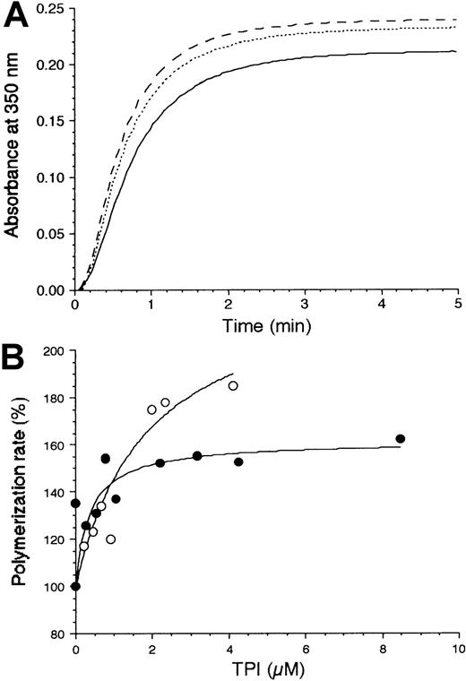
The consequences of the heteroassociation on the dynamics of MT assembly were tested by turbidity measurements. Figure1A shows typical polymerization curves of tubulin and the stimulating effect of the wTPI at 2 different enzyme concentrations. The stimulating effect of TPI is unique among the glycolytic enzymes because inhibition, no effect, or bundling have been observed so far with other enzymes.28 Similar stimulating effects on tubulin polymerization were detected by the addition of normal brain or neuroblastoma cell extracts but not with muscle cell extracts.29 Differences in the enhancement of tubulin polymerization by wild-type and mutant enzymes are documented in Figure1B. The mutant enzyme is more efficient in augmenting the MT assembly. The physiologic/pathological relevance of this phenomenon is unclear. The finding that the TPIs of hemolysates can associate with MTs in the presence of high excess of other proteins may suggest that in neuronal cell the MTs, the major component of the axon, is a target of TPI.
Effect of TPIs on tubulin polymerization kinetics.
(A) The assembly of MTs was monitored by turbidimetry at 37°C after initiating the polymerization of 10 μM tubulin by 20 μM paclitaxel alone (solid line) or in the presence of wTPI at a concentration of 0.53 μM (dotted line) or 2.12 μM (dashed line). (B) The dependence of the tubulin polymerization rate on TPI concentration is shown for mTPI (○) and wTPI (●), respectively. Data points represent the maximal slope of the turbidity traces shown in panel A, relative to the control (10 μM tubulin alone).
Stability studies
As shown above, the activity of the mTPI is reduced by heteroassociations with components present in the hemolysate or with cell membrane or MTs. Because the mutation of amino acid residues frequently leads to instability of the proteins, in the present study 2 different techniques were used to compare the heat stability of the normal and mTPIs.
Circular dichroism measurements.
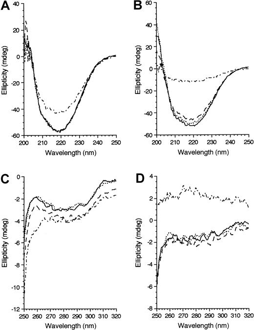
Thermal stability of recombinant TPIs was monitored by far-UV as well as by near-UV CD spectroscopy. Alteration in secondary structure due to mutation could be also detected by far-UV CD. As Figure2A and 2B show, the α-helix content of mTPI (38.4%) was slightly lower than that of wTPI (43.7%) at 30°C. The negative CD signal characteristic for the α-helix content was almost completely abolished with mTPI at 60°C, while much less reduction of α-helix content of wTPI (about 30%) was detected.
Spectra dependence.
Dependence of far-UV (A,B) and near-UV (C,D) CD spectra of recombinant wTPI (A,C) and mTPI (B,D) on temperature. Protein concentration was 7.5 μM. The spectra were recorded at 30°C (solid lines), 40°C (dotted lines), 50°C (dashed lines), and 60°C (alternating dots and dashes).
Spectra dependence.
Dependence of far-UV (A,B) and near-UV (C,D) CD spectra of recombinant wTPI (A,C) and mTPI (B,D) on temperature. Protein concentration was 7.5 μM. The spectra were recorded at 30°C (solid lines), 40°C (dotted lines), 50°C (dashed lines), and 60°C (alternating dots and dashes).
The near-UV CD spectra in the range of 250 to 320 nm show that at 60°C only the mTPI lost the characteristic maxima and minima detected at lower temperatures (Figure 2C,D), indicating the reduced stability of its tertiary structure.
Inactivation measurements.
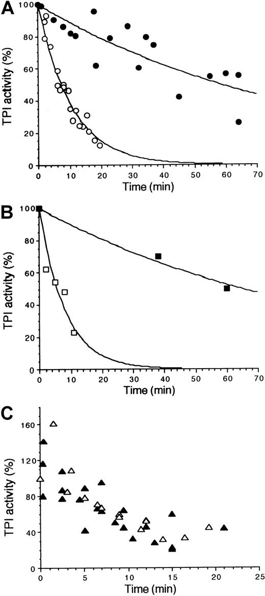
The time-dependent activity decreases of the recombinant TPIs and those in hemolysates were determined with and without MTs at 52°C. On the basis of the CD experiments, significant difference could be expected at this temperature in the heat stability of wTPIs and mTPIs. As shown in Figure 3, the activity of the mTPI was dramatically reduced, as compared with that of wTPI, independently of whether the enzymes were in purified form (Figure 3A) or in hemolysates (Figure 3B). By fitting the experimental points with a single exponential, the t0.5 values were estimated as 51.1 ± 7.3 minutes or 63.5 ± 5.8 minutes for the wTPIs and 7.0 ± 0.4 minutes or 6.3 ± 1.4 minutes for mTPIs for purified enzymes and hemolysates, respectively. Therefore, the stability of the mutant enzyme was significantly reduced (t0.5 values were decreased) independently of the presence of other enzymes in the hemolysate.
Thermal stability of TPI at 52°C detected by enzyme activity. Effect of MTs and IOVs on the stability of mTPI.
Decays were fitted by single exponentials as shown by solid lines. (A) Recombinant mTPI (○) and wTPI (●), characterized by t0.5 = 7.0 ± 0.4 minutes and t0.5 = 51.1 ± 7.3 minutes, respectively. (B) TPI activities of hemolyzates from the propositus (■) and control (▪), characterized by t0.5 = 6.3 ± 1.4 minutes and t0.5 = 63.5 ± 5.8 minutes, respectively. (C) Activity of mTPI in the presence of MTs (▵) or IOVs (▴); activity is the reduced activity of mTPI due to the binding to MTs or IOVs.
In another set of experiments, the effect of TPI's binding to either IOVs or MTs was assayed. While the slow inactivation of the recombinant wTPI was further reduced by the binding to IOVs or MTs (data not shown), more complex (not a single exponential) curves were obtained with mTPI (Figure 3C). The complex character of the binding/inactivation processes may be indicative of the heat-sensitive binding of TPI to the subcellular particles.
MD simulations on truncated heterodimeric TPI
The possibility that the presence of the truncated peptide, occurring likely in reduced amount in the deficient cells of the Hungarian compound heterozygotes, could cause the unexpected decrease of enzyme activity via formation of a heterodimeric TPI was tested in silico. To predict whether this heterodimer is a stable species, MD modeling has been carried out based upon the crystallographic data of human recombinant wTPI.2
The x-ray crystal structure of the recombinant human TPI complexed with the transition-state analog 2-PG showed an asymmetric unit containing a complete dimer of 53 kd, with only one of the subunits binding the 2-PG. The flexible loop, comprising the residues 168 to 175, is in its “closed” conformation in the subunit that binds 2-PG (M-2) and in the “open” conformation in the other subunit without 2-PG (M-1). Two situations when M-1 or M-2 associated with the truncated fragment were modeled by means of MD calculations. In the first run (MD1) the N-terminal 144 amino acid residues fragment of TPI (all other residues were deleted) is complexed with M-1, and in the second run (MD2) the 144-fragment binds to M-2.
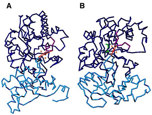
Both MD runs converged to an equilibrium state, and the structures averaged on equilibrated trajectories were used for further analysis. The position and the 3-dimensional structure of the flexible loop show differences depending on the presence or absence of 2-PG within the monomer. Therefore, the constellation of this loop was considered as a “sensor” to monitor the conformational alteration that occurred due to the truncation of one of the monomers. The distances of 2 residues of the flexible loop (Gly171 or Thr172) from the Ala73 localized in the loop 3 of the other monomer (truncated fragment), which is extensively involved in the formation of the contact surface of the 2 subunits, were determined when the intact monomer does (closed) or does not (open) bind 2-PG. Our calculations showed that the distances of the Cα atoms of Ala73 and Gly171 or Thr172 (compare Figure 4and Table 3) are longer in the open than in the closed conformers, similarly to the native dimer. Therefore, the flexible loop movement that exists in the case of the native dimer also exists in the artificial heterodimer, which maintains its substrate binding ability.
MD structures of the truncated heterodimers.
The intact monomer is blue, the truncated one is cyan, the substrate is green, and the flexible loop is magenta. (A) MD1 calculation; the truncated monomer is complexed with an intact monomer without substrate. (B) MD2 calculation; the truncated monomer is complexed with an intact one with substrate bound. The distances between the Cα (nearest) atoms of Ala73 of the truncated monomer and Cα atoms of Gly171 or Thr172 of the loop in the intact monomer are indicated by A and B, respectively.
Distances in open and closed TPI conformers
| . | . | Open . | Closed . |
|---|---|---|---|
| MD simulation | A | 0.755 nm (7.55 Å) | 0.735 nm (7.35 Å) |
| (truncated heterodimer) | B | 1.057 nm (10.57 Å) | 0.590 nm (5.90 Å) |
| X-ray data | A | 1.142 nm (11.42 Å) | 0.839 nm (8.39 Å) |
| (native) | B | 1.508 nm (15.08 Å) | 1.214 nm (12.14 Å) |
Distances in open and closed TPI conformers between the Cα (nearest) atoms of Ala93 of one of the subunits and Cα atoms of Gly171 or Thr172 of the “flexible loop” of the other subunit are indicated by A and B, respectively.
Discussion
Many inherited glycolytic enzymopathies were found in RBCs, resulting in hemolysis and other aberrations.3 In homozygotes and compound heterozygotes of TPI deficiency, chronic hemolytic anemia is associated with progressive neurologic dysfunction and death in early childhood.30 A complete list of all known TPI mutations was recently published with the observed genotypes.4 All but one of the homozygotes reported carried the Glu104Asp mutation. Several compound heterozygotes had been identified, most in connection with Glu104Asp. The compound heterozygote Hungarian brothers with their missense (Phe240Leu) and nonsense (Glu145stop codon) mutations are unique at least from 2 aspects: (1) the elder brother is free of neurologic manifestations and (2) both, even the propositus with neurologic symptoms, are beyond 20 years.
In this paper, the catalytic, thermodynamic, as well as associative properties of recombinant wTPI and (Phe240Leu) mTPI were characterized and compared with those of TPIs in hemolysates from normal and deficient cells. The mTPI showed both in recombinant form or in hemolysate reduced thermal stability as compared with that of wTPI, which was partially retained by MTs. Similar differences in thermal stability between normal and deficient lymphocyte lysates6and hemolysates5 were reported earlier. A possible cause of the reduced heat stability is a mutation-facilitated conformational change coupled with inactivation as shown by our CD measurements.
We have pointed out in previous studies that TPI activity in hemolysate is not as low as to be responsible for the high DHAP levels and suggested that further effects may cause catalytic reduction of TPI.14 In the present work we provide evidence for the role of attachment of mTPIs to either IOVs or MTs in reducing further the catalytic activity of the enzyme. Although the binding of the mutant enzyme to MTs decreased its activity only to a slightly greater extent than observed with the wild-type enzyme, in the former case the consequence of the heteroassociation is significant because the further inactivation of the partially active mTPI, by reaching a threshold value, leads to an extremely high DHAP level. A similar mechanism could be responsible for the extremely high DHAP level in the case of the British homozygote (Glu104Asp) as well: The about 3% TPI activity is further reduced due to the attachment of the mutant enzyme to the membrane. The level of DHAP in platelets appears to be only slightly higher than the normal values. A plausible explanation for this is that while in the mature RBCs the DHAP is a dead-end product, in platelets and in most other cells it is consumed by the action of glycerol-3-phosphate dehydrogenase and the product of this reaction can be further converted. Our data seem to verify the hypothesis suggested by Schneider.4
In the crystal structure, Phe240 is not directly part of the active site2,6 but, given its localization near to the active site, it was suggested to be an essential factor in maintaining the active site geometry. Our comparative data with recombinant enzymes and hemolysates allowed us to quantify the differences originating from the replacement of Phe240 by Leu. We found that the recombinant mutant enzyme displays significantly higher activity (30%) as expected from the data obtained with the hemolysate of the propositus (3%). The propositus and his brother are compound heterozygotes, having the Phe240Leu mutation inherited from their mother and another mutant form of TPI from their father. This mutation (Glu145stop codon) results in a TPI fragment (roughly half of the intact TPI monomer), which is catalytically inactive because it does not contain all of the active site residues. Therefore, at least 15% activity in the hemolysate of the propositus ought to be detected instead of 3%. These data together with the observation that the TPI activity in the mother's hemolysate, who carries the same mutation (Phe240Leu) on one allele, measured around 40% of normal5 (Table 2) (the calculated TPI activity should be around 65%, taking into account that one allele is normal and the mutant allele [Phe240Leu] displays 30% activity) strongly suggests the presence of additional inherited or epigenetic factor(s) in the hemolysate of the TPI-deficient cells.
Interestingly, these discrepancies cannot be seen with platelets. Nevertheless, enhanced association of TPI from lysates of platelets of deficient cells was also detected.
Our MD simulation provided in silico evidence for the possibility of formation of a heterodimeric TPI, which is composed from a monomer and the truncated fragment. This artificial species, if it exists, may display reduced or no catalytic activity and enhanced associative/aggregative properties in respect to the intact, active homodimeric TPI. The evaluation of how the “stickiness” of mTPI and other factors involved in the reduction of activity and instability of TPI lead to neurodegeneration requires further investigation at the atomic, molecular, and cellular levels.
The excellent technical contributions of Mimi Nuridsány and Emma Hlavanda are gratefully acknowledged. We express our thanks to Prof D. M. Layton and Dr R. Arya for providing us a blood sample of an English patient homozygous for the Glu104Asp mutation.
Supported by grants from the Hungarian National Science Foundation OTKA (T-025291 and T-031892 to J.O., T-029924 and T-035019 to F.O., T-033138 to S.H., and T-30044 to G.M.K.), the Hungarian Ministry of Education (NKFP-1/047/2001 to J.O.), the Hungarian Medical Research Council (ETT-408 to S.H.), the James Stewardson TPI Research Trust (S.H.), and ESA-ESTEC (contract No. 12987/98/NL/VJ(IC)-Prodex to J.A.M.)
The publication costs of this article were defrayed in part by page charge payment. Therefore, and solely to indicate this fact, this article is hereby marked “advertisement” in accordance with 18 U.S.C. section 1734.
References
Author notes
Judit Ovádi, Institute of Enzymology, Biological Research Center, Hungarian Academy of Sciences, H-1518 Budapest, PO Box 7, Hungary; e-mail: ovadi@enzim.hu.


This feature is available to Subscribers Only
Sign In or Create an Account Close Modal