Abstract
Congenital afibrinogenemia is a rare coagulation disorder with autosomal recessive inheritance, characterized by the complete absence or extremely reduced levels of fibrinogen in patients' plasma and platelets. Eight afibrinogenemic probands, with very low plasma levels of immunoreactive fibrinogen were studied. Sequencing of the fibrinogen gene cluster of each proband disclosed 4 novel point mutations (1914C>G, 1193G>T, 1215delT, and 3075C>T) and 1 already reported (3192C>T). All mutations, localized within the first 4 exons of the Aα-chain gene, were null mutations predicted to produce severely truncated Aα-chains because of the presence of premature termination codons. Since premature termination codons are frequently known to affect the metabolism of the corresponding messenger RNAs (mRNAs), the degree of stability of each mutant mRNA was investigated. Cotransfection experiments with plasmids expressing the wild type and each of the mutant Aα-chains, followed by RNA extraction and semiquantitative reverse-transcriptase–polymerase chain reaction analysis, demonstrated that all the identified null mutations escaped nonsense-mediated mRNA decay. Moreover, ex vivo analysis at the protein level demonstrated that the presence of each mutation was sufficient to abolish fibrinogen secretion.
Introduction
All eukaryotes possess the ability to detect and selectively degrade messenger RNAs (mRNAs) harboring premature termination codons. It has been suggested that this mechanism, known as nonsense-mediated mRNA decay, participates in the control of gene expression by regulating the stability of selected physiological transcripts and that it modulates the phenotypic severity of a number of genetic diseases.1-3 In this respect, the relatively small size of the β-globin gene, together with the wide range of nonsense mutations associated to this locus, represented a widely used model for investigating the effects of premature termination codons on mRNA metabolism. These studies demonstrated that nonsense-mediated mRNA decay requires at least 1 intron 3′ of the nonsense codon and a minimum interval of about 50 to 55 nucleotides between the premature termination codon and the 3′-most exon-intron junction.4 5
Fibrinogen is a complex glycoprotein that consists of 2 sets of 3 different polypeptide chains (Aα, Bβ, and γ) and that participates in the final stages of the blood coagulation process.6,7 The 3 chains are encoded by 3 different genes clustered on chromosome 4q28 in the sequence γ, Aα, and Bβ.8,9 The coordinated synthesis of the 3 chains is regulated mainly at the transcriptional level.10 The lack of one fibrinogen chain has been demonstrated to be sufficient to abolish the assembly of the hexameric molecule, thus preventing fibrinogen secretion.11 Among fibrinogen congenital abnormalities, which include dysfibrinogenemia, hypofibrinogenemia, and afibrinogenemia, congenital afibrinogenemia (CAF) (Mendelian Inheritance in Man No. 202 400) refers to the complete absence of clottable plasma fibrinogen, even though trace amounts of the molecule can be usually measured by sensitive immunoassays.12 This rare disease is inherited as an autosomal recessive trait and is characterized by a mild to moderate bleeding tendency.13 Although the clinical picture of afibrinogenemia is usually not as dramatic as could be expected on the basis of complete absence of clottable fibrinogen,14severe central nervous system bleeding15,16 and recurrent abortions17 18 have been described.
Mutations in the 3 fibrinogen genes responsible for this disease have been recently described. Apart from an 11-kilobases (kb) deletion in the Aα-chain gene,19 all the other known mutations are point mutations leading to severe truncations of the corresponding polypeptide chain.20-26 The only exceptions are represented by 2 missense mutations in the Bβ-chain gene that impair fibrinogen secretion.12
In this study, 5 point mutations (4 novel and 1 previously reported) were found in 8 afibrinogenemic patients. All mutations were located within the first 4 exons of the Aα-chain gene and produced premature termination codons. Since premature termination codons are often associated with allele-specific reduction in levels of mRNA, the role of nonsense-mediated mRNA decay in the pathogenesis of CAF was investigated. The 5 truncated fibrinogen Aα-chains were also demonstrated to cause a complete lack of fibrinogen secretion when coexpressed with Bβ- and γ-polypeptides in COS-1 cells.
Materials and methods
Materials
Full-length Bβ and γ complementary DNAs (cDNAs), cloned in the mammalian expression vector pRSV-Neo, were kindly provided by Dr C. M. Redman (Lindsley Kimball Research Institute, New York Blood Center, New York, NY).27 28 Rabbit polyclonal antibodies to human fibrinogen were from Dako (Copenhagen, Denmark); restriction endonucleases from Roche (Basel, Switzerland); and oligonucleotides from Life Technologies (Inchinnan, Paisley, United Kingdom).
Blood collection and coagulation tests
Approval for this study was obtained from the Institutional Review Board of the University of Milan. All examined subjects signed appropriate informed consent before blood withdrawal. Venous blood was collected in 1:10 volume of 0.125 M trisodium citrate, pH 7.3. Plasma was obtained by blood centrifugation at 2000g for 10 minutes.
Fibrinogen was measured in plasma by a functional assay based on fibrin polymerization time with the use of a commercial kit (Laboratoire Stago, Asnières, France) and by an enzyme-linked immunosorbent assay.29 The normal range for both tests was 160 to 400 mg/dL; the sensitivities of the functional assay and of the immunoassay were 5 mg/dL and 0.02 mg/dL, respectively. The immunoassay was performed for each proband in at least triplicate on the same plasma samples.
DNA sequence analysis
Genomic DNA was extracted from whole blood by means of the Nucleon BACC1 Kit (Amersham Pharmacia Biotech, Uppsala, Sweden). Plasmid DNA was isolated by QIAprep Spin Miniprep Kit (Qiagen, Hilden, Germany). DNA sequencing was performed on both strands, either directly on purified polymerase chain reaction (PCR) products or on plasmids, by means of the BigDye Terminator Cycle Sequencing Kit and an automated Abi-310 Genetic Analyzer (Applied Biosystems, Foster City, CA). Primers used for sequencing were designed on the basis of the known sequences of the 3 fibrinogen genes and intergenic regions (GenBank accession numbers: M64982, M64983, M10014, U36478, and AF229198). Details on primers and PCR conditions will be supplied on request. Factura and Sequence Navigator softwares (Applied Biosystems) were used for mutation detection.
Haplotype analysis
Haplotype analysis was performed with the use of the following Aα-chain gene polymorphic markers: α −128 C>G and α −58 G>A polymorphisms (both single-base substitutions),30FGA-i3 microsatellite (a TCTT repeat),31 andTaqI polymorphism (a 28–base pair [bp] duplication).32 Analyses were carried out as previously described.12 30
Construction of expression vectors and mutagenesis
A region of human fibrinogen Aα-chain gene, spanning from exon 1 to intron 5, was inserted as a 5.4-kb PCR-amplified fragment into the mammalian expression vector pTARGET (Promega, Milan, Italy). This fragment was amplified from genomic DNA with the use of the primer couple FGA-Ex1-F (5′-TAGGAGCCAGCCCCACCCTAGA-3′) and FGA-In5-R (5′-GTCATGGCTCTGTACTGTTAGGCA-3′). PCR was carried out in a 50-μL reaction mixture containing 100 ng genomic DNA (from a healthy control individual), by means of the Expand Long 20kb-plus Kit (Roche), in a PTC-100 thermal cycler (MJ-Research, Watertown, MA). The sample was subjected to 10 cycles of denaturation at 94°C for 10 seconds, and elongation at 68°C for 6 minutes, followed by 20 cycles of denaturation at 94°C for 10 seconds, and elongation at 68°C for 6 minutes plus a time increment of 10 seconds for each cycle. A final extension step of 10 minutes at 68°C was added after the last cycle. This PCR product was A-tailed by a modification of the method described by Marchuck et al33 (with the use of deoxyadenosine 5′-triphosphate instead of deoxythymidine 5′-triphosphate) and was cloned with the use of the pTARGET Mammalian Expression T-Vector System Kit (Promega). The pT-Aα–wild-type (wt) recombinant plasmid was checked by sequencing. The 5 identified mutations were independently introduced in pT-Aα-wt by the QuickChange Site-Directed Mutagenesis Kit (Stratagene, La Jolla, CA), according to the manufacturer's instructions. Each mutant plasmid (pT-Aα-mut, with “mut” indicating either Gly13stop, frameshift [FS]–51stop, Ser100stop, Arg110stop, or Arg149stop mutation) was checked by sequencing.
“Fluorescent hot-stop” PCR
Quantitation of allele ratio was carried out by a modification of the “hot-stop” PCR technique34 (with the use of a fluorochrome-labeled primer instead of a radiolabeled one). Accuracy of the quantitation by the “fluorescent hot-stop” PCR was verified by performing PCR amplifications on DNA mixtures, containing known molar ratios of pT-Aα-wt and pT-Aα-Arg149stop plasmids (1:1, 1:4, 1:7, 1:9, 9:1, 7:1, and 4:1). The total amount of template was 0.001 ng in each experiment. Both the wild-type and the mutant alleles were PCR-amplified with the use of the primer couple 5′-TGTTAGAGCTCAGTTGGTTGATATGTAA-3′ (sense mutagenic primer introducing an allele-specific MaeIII restriction site) and 5′-CAGAGGTGTGGTGATGTAATG-3′ (antisense primer) under standard PCR conditions. The 309-bp–amplified product was subjected to one further amplification cycle after the addition of 20 pmol antisense primer, 5′-labeled with 6-Fam. The labeled fragments from each PCR assay were digested with MaeIII, according to the manufacturer's instructions. The mutant uncut (309 bp) and the wild-type digested (280 bp) products were separated on an Abi-310 Genetic Analyzer, and peak areas were measured by means of GeneScan Analysis software 3.1 (Applied Biosystems).
mRNA analysis
The African green monkey kidney cell line COS-1 was cultured in Dulbecco modified Eagle medium containing 10% fetal calf serum, antibiotics (100 IU/mL penicillin and 100 μg/mL streptomycin), and glutamine (1%). Cells were grown at 37°C in a humidified atmosphere of 5% CO2 and 95% air. Semiconfluent COS-1 cells were transfected with equimolar amounts (2 pmol each) of pT-Aα-wt and each of the pT-Aα-mut plasmids, by means of Cellfectin (Life Technologies). Equimolarity of cotransfected plasmids was verified by fluorescent hot-stop PCR as described above. At 5 hours after transfection, media were replaced with fresh ones, and cells were incubated for 60 additional hours. Cells were then washed twice with phosphate-buffered saline, and total RNA was extracted by means of the RNAWIZ Kit (Ambion, Austin, TX). First-strand cDNA synthesis, starting from 200 ng total RNA, was performed with the use of random nonamers and Enhanced Avian RT-PCR Kit (Sigma, St Louis, MO). We used 5 μL of 20 μL as template to PCR-amplify DNA fragments containing the corresponding mutation. A mutagenic PCR strategy enabling allele-specific restriction enzyme digestion was used to distinguish between the wild-type and mutant transcripts (Table1). All mutagenic PCRs were performed by means of the fluorescent hot-stop technique. The labeled fragments, after digestion with the appropriate restriction endonuclease (according to the manufacturer's recommendations), were detected and measured as described above.
Mutagenic polymerase chain reaction strategy and allele-specific restriction enzyme digestions used to perform messenger RNA analysis
| Amino acid change (nucleotide change) . | Primer couple* 5′.3′ . | Position† . | Exon . | PCR product (bp)‡ . | Restriction enzyme . | Fragment size wt/mut (nt)1-153 . |
|---|---|---|---|---|---|---|
| Gly13stop | F:AGATGTTTTCCATGAGGATCGTCTGCCTAGT | 29 -59 | 1 | 126 | FokI | 106+20/126 |
| (1193G>T) | R:TTTCCACAACCCTTGGGCCACGCACGC ATC | 1223 -1194 | 2 | |||
| FS51stop | F:AGATGTTTTCCATGAGGATCGTCTGCCTAGT | 29 -59 | 1 | 388/387 | BstXI | 388/269+118 |
| (1215delT) | R:CGGTTGTAGGTATTATCACGGTTATTGGCT | 3076 -1915 | 3, 4 | |||
| Ser100stop | F:GCAGGATGAAAGGGTTGATTGATGAAGTCA | 1761 -1790 | 3 | 184 | FokI | 135+44/179 |
| (1914C>G) | R:CGGTTGTAGGTTTATCACGGTTATTGG AT | 3076 -1915 | 3, 4 | |||
| Arg110stop | F:AGATGTTTTCCATGAGGATCGTCTGCCTAGT | 29 -59 | 1 | 417 | HpaII | 386+31/417 |
| (3075C>T) | R:CAATTCTGCTTCTCAGATCCTCTGACAC CC | 3105 -3076 | 4 | |||
| Arg149stop | F:TGTTAGAGCTCAGTTGGTTGATATG TAA | 3164 -3191 | 4 | 147 | MaeIII | 118+29/147 |
| (3192C>T) | R:ACCTGTTCAAGTTGCTTCTGCTG | 3895 -3873 | 5 |
PCR indicates polymerase chain reaction; bp, base pairs; wt, wild type; nt, nucleotide; mut, mutation; and FS, frameshift.
Mutated nucleotides are double-underlined.
Numbering according to the fibrinogen Aα-chain gene sequence (GenBank accession number M64982).
Product lengths refer to PCR amplifications performed on complementary DNA.
Lengths of the restriction fragments detectable by the automated DNA sequencer are underlined.
Protein analysis
Semiconfluent COS-1 cells were transfected with equimolar amounts (1.5 pmol) of pRSV-Neo-Bβ, pRSV-Neo-γ, and pT-Aα plasmids, the latter being either wild type or mutant, by means of Cellfectin. At 5 hours after transfection, media were replaced with fresh ones. At 16 hours later, the cells were fed fresh media without serum and cultured for an additional 48 hours. Conditioned media were collected and a protease inhibitor mixture was added (Complete; Roche). Then, 10 mL medium from each plate was centrifuged to remove cell debris and was concentrated with the use of a Centricon Plus-20 column (Millipore, Bedford, MA). Untransfected human hepatoma HepG2 cells, cultured as previously described,12 were used as control. The concentrated samples were analyzed by sodium dodecyl sulfate–polyacrylamide gel electrophoresis (SDS-PAGE), essentially as described by Laemmli.35 Polyacrylamide gels were electroblotted onto polyvinylidene difluoride (PVDF) membranes (Hybond-P; Amersham Pharmacia Biotech) for 1 hour at 200 mA by means of a Biometra Fast Blot (Biometra, Göttingen, Germany). PVDF membranes were soaked in a blocking buffer containing 5% skim milk in TBST (10 mM Tris-HCl pH 7.4, 150 mM NaCl, 0.05% Tween20) at 4°C for 16 hours and incubated with rabbit antihuman fibrinogen antibodies (1:1000 dilution) in TBST buffer at 4°C for 2 hours. The membranes were washed 3 times for 5 minutes with TBST buffer and incubated for 1 hour at room temperature with horseradish peroxidase–conjugated antirabbit immunoglobulin (1:1500 dilution) (Envision; Dako). After 3 washings with TBST buffer, immunoreactive bands were visualized with an enhanced chemioluminescence kit (ECLplus; Amersham Pharmacia Biotech).
Results
Patient data
Eight afibrinogenemic patients, 6 Italian, 1 Iranian, and 1 from Barbados, were studied. The Iranian patient was the only patient belonging to a family with overt consanguinity. All the patients had unmeasurable functional plasma fibrinogen levels, but very low fibrinogen levels were detected by immunoassay (ranging from 0.015 to 4.25 mg/dL) (Table 2). The patients, whose main clinical features are summarized in Table 2, did not suffer from concomitant coagulation disorders. All probands' parents were asymptomatic and had approximately half the normal fibrinogen levels, with good concordance between functional and immunologic values (data not shown).
Characteristics of patients with congenital afibrinogenemia
| Patient characteristics and treatment . | B1 . | C9 . | F5 . | I3 . | P4 . | R3 . | S2 . | S3 . |
|---|---|---|---|---|---|---|---|---|
| Patient characteristics | ||||||||
| Origin | Barbados | Cagliari | Florence | Iran | Palermo | Rome | Sassari | Sassari |
| Sex | M | M | M | M | F | M | M | M |
| Present age, y | Adult | 36 | 43 | 6 | 4 | 28 | 37 | 41 |
| Main symptoms | Hemarthroses | Posttraumatic hematomas | Mouth and neck bleeding | Hemoperitonem | Asymptomatic | Posttraumatic hematomas | Intracranial hemorrhage | Posttraumatic hematomas |
| Treatment | a | a, b, c | a | b, c | None | a | a | None |
| Plasma fibrinogen levels | ||||||||
| Functional (mg/dL) | < 5 | < 5 | < 5 | < 5 | < 5 | < 5 | < 5 | < 5 |
| Immunologic (mg/dL) | 0.041 | 1.73 | 0.037 | 4.25 | 0.015 | 1.43 | 1.2 | 0.72 |
a indicates fibrinogen concentrates; b, whole blood; and c, plasma.
Sequence analysis
Sequencing of the entire coding region as well as of the splicing junctions and of about 500 bp of the promoter region of each fibrinogen gene, performed in all probands, disclosed 5 mutations, all located within the first 4 exons of the fibrinogen Aα-chain gene. Three mutations were in the homozygous state: 1914C>G (in proband I3); 3075C>T (in probands P4 and B1); and 3192C>T (in probands C9, S2, and S3). The remaining 2 mutations were in the heterozygous state: 1193G>T (in proband F5) and 1215delT (in proband R3). In patients F5 and R3, we could not detect, in the sequenced regions, the second mutation that is expected to contribute to the observed afibrinogenemic phenotype. Southern blot analysis and long PCR amplification of each fibrinogen gene excluded the presence of gross deletions in the fibrinogen cluster (data not shown).
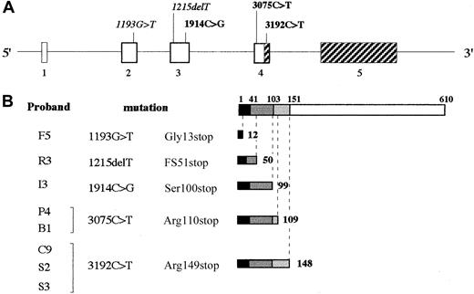
Figure 1 shows the position of the identified mutations in the Aα-chain gene, together with the predicted effects on the Aα-chain length. All mutations give rise to severely truncated Aα-chain polypeptides because of the presence of premature termination codons. In particular, 1193G>T, 1914C>G, 3075C>T, and 3192C>T nucleotide substitutions (corresponding to Gly13stop, Ser100stop, Arg110stop, and Arg149stop mutations, respectively) would determine the synthesis of truncated Aα-chain mature molecules of 12, 99, 109, and 148 amino acids, respectively. The 1215delT deletion would introduce a frameshift followed by a premature stop at codon 51 (FS51stop), preceded by an abnormal sequence of 31 amino acids.
Mutations in the fibrinogen Aα-chain gene identified in the analyzed patients with congenital afibrinogenemia.
(A) Schematic representation of the fibrinogen Aα-chain gene showing the positions of the identified mutations. Heterozygous mutations are shown in italics; homozygous mutations are shown in bold. Exons (numbered) and introns are indicated by boxes and narrow lines, respectively, and are drawn to scale. The shaded portions of exons represent the regions where a premature termination codon should not induce nonsense-mediated mRNA decay. Exon 6 of Aα-chain gene, which is present only in 2% of transcripts, has been omitted. (B) Predicted effects of the identified mutations at the protein level. Schematic representations of the fibrinogen Aα-chain (omitting the signal peptide encoded by exon 1) and of the predicted mutant truncated polypeptides are shown. Portions of Aα-chain encoded by different exons are drawn to scale and shaded in different grays. Numbers above the schematic drawing of the mature Aα-chain refer to amino acid positions corresponding to exon/exon boundaries; numbers beside the truncated polypeptides refer to their amino acid lengths.
Haplotype analysis
In order to check for the existence of a common ancestor, haplotype analysis for markers located in the Aα-chain gene was performed in probands showing the occurrence of the same mutation (Arg110stop in patients P4 and B1; Arg149stop in patients C9, S2, and S3). All analyzed probands were homozygous for each marker (Table3). Probands C9, S2, and S3 from Sardinia, a relatively isolated Italian island, shared the same haplotype. In contrast, patients P4 and B1, originating from Palermo and Barbados, respectively, showed different haplotypes.
Haplotypes of probands C9, S2, S3, P4, and B1 for markers located in the Aα-chain gene
| Marker . | Location . | Haplotype . | ||||
|---|---|---|---|---|---|---|
| C93-150 . | S23-150 . | S33-150 . | P43-151 . | B13-151 . | ||
| α − 128 C>G | promoter | C | C | C | C | C |
| α − 58 G>A | promoter | A | A | A | G | G |
| FGA-i33-152 | intron 3 | 204 | 204 | 204 | 192 | 208 |
| TaqI3-153 | 3′UTR | − 28 bp | − 28 bp | − 28 bp | − 28 bp | − 28 bp |
FGA indicates fibrinogen alpha-chain gene; bp, base pairs; UTR, untranslated region.
Probands carrying the Arg149stop mutation.
Probands carrying Arg110stop mutation.
Containing a TCTT repeat.
Owing to a 28-bp duplication; −28 bp indicates the absence of the duplication.
mRNA analysis by fluorescent hot-stop PCR
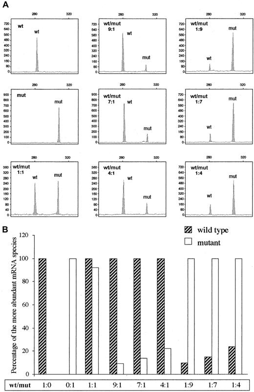
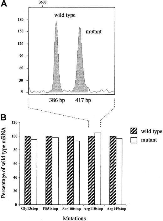
In order to evaluate mutant mRNA stability, a 5.4-kb fragment of fibrinogen Aα-chain gene, spanning from exon 1 to intron 5, was inserted into the pTARGET expression vector giving rise to the pT-Aα-wt plasmid containing the normal Aα-chain. This construct was used to produce, by site-directed mutagenesis, 5 recombinant vectors each containing one of the identified mutations. Equal amounts of plasmids expressing wild type and each mutant fibrinogen Aα mRNA were transiently cotransfected in COS-1 cells (not expressing fibrinogen). Expression levels of mutant and wild-type transcripts were compared in each experiment, using a semiquantitative reverse-transcriptase (RT)–PCR analysis. RT-PCR assays were performed with primers chosen in adjacent exons, in order to avoid the amplification of contaminating genomic DNA. Moreover, to distinguish between mRNAs transcribed from mutant and wild-type alleles, a single mismatch in one primer of each couple was introduced, thereby creating an allele-specific restriction endonuclease site (Table 1). The only exception was represented by the 1215delT mutation, whose presence creates a restriction site for BstXI enzyme. The heteroduplex formation that under these conditions usually skews the results of restriction digestion of PCR products was circumvented by performing the RT-PCR assays by means of the hot-stop PCR technique.34 This recently described assay involves the addition, at the final PCR cycle, of a radiolabeled PCR primer that excludes heteroduplexes from quantitation, since heteroduplexes do not contain the labeled primer. We used a slight modification of this technique, consisting of the use of a fluorochrome-labeled primer instead of a radiolabeled one and enabling allele quantitation by means of an automated DNA sequencer (fluorescent hot-stop PCR). This modification was first tested by 7 independent PCR assays on varying ratios of pT-Aα-wt and pT-Aα-Arg149stop plasmids (the latter containing the 5.4-kb fragment of Aα-chain gene carrying the Arg149stop mutation). Quantitation of 6-Fam–labeledMaeIII-digested PCR products (see “Materials and methods”) showed that the actual allele ratios were equal to the expected ones (Figure 2), demonstrating the accuracy and linearity of the fluorescent hot-stop PCR. This method was therefore used to quantitate the relative amount of wild-type and mutant transcripts in RNA from cells transfected with equal amounts of wild-type and mutant plasmids. A mutant–wild-type ratio of approximately 1 was estimated for each mutation (Figure3). As control, a similar experiment was performed on an aliquot of the plasmid DNA mixtures used to carry out the cotransfection experiments and resulted in a mutant–wild-type ratio of approximately 1, as expected (data not shown).
Fluorescent hot-stop PCR.
Accuracy of the quantitation of the relative abundance of 2 DNA species by the newly developed fluorescent hot-stop PCR was verified by performing a number of PCR amplifications using different molar ratios of pT-Aα-wt and pT-Aα-Arg149stop plasmids as template. Labeled PCR products were subjected to allele-specific digestion withMaeIII and quantitated on an Abi-310 Genetic Analyzer. Areas of fluorescence peaks corresponding to the mutant and wild-type restriction fragments were measured by GeneScan Analysis software 3.1. (A) GeneScan Analysis windows showing fluorescence peaks corresponding to wild-type (280 nt) and Arg149stop mutant (309 nt) single-stranded fragments. The x-axis represents GeneScan data points and the y-axis represents fluorescence units (FUs). (B) Results of the semiquantitative analysis. The reported values correspond to the peak areas, setting the more abundant mRNA species of each experiment equal to 100%.
Analysis of the relative abundance of wild-type and mutant mRNAs.
(A) Example of GeneScan analysis window showing fluorescence peaks corresponding to wild-type and mutant fragments for Arg110stop mutation. These fragments, whose lengths are indicated below each peak, were obtained by a suitable allele-specific digestion subsequent to an RT-PCR performed on RNA extracted from transfected COS-1 cells, coexpressing wild type and each mutant Aα-chain (see “Materials and methods”). The x-axis represents GeneScan data points, and the y-axis represents FUs. (B) Comparison of wild-type and mutant mRNAs levels. The reported values correspond to the peak area measured by means of GeneScan analysis software, with the wild-type mRNA level set equal to 100%.
Protein studies
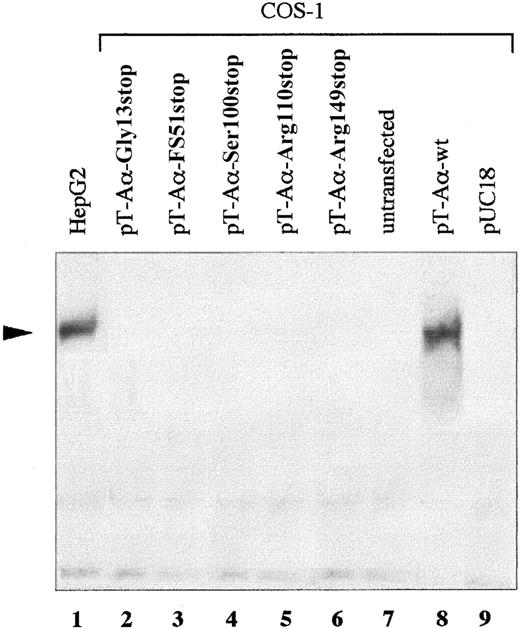
To demonstrate the causal role of the identified mutations in affecting fibrinogen secretion, each expression vector pT-Aα-mut was independently cotransfected with equimolar amounts of pRSV-Neo-Bβ and pRSV-Neo-γ plasmids (expressing the wild-type fibrinogen Bβ- and γ-chains) in COS-1 cells. As positive controls, COS-1 cells transfected with plasmids expressing the 3 wild-type chains and untransfected fibrinogen-expressing HepG2 cells were used. The presence of fibrinogen in conditioned media was checked by SDS-PAGE under nonreducing conditions, followed by Western blot analysis with the use of polyclonal antihuman fibrinogen antibodies. Secreted fibrinogen was detected only in HepG2-conditioned medium and in the supernatant of COS-1 cells expressing the 3 wild-type chains (Figure 3, lanes 1 and 8). In contrast, in culture media of cells expressing each mutant fibrinogen and in conditioned medium of mock transfected (pUC18) COS-1 cells used as negative control, the secreted molecule was undetectable (Figure 4, lanes 2-7).
Expression of wild-type and mutant fibrinogens.
Fibrinogens secreted from COS-1 cells, transfected with wild-type Bβ- and γ-chains, together with each mutant (Gly13stop, FS51stop, Ser100stop, Arg110stop, and Arg149stop) Aα-chain were analyzed as shown. Transfections, untransfected HepG2 controls, SDS-PAGE, and Western blot were carried out as described in “Materials and methods.” Proteins were separated on 4% SDS-PAGE under nonreducing conditions. The arrowhead indicates the 340-kd normal fibrinogen.
Discussion
In this study, we analyzed 8 afibrinogenemic patients, whose bleeding manifestations ranged from the complete absence of symptoms to severe intracranial hemorrhages. Sequence analysis performed on the 3 fibrinogen genes disclosed 4 nonsense mutations and 1 single-base deletion, all located in the Aα-chain gene and leading to premature translation termination. Patients carrying the same mutation (ie, Arg110stop or Arg149stop) actually showed completely different symptoms from one another. In particular, the Arg110stop mutation was found both in a patient from Barbados, who suffered from severe hemarthroses, and in a Palermitan young girl who is still asymptomatic. The Arg149stop mutation was identified in the 3 Sardinian patients, and it was associated with symptoms ranging from intracranial hemorrhage in patient S2 to posttraumatic hematomas in patients C9 and S3. The Arg149stop mutation has been very recently described in a Norwegian patient with life-threatening gastrointestinal bleeding.22Genotyping of the 3 patients carrying the Arg149stop mutation enabled demonstration of a possible common origin for this mutation, suggesting a founder effect in the isolated Sardinian community. Nevertheless, the occurrence of the same Arg149stop mutation in the Norwegian patient22 also suggests the possibility of the presence in the Aα-chain gene of a mutational “hot spot” at position 3192. The same hypothesis can be put forward for the Arg110stop mutation, since it seems to have appeared twice in 2 different alleles. It is noteworthy that both the Arg110stop and the Arg149stop mutations occurred in correspondence of a CpG dinucleotide, which is often located at hot spots for pathogenic mutations in coding DNA.36
The Gly13stop and FS51stop mutations were found, in the heterozygous state, in patients F5 and R3, respectively. In both cases, one parent (the mother of F5 and the father of R3), with half normal functional and immunoreactive plasma fibrinogen levels, was heterozygous for the same mutation present in the corresponding affected offspring. These data demonstrate that Gly13stop and FS51stop mutations are not sufficient, in the heterozygous state, to cause congenital afibrinogenemia. Both the remaining parents (the father of F5 and the mother of R3) were found to be homozygous normal at the corresponding nucleotide position. As they were phenotypically heterozygous, the existence of a second, yet undiscovered, mutation is postulated. The identity of these mutations could not be determined after mutational screening of the 3 fibrinogen genes. Possibly they are located in the intronic regions of the fibrinogen cluster not covered by sequencing.
Since premature termination codons are frequently known to affect the metabolism of the corresponding mRNAs, stability of mutant mRNAs was evaluated by coexpressing each mutant mRNA in COS-1 cells, together with the wild-type transcript, and by performing a hot-stop PCR for linear quantitation of allele ratio. This assay was modified by using a fluorochrome-labeled primer (fluorescent hot-stop PCR) and an automated DNA sequencer to separate and quantitate alleles, a simpler method than that originally proposed by Uejima et al.34 Allele quantitations demonstrated that none of the identified mutations induced a selective degradation of the corresponding mRNA. These results increase the number of cases in which the nonsense-mediated mRNA decay surveillance system is bypassed.37-40 The mechanisms by which transcripts carrying premature termination codons escape nonsense-mediated mRNA decay have not been completely elucidated. It has been demonstrated that premature termination codons located within a distance of 50 to 55 nucleotides from the 3′-most exon-intron junction do not trigger mRNA degradation by nonsense-mediated mRNA decay.1-5 This condition is fulfilled only by the Arg149stop mutation, which is due to a C>T transition at position 3192, which is 9 nucleotides upstream of the donor splice site of intron 4 (Figure 1). This exon-intron junction represents the 3′-most one for the large majority of Aα-chain transcripts, exon 6 being included only in 2% of Aα-chain mRNA molecules.41 Since only the Arg149stop mutation is expected to escape nonsense-mediated mRNA decay on the basis of the premature termination codon position, the remaining identified mutations (Gly13stop, FS51stop, Ser100stop, and Arg110stop) must use other mechanisms to avoid degradation. In some cases, nonsense mutations have been demonstrated to induce skipping of constitutive exons, which in turn interferes with the mRNA decay.37,38Another proposed mechanism involves the presence of a translation initiation codon downstream of the premature termination codon, which would determine the reinitiation of translation.39 In the fibrinogen Aα-chain gene, an AUG initiation codon is located at amino acid positions 139 to 140 (exon 4). This AUG is located downstream of the Arg110stop mutation, and is preceded by a nucleotide sequence (AGAAAA) similar to the 5′-flanking region of the canonical Aα-chain initiation codon (AGAAAAG). Possibly, this AUG may induce a reinitiation of translation, determining the nonsense-mediated mRNA decay escape of transcripts carrying Gly13stop, FS51stop, Ser100stop, and Arg110stop mutations, together with the possible production of small nonfunctional and easily degradable peptides.
Owing to the lack of mutant mRNA degradation, the effect of severely truncated Aα-chain on fibrinogen secretion was investigated. The complete absence of one fibrinogen chain was shown to abolish assembly of the hexameric molecule, preventing its secretion.11Moreover, in vitro expression of a fibrinogen molecule truncated at residue 252 of the Aα-chain has been reported to result in a 5- to 10-fold decrease in the secretion levels of the mutant fibrinogen.42 All mutations described here are expected to determine the synthesis of severely truncated Aα-chains, with all truncations affecting the coiled coil domain of the molecule (residues 45 to 165). This domain plays a key role in the initial stage of the fibrinogen assembly,43 and its deletion is likely to result in unstable fibrinogen molecules that are retained in the hepatocytes. To assess whether Gly13stop, FS51stop, Ser100stop, Arg110stop, or Arg149stop mutations could be responsible for the observed phenotype, each mutant fibrinogen was expressed in COS-1 cells. Transfection experiments demonstrated that the presence of each severely truncated fibrinogen Aα-chain is sufficient to abolish fibrinogen secretion. Interestingly, a heterozygous missense mutation in the γ-chain gene (Gly284Arg) has been demonstrated to cause retention of the mutant protein in hepatocyte inclusion bodies and to predispose to the development of chronic liver cirrhosis.44 Since 3 different morphological types of fibrinogen inclusion bodies have been described in liver biopsies, it has been postulated that different fibrinogen genetic abnormalities could produce these morphological variants in fibrinogen inclusions.45 The demonstration that fibrinogen Aα-chain null alleles do not trigger mRNA decay and that truncated Aα-chains prevent fibrinogen secretion raises the question of whether some nonsense mutations can result in an intracellular accumulation of mutant fibrinogen.
In summary, besides confirming that the majority of cases of congenital afibrinogenemia are due to Aα-chain truncations,20 our results strengthen the emerging allele heterogeneity of this disease. It was demonstrated that all identified mutations (Gly13stop, FS51stop, Ser100stop, Arg110stop, and Arg149stop) escaped nonsense-mediated mRNA decay and were responsible for an impaired secretion of the hexameric molecule, rather than for a selective degradation of the corresponding mutant mRNA.
We thank Dr C. M. Redman for kindly providing Bβ- and γ-chain–expressing plasmids. We wish to thank Dr P. L. F. Giangrande (Oxford Haemophilia Centre, The Churchill, Oxford, United Kingdom), Prof. G. Mancuso (Department of Paediatry, Haemofilia Centre, Ospedale dei Bambini, Palermo, Italy), Dr M. G. Mazzucconi (Department of Cellular Biotechnology and Hematology, Hemophilia and Thrombosis Center, Rome, Italy), and Dr M. Morfini (Department of Hematology and the Hemophilia Center, Azienda Ospedaliera Careggi, Florence, Italy) for the clinical identification of the probands, family history, and blood collection. We also thank all family members for their participation in this study.
Supported by the Ministero dell'Università e della Ricerca Scientifica e Tecnologica (MURST 60%), by Progetto Giovani (1999 and 2000), and by IRCCS Maggiore Hospital, Milan, Italy.
The publication costs of this article were defrayed in part by page charge payment. Therefore, and solely to indicate this fact, this article is hereby marked “advertisement” in accordance with 18 U.S.C. section 1734.
References
Note added in proof
Neerman-Arbez et al26 report the identification of a frameshift mutation (g1185delT) which is identical to 1215delT.
Author notes
Maria Luisa Tenchini, Department of Biology and Genetics for Medical Sciences, via Viotti, 3/5-20133 Milan, Italy; e-mail: marialuisa.tenchini@unimi.it.


This feature is available to Subscribers Only
Sign In or Create an Account Close Modal