Key Points
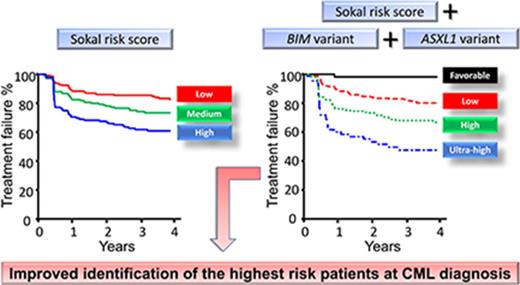
Germ line variants in ASXL1 and BIM are strong biomarkers of response to imatinib in chronic phase CML.
A combined Sokal risk and ASXL1 and BIM variant model identified a subgroup of patients with the greatest risk of treatment failure.
Abstract
Scoring systems used at diagnosis of chronic myeloid leukemia (CML), such as Sokal risk, provide important response prediction for patients treated with imatinib. However, the sensitivity and specificity of scoring systems could be enhanced for improved identification of patients with the highest risk. We aimed to identify genomic predictive biomarkers of imatinib response at diagnosis to aid selection of first-line therapy. Targeted amplicon sequencing was performed to determine the germ line variant profile in 517 and 79 patients treated with first-line imatinib and nilotinib, respectively. The Sokal score and ASXL1 rs4911231 and BIM rs686952 variants were independent predictors of early molecular response (MR), major MR, deep MRs (MR4 and MR4.5), and failure-free survival (FFS) with imatinib treatment. In contrast, the ASXL1 and BIM variants did not consistently predict MR or FFS with nilotinib treatment. In the imatinib-treated cohort, neither Sokal or the ASXL1 and BIM variants predicted overall survival (OS) or progression to accelerated phase or blast crisis (AP/BC). The Sokal risk score was combined with the ASXL1 and BIM variants in a classification tree model to predict imatinib response. The model distinguished an ultra-high-risk group, representing 10% of patients, that predicted inferior OS (88% vs 97%; P = .041), progression to AP/BC (12% vs 1%; P = .034), FFS (P < .001), and MRs (P < .001). The ultra-high-risk patients may be candidates for more potent or combination first-line therapy. These data suggest that germ line genetic variation contributes to the heterogeneity of response to imatinib and may contribute to a prognostic risk score that allows early optimization of therapy.
Introduction
The majority of chronic myeloid leukemia (CML) patients are now effectively treated using BCR-ABL1 tyrosine kinase inhibitor (TKI) therapy. The first-generation TKI imatinib induces high rates of molecular response (MR) and improved survival and has a well-documented safety profile.1,2 The second-generation TKIs nilotinib and dasatinib lead to more rapid molecular responses than imatinib and may be associated with a decreased risk of blastic transformation. However, they are associated with idiosyncratic toxicities, and there are no overall survival (OS) differences among the 3 TKIs.3,4 All 3 drugs are currently approved for first-line therapy, and the optimal agent for each individual patient is selected after balancing the risk of disease progression and expected toxicities.5,6 No current algorithm can accurately determine the risk of disease progression at diagnosis.
Time-dependent milestone molecular responses have excellent correlation with long-term outcomes regardless of the TKI used: the BCR-ABL1 value after 3, 6, or 12 months of TKI treatment can determine whether therapy has failed, which mandates an early change of treatment in an attempt to reduce the risk of disease progression and death.5,6 However, response-based molecular monitoring is limited because it does not aid selection of first-line therapy, and there is a lack of evidence that salvage approaches in this setting are effective.
Currently, clinical scoring systems at the time of CML diagnosis, such as Sokal, Hasford, European Treatment and Outcome Study (EUTOS), or European Long-Term Survival (ELTS) risk scores, provide important prognostic information, are used to stratify risk,7-10 and are integral to any risk-adapted treatment strategy.11 However, these scoring systems by themselves are of limited sensitivity and specificity. Their predictive power might be improved in combination with emerging prognostic biomarkers, which may further aid rational selection of optimal first-line treatment and increase optimal responses to TKI therapy.
Biological factors, such as a patient’s germ line genetic variation, may play a role in the kinetics of leukemic cell decline and thus affect an individual’s response to TKIs. Recently, extensive efforts have been made to identify molecular biomarkers for improved prediction of early response. Indeed, an inherited deletion polymorphism in BIM (BCL2L11), which is common in East Asians, has been reported to be associated with intrinsic imatinib resistance and inferior clinical responses, which may be overcome by more potent inhibitors.12 BIM is a pro-apoptotic member of the BCL2 intrinsic apoptosis pathway. Treatment with TKIs leads to upregulation of proapoptotic proteins, of which BIM is a key protein, and initiation of leukemic cell death.13-15 In addition, a common synonymous variant in the BH3 functional domain of BIM has been associated with inferior responses to imatinib and delayed decline of BCR-ABL1.16 Interestingly, this variant has also been associated with lower OS for children with acute lymphoblastic leukemia in which BIM mediates apoptosis of leukemic cells induced by corticosteroids.17 The variant favored the generation of an isoform that lacked the proapoptotic BH3 domain.
Germ line genetic variation in genes in other biological pathways have also been associated with response to TKI treatment, including hedgehog signaling (SMO), and pharmacogenetic processing of imatinib, such as SLC22A1 (OCT1), ABCG2, ABCB1 (MDR1), and CYP3A5.18-22 To date, none of these genetic markers of response have translated into routine clinical management of CML patients. Moreover, clinically relevant genes with prognostic relevance, such as ASXL1,23 TET2,24 RUNX1,25 IKZF1,26 and TP53,27 predict response to therapy when somatically altered in hematopoietic diseases. These genes may also harbor germ line and somatic variants that have prognostic relevance for CML patients treated with TKIs.
Next-generation sequencing (NGS) technologies now facilitate large-scale multiplexing, which allows simultaneous directed amplification and sequencing of genomic regions of interest. We used targeted NGS to investigate germ line genetic variation in BCL2 family members of the apoptotic pathway, drug metabolism genes, and genes implicated in hematologic malignancies to determine association with response to first-line imatinib and nilotinib treatment. Two germ line variants, 1 in BIM and another in ASXL1, were identified that when combined with the Sokal risk score, more precisely predicted outcome than Sokal alone.
Methods
Patients
Consecutive patients treated with first-line imatinib (n = 528) or nilotinib (n = 84) who were molecularly monitored at our laboratory were evaluated for inclusion in the study. The median follow-up (estimated by reverse Kaplan-Meier) of the patients treated with first-line imatinib or nilotinib was 45 months (range, 1-135 months) and 23 months (range, 1-25 months), respectively. This study was approved by the Royal Adelaide Hospital Human Research Ethics Committee and was conducted in accordance with the Declaration of Helsinki.
Molecular analysis
BCR-ABL1 transcript levels were measured by using real-time quantitative polymerase chain reaction, as previously published.28 Genotype analysis was performed for patients with archived samples available for testing (517 of 528 imatinib-treated patients and 79 of 84 nilotinib-treated patients). Posttreatment samples at various time points (with undetectable or minimal BCR-ABL1) were selected for molecular analysis. Somatic variation was detected by using this method; however, the predictive value of somatic variants was not assessed in this study.
Early MR (EMR) was defined as BCR-ABL1 ≤10% at 3 months. Confirmed major MR (MMR), MR4, and MR4.5 were defined as BCR-ABL1 values of ≤0.10%, ≤0.01%, and ≤0.0032% on the International Scale, respectively, at 2 consecutive measurements. The time to MMR, MR4, and MR4.5 was the duration between the start of therapy and the month of the first of the 2 consecutive measurements that met the response criteria.
The primer panel was designed by using the Ion AmpliSeq Designer. Libraries were prepared by using the Ion AmpliSeq Library Kit 2.0 and IonXpress barcodes. The Ion Proton System and Torrent Suite v4.4 were used for sequencing and variant calling, respectively (Thermo Fisher Scientific). The Sequenom iPLEX absorption, distribution, metabolism, excretion (ADME), and/or toxicity PGx Panel v1.0 (Agena Bioscience) was performed per the manufacturer’s instructions.
Development and validation of a targeted NGS panel for variant genotyping
To identify germ line variants of potential prognostic value, a custom sequencing panel of 474 amplicons targeting 39 genes (Table 1) involved in the intrinsic apoptotic pathway, drug metabolism, and hematologic malignancy was designed (supplemental Tables 1 and 2).
The genes and regions targeted by the sequencing panel
| Gene . | Region . |
|---|---|
| Intrinsic apoptosis* | |
| BCL2A1 (A1) | All exons and splice sites, 3′ and 5′ UTR regions |
| BAD | |
| BAK1 | |
| BAX | |
| BCL2 | |
| BCL2L2 (BCLW) | |
| BCL2L1 (BCLX) | |
| BCL2L11 (BIM) | |
| BMF | |
| MCL1 | |
| Hematologic malignancy* | |
| ASXL1 | Exon 12 |
| CBL | Exons 8 and 9 |
| CDKN2A | All exons |
| GATA2 | Exon 5 |
| IDH1 | Exon 4 |
| IDH2 | Exon 5 |
| IKZF1 | Exons 3-6 and 3′ UTR |
| KRAS | Exon 2 |
| NRAS | Exon 2 |
| PAX5 | Exon 3 |
| PDGFRA | Exons 12, 14, 18 |
| RB1 | All exons |
| RUNX1 | All exons |
| TET2 | All exons |
| TP53 | All exons |
| WT1 | All exons |
| Variant association* | |
| IFNG | rs2069705 |
| IL15 | rs10519613 and rs17007695 |
| MDM2 | rs2279744 |
| PTCH1 | rs41316950 |
| SMO | rs1061285 |
| Drug metabolism* | |
| ABCG2 | rs2231142 |
| CYP2C19 | rs3758581 |
| CYP3A5 | rs776746 |
| GSTM1 | rs74837985 |
| NAT2 | rs1799930 |
| SLC22A1 (OCT1) | rs628031 and rs35191146 |
| SLCO1B1 | rs4149056 |
| UGT2B7 | rs7668258 |
The genomic coordinates of the targeted regions are provided in supplemental Table 1.
Genotypes were called at variant sites with minor allele frequencies >1%, as reported by the Single Nucleotide Polymorphism Database (dbSNP) (n = 267). Strict filtering was undertaken to eliminate uninformative or poor-quality genotype data, leaving 113 variants for association testing (supplemental Table 3). To validate the genotype data, the AmpliSeq and ADME panel genotyping results for 7 variants in 150 patients which overlapped between studies were compared. Genotype calls were, on average, 99.2% (range, 98.2%-100%) concordant between the methodologies (supplemental Table 4), confirming the high quality of the genotype data.
Statistical analysis
All statistical analyses were completed in R. Linkage disequilibrium (LD) analysis was performed by sing the LDheatmap package, and tests for Hardy-Weinberg equilibrium used the HWE.chisq function from the genetics package. Haplotype blocks were determined by using HapMap data (version 3, release R2) in Haploview v4.2.29 Pearson’s χ2 test was used to assess the frequency distribution of variant genotypes between groups.
Fine and Gray models were used to assess the cumulative incidence of MRs (for which achievement of the response was the event of interest, and competing risks were any event that led to permanent cessation of first-line imatinib or nilotinib30,31 ) and also progression to accelerated phase or blast crisis (AP/BC) in which death in chronic phase was the competing risk. Associations of variables with molecular outcomes at univariate and multivariate levels were examined by using the cmprsk package in R, with significance determined by the Wald test. Akaike information criterion was used for model selection in the multivariate Fine and Gray regression.
Failure-free survival (FFS), in which failure events included lack of milestone responses at 3, 6, and 12 months; loss of MR, hematologic response, or cytogenetic response; acquisition of BCR-ABL1 mutations; clonal chromosomal abnormalities in Philadelphia chromosome cells; progression to AP/BC; death5 ; and OS were analyzed by using the Kaplan-Meier method. Both FFS and OS were measured from the date of TKI commencement. The log-rank test was used to assess the significance of differences in survival distributions. Cox proportional hazards were calculated, and proportionality assumptions were checked by using the coxph and cox.zph functions from the survival package.
Time point estimates were made at 12 months for MMR and at 48 months for MR4, MR4.5, FFS, progression to AP/BC, and OS in imatinib-treated patients and at 12 months for MMR and at 24 months for MR4, MR4.5, and FFS in nilotinib-treated patients.
Landmark analysis assessed rates of MMR, MR4, MR4.5, and FFS among patients grouped by BCR-ABL1 at 3 months (≤10% and >10%) or by classification tree risk group. Patients with the event of interest by 3 months were excluded from the landmark analyses of MMR, MR4, or MR4.5. Landmark analysis of FFS excluded patients with an event and those who were censored by 3 months.
Random forests analysis was implemented using the rfsrc function from the randomForestSRC package.32,33 The classification tree model was produced by using the rpart package in R was and pruned to optimize the prediction accuracy in 10-fold cross-validation. Bootstrap subsampling cross-validation was used to quantify the predictive discrimination of each model using the concordance index (C-index).
Variant functional prediction analysis
SNPnexus software was used to investigate the potential functional significance of the ASXL1 and BIM variants and variants in linkage disequilibrium with the risk variants.34,35 TargetScan (v7.0) prediction software was used to identify loss/gain/modification of microRNA (miRNA) binding sites at ASXL1 3′ untranslated region (UTR) sites.36 Noncoding RNA profiling by array data sets publicly available on the Gene Expression Omnibus database were analyzed by using GEO2R differential expression analysis of mature miRNAs.37,38
Results
Sokal risk score was predictive of imatinib response
Baseline predictors were assessed for prediction of EMR, time to MMR, MR4, MR4.5, FFS, progression to AP/BC, and OS. The Sokal score was the only baseline variable that consistently predicted for EMR, MMR, MR4, MR4.5, and FFS (Table 2). Female sex predicted for MMR (P = .026) and MR4 (P = .009), but not EMR, MR4.5, or FFS. BCR-ABL1 transcript type (e14a2) predicted only for MR4.5 (P = .018; Table 2). Imatinib starting dose predicted only for EMR (P = .005). None of the assessed baseline variables predicted for progression to AP/BC or OS (Table 2).
Univariate analysis of baseline factors and the ASXL1 rs4911231 and BIM rs686952 variants in imatinib-treated patients
| Factor . | No. of patients . | 3-mo EMR . | 12-mo MMR . | 48-mo MR4 . | 48-mo MR4.5 . | 48-mo FFS . | 48-mo progression to AP/BC . | 48-mo OS . | |||||||
|---|---|---|---|---|---|---|---|---|---|---|---|---|---|---|---|
| 3% . | P . | % . | P . | % . | P . | % . | P . | % . | P . | % . | P . | % . | P . | ||
| Overall response | 528 | 77 | 48 | 50 | 31 | 74 | 4 | 95 | |||||||
| Sokal risk group | <.0001 | <.0001 | .011 | .005 | <.0001 | .259 | .677 | ||||||||
| Low | 215 | 87 | 56 | 55 | 37 | 83 | 2 | 96 | |||||||
| Intermediate | 161 | 74 | 47 | 50 | 32 | 72 | 6 | 94 | |||||||
| High | 130 | 65 | 38 | 43 | 20 | 59 | 6 | 94 | |||||||
| Sex | .12 | .026 | .009 | .074 | .311 | .210 | .058 | ||||||||
| Female | 225 | 77 | 57 | 56 | 34 | 75 | 3 | 97 | |||||||
| Male | 303 | 77 | 42 | 46 | 28 | 71 | 5 | 93 | |||||||
| BCR-ABL1 transcript type | .212 | .701 | .068 | .018 | .0741 | .782 | .393 | ||||||||
| e13a2 | 198 | 79 | 43 | 47 | 25 | 78 | 4 | 96 | |||||||
| e14a2 | 229 | 76 | 54 | 54 | 38 | 73 | 3 | 95 | |||||||
| e13a2 and e14a2 | 96 | 77 | 45 | 54 | 25 | 65 | 6 | 92 | |||||||
| Median age, y | .43 | .24 | .38 | .12 | .672 | .520 | .523 | ||||||||
| ≤48 | 269 | 77 | 46 | 49 | 27 | 73 | 5 | 95 | |||||||
| >48 | 259 | 77 | 51 | 51 | 34 | 73 | 3 | 94 | |||||||
| Imatinib starting dose, mg | .005 | .682 | .088 | .264 | .054 | .797 | .164 | ||||||||
| 400 | 90 | 67 | 43 | 50 | 24 | 71 | 2 | 98 | |||||||
| 600 | 313 | 80 | 49 | 55 | 34 | 75 | 4 | 92 | |||||||
| 800 | 125 | 79 | 51 | 40 | 29 | 71 | 5 | 96 | |||||||
| ASXL1 rs4911231 genotype | .029 | .012 | .013 | <.0001 | .002 | .200 | .622 | ||||||||
| Good risk | 155 | 85 | 56 | 59 | 43 | 83 | 2 | 96 | |||||||
| Poor risk | 353 | 75 | 46 | 47 | 26 | 71 | 4 | 94 | |||||||
| BIM rs686952 genotype | .002 | <.0001 | <.0001 | <.0001 | .035 | .057 | .347 | ||||||||
| Good risk | 249 | 84 | 55 | 60 | 41 | 80 | 2 | 96 | |||||||
| Poor risk | 259 | 73 | 44 | 43 | 22 | 70 | 5 | 93 | |||||||
Identification of germ line variants important for prediction of response to imatinib
Random forests analysis was undertaken to identify germ line variants that could be combined with the Sokal score to predict response to imatinib at diagnosis. The Sokal score and 4 variants (BIM rs686952, ASXL1 rs4911231, ASXL1 rs2295763, and ASXL1 rs2295764) were the only variables that had high association with outcomes in multiple models (EMR, MMR, MR4, MR4.5, and FFS; supplemental Table 5). The 3 ASXL1 variants were in high linkage disequilibrium (r2, 0.93-0.99; supplemental Figure 2). The variant minor allele frequencies detected in the imatinib-treated cohort were BIM rs686952 (A, 28.9%), ASXL1 rs4911231 (C, 43.8%), ASXL1 rs2295763 (C, 42.2%), and ASXL1 rs2295764 (G, 42.3%), which were similar to those in the dbSNP 147 database (24.3%, 40.5%, 40%, and 40%, respectively). The marker ASXL1 variant with the strongest association with imatinib response (ASXL1 rs4911231) was chosen for subsequent analyses.
Germ line variants in BIM and ASXL1 were predictive of response to imatinib but not nilotinib
To further examine the association of BIM rs686952 and ASXL1 rs4911231 variants with outcome, patients were stratified into good- vs poor-risk groups on the basis of genotyping information in univariate analyses. The T/T genotype of ASXL1 rs4911231 and the A/A and A/C genotypes of BIM rs686952 were associated with superior achievement of MR4.5 (Figure 1), and were condensed into good-risk genotypes and compared with poor-risk genotypes for all subsequent comparisons. Univariate analysis of achievement of the MRs and FFS demonstrated that the BIM and ASXL1 good-risk variants were both significantly associated with superior achievement of MR and FFS (Table 2). Neither variant predicted for progression to AP/BC or OS (Table 2).
Good- and poor-risk genotypes were identified for the ASXL1 rs4911231 and BIM rs686952 variants. Univariate analysis of the achievement of MR4.5 as stratified by (A) the ASXL1 rs4911231 and (B) the BIM rs686952 variant genotype identified good- and poor-risk genotypes which were strongly associated with MR4.5.
Good- and poor-risk genotypes were identified for the ASXL1 rs4911231 and BIM rs686952 variants. Univariate analysis of the achievement of MR4.5 as stratified by (A) the ASXL1 rs4911231 and (B) the BIM rs686952 variant genotype identified good- and poor-risk genotypes which were strongly associated with MR4.5.
A median of 23 months of follow-up data were available for a cohort of 83 patients treated with first-line nilotinib, among whom 79 were evaluable for outcome assessment according to the BIM and ASXL1 variant genotypes. In contrast to the imatinib cohort, a marginally significant difference was detected only for MR4 for the ASXL1 variant (P = .048) and no differences were detected for the BIM variant (Table 3). There was no difference in genotype frequencies between the variant and nilotinib-treated patients for the BIM (28.9% vs 27.0%; P = .431) or the ASXL1 variant (43.8% vs 42.3%; P = .968). These results, although they were in a smaller patient cohort, suggest that the poor risk associated with the BIM and ASXL1 variants may be abrogated by treating with a more potent TKI such as nilotinib.
Univariate analysis of the ASXL1 rs4911231 and BIM rs686952 variants in nilotinib-treated patients
| Factor . | No. of patients . | 12-mo MMR . | 24-mo MR4 . | 24-mo MR4.5 . | 24-mo FFS . | ||||
|---|---|---|---|---|---|---|---|---|---|
| % . | P . | % . | P . | % . | P . | % . | P . | ||
| ASXL1 rs4911231 genotype | .28 | .048 | .31 | .537 | |||||
| Good risk | 23 | 78 | 66 | 32 | 86 | ||||
| Poor risk | 55 | 67 | 42 | 20 | 92 | ||||
| BIM rs686952 genotype | .52 | .77 | .17 | .877 | |||||
| Good risk | 33 | 70 | 45 | 31 | 91 | ||||
| Poor risk | 43 | 70 | 50 | 16 | 88 | ||||
Combining the Sokal score with the BIM and ASXL1 variants showed superior predictive power to that of Sokal alone
To assess the prognostic power of combining the BIM and ASXL1 variants with the Sokal risk group, multivariate models were developed for the prediction of EMR, MMR, MR4, MR4.5, and FFS. The Sokal risk group and the BIM and ASXL1 variants each maintained significance (P < .05) in multivariate models for the prediction of MMR, MR4, MR4.5, and FFS (Table 4), demonstrating that these variables are independent predictors of long-term response to imatinib at diagnosis. Importantly, the combination of the Sokal score with the ASXL1 and BIM variants improved prediction of imatinib response at diagnosis (supplemental Table 6).
Multivariate analysis of Sokal risk score, ASXL1 rs4911231, and BIM rs686952
| . | EMR . | MMR . | MR4 . | MR4.5 . | FFS . | |||||
|---|---|---|---|---|---|---|---|---|---|---|
| RR . | P . | RR . | P . | RR . | P . | RR . | P . | HR . | P . | |
| Sokal risk group* | ||||||||||
| Low | 1 | 1 | 1 | 1 | 1 | |||||
| Intermediate | 0.8 | .078 | 0.7 | .006 | 0.87 | .35 | 0.81 | .28 | 1.62 | .042 |
| High | 0.57 | <.001 | 0.62 | <.001 | 0.65 | .01 | 0.5 | .004 | 2.81 | <.001 |
| ASXL1 rs4911231 | ||||||||||
| Good risk | 1 | 1 | 1 | 1 | 1 | |||||
| Poor risk | 0.85 | .15 | 0.75 | .013 | 0.75 | .041 | 0.61 | .005 | 1.88 | .006 |
| BIM rs686952 | ||||||||||
| Good risk | 1 | 1 | 1 | 1 | 1 | |||||
| Poor risk | 0.72 | .002 | 0.71 | .001 | 0.6 | <.001 | 0.52 | <.001 | 1.45 | .046 |
Relative risks (RRs) and hazard ratios (HRs) are compared with the Sokal low-risk group.
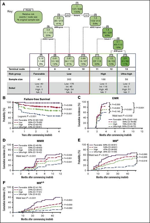
To further explore the interactions between the Sokal risk score and the ASXL1 and BIM variants, we applied a classification tree model for prediction of FFS. Sokal and the BIM and ASXL1 variants were retained in the final classification model after minimization of the 10-fold cross-validation error. The classification tree produced 7 terminal nodes which could readily be grouped into 4 risk categories: favorable, low, high, and ultra-high (Figure 2A). Kaplan-Meier analysis of these 4 risk groups showed highly significant stratification between these risk categories for the prediction of FFS as expected (97%, 80%, 67%, and 44%, respectively; nominal P < .001; Figure 2B). Importantly, this model also strongly predicted for EMR (P = .002; Figure 2C), MMR (P < .001; Figure 2D), MR4 (P = .012; Figure 2E), and MR4.5 (P < .001; Figure 2F), demonstrating that the model retained significance across clinical outcomes.
A risk classification tree model identified a subgroup of ultra-high-risk patients with the highest risk of experiencing therapy failure and disease progression. (A) A classification tree based on Sokal risk score and the BIM and ASXL1 variant genotypes to predict 48-month FFS was generated by recursive partitioning using the Rpart package. The final classification tree produced 7 terminal nodes, which were readily distinguished into 4 risk categories; favorable, low, high and ultra-high. Each node box displays the relative risk of the node compared with the whole population, the number of events/sample size at that node, and the percentage of observations used at that node. (B) Kaplan-Meier FFS survival plot based on the classification tree risk categories. (C-F) Cumulative incidence of (C) EMR, (D) MMR, (E) MR4, and (F) MR4.5 as stratified by the classification tree risk groups, as defined in panel A. CI, 95% confidence interval.
A risk classification tree model identified a subgroup of ultra-high-risk patients with the highest risk of experiencing therapy failure and disease progression. (A) A classification tree based on Sokal risk score and the BIM and ASXL1 variant genotypes to predict 48-month FFS was generated by recursive partitioning using the Rpart package. The final classification tree produced 7 terminal nodes, which were readily distinguished into 4 risk categories; favorable, low, high and ultra-high. Each node box displays the relative risk of the node compared with the whole population, the number of events/sample size at that node, and the percentage of observations used at that node. (B) Kaplan-Meier FFS survival plot based on the classification tree risk categories. (C-F) Cumulative incidence of (C) EMR, (D) MMR, (E) MR4, and (F) MR4.5 as stratified by the classification tree risk groups, as defined in panel A. CI, 95% confidence interval.
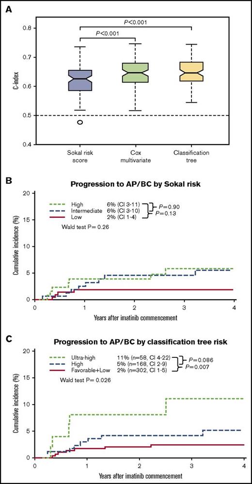
Bootstrap cross-validation was undertaken to assess and compare the predictive power of the multivariate models combining Sokal, BIM rs686952, and ASXL1 rs4911231 variants. The Sokal score univariate model provided a modest discriminatory power (C-index, 0.62; range, 0.47-0.74). Importantly, the addition of the BIM and ASXL1 variants to the Sokal score significantly improved the predictive power in cross-validation assessments of predictive performance score in Cox multivariate and classification tree models for FFS (Figure 3A). Moreover, prediction of progression to AP/BC was improved by addition of the risk variants to Sokal (Figure 3B-C).
Comparison of the predictive power of Sokal and ASXL1 and BIM variant genotypes and their combinations. (A) The data set was randomly split into training and test sets 100 times and the concordance index (C-index) was calculated. The C-index is a nonparametric measure to quantify the discriminatory power of a predictive model, in which a C-index of 0.5 indicates no predictive discrimination, and a C-index of 1 indicates perfect predictive accuracy. For each model, during each of the 100 times of random splitting, 80% of the total samples were used to train the model, and the remaining 20% were used as the test set for C-index calculations. Both the Cox multivariate and classification tree multivariate models showed performance superior to that of the Sokal risk score univariate model (one-sided Wilcoxon signed rank test P < .001). The boundaries of the box mark the first and third quartile, with the median in the center and whiskers extending to 1.5 interquartile range from the boundaries. The horizontal black dashed line marks the C-index equivalent to random guess (C-index = 0.5). Cumulative incidence of progression to AP/BC, stratified by (B) Sokal risk group and (C) classification tree risk group.
Comparison of the predictive power of Sokal and ASXL1 and BIM variant genotypes and their combinations. (A) The data set was randomly split into training and test sets 100 times and the concordance index (C-index) was calculated. The C-index is a nonparametric measure to quantify the discriminatory power of a predictive model, in which a C-index of 0.5 indicates no predictive discrimination, and a C-index of 1 indicates perfect predictive accuracy. For each model, during each of the 100 times of random splitting, 80% of the total samples were used to train the model, and the remaining 20% were used as the test set for C-index calculations. Both the Cox multivariate and classification tree multivariate models showed performance superior to that of the Sokal risk score univariate model (one-sided Wilcoxon signed rank test P < .001). The boundaries of the box mark the first and third quartile, with the median in the center and whiskers extending to 1.5 interquartile range from the boundaries. The horizontal black dashed line marks the C-index equivalent to random guess (C-index = 0.5). Cumulative incidence of progression to AP/BC, stratified by (B) Sokal risk group and (C) classification tree risk group.
Predictive models incorporating the Sokal score and the ASXL1 and BIM variants identified a subgroup of patients with the greatest risk of treatment failure
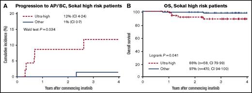
The FFS of the 130 Sokal high-risk patients was 59%. Among these, additional information from the 2 variants allowed further risk stratification: 34 patients were reclassified as low risk, 45 as high risk, and 51 as ultra high risk with 75%, 64%, and 44% FFS, respectively (nominal P = .012). We could thus identify a subgroup of patients with high Sokal scores who had the greatest risk of treatment failure. Furthermore, these patients classified as ultra high risk were significantly more likely to progress to AP/BC compared with all other patients (12% vs 1%; P = .034; Figure 4A) and had inferior OS (88% vs 97%; P = .041; Figure 4B). These results demonstrate a further refinement for risk stratification when variant genotyping information is added to Sokal risk scores.
The classification tree risk model identified a subgroup of ultra-high-risk patients with the highest risk of experiencing therapy failure and disease progression. (A) Progression to AP/BC and (B) OS was assessed in Sokal high-risk patients (n = 130), as stratified by ultra-high-risk group and the “other” risk groups (combined high-, low-, and favorable-risk groups as a result of a low number of events).
The classification tree risk model identified a subgroup of ultra-high-risk patients with the highest risk of experiencing therapy failure and disease progression. (A) Progression to AP/BC and (B) OS was assessed in Sokal high-risk patients (n = 130), as stratified by ultra-high-risk group and the “other” risk groups (combined high-, low-, and favorable-risk groups as a result of a low number of events).
The combined Sokal, ASXL1 variant, and BIM variant classification tree risk model identified patients at ultra high risk among those with subsequent EMR
Numerous studies have demonstrated the strong association between EMR and long-term outcomes. In our study, patients who achieved EMR at 3 months of imatinib treatment was a very strong predictor of MMR (P < .001), MR4 (P < .001), MR4.5 (P < .001), FFS (P < .001), progression to AP/BC (P < .001), and OS (P < .001) compared with patients who did not achieve EMR. Interestingly, the majority (31 [62%] of 51 evaluable patients) of the ultra-high-risk patients identified in the risk classification tree achieved the EMR milestone. EMR achievers (n = 410) were stratified by their baseline classification tree risk group. The ultra-high-risk patients by definition had significantly inferior achievement of long-term outcomes: 12-month MMR (P = .009), 48-month MR4 (P = .004), MR4.5 (P = .002), FFS (P < .001), and progression to AP/BC (P = .026) (supplemental Figure 3). Multivariate analysis demonstrated that the classification tree risk model of variables assessed at baseline was an independent predictor of outcome (Table 5).
Multivariate analysis of the classification tree risk groups and EMR status at 3 months
| . | MMR . | MR4 . | MR4.5 . | FFS . | ||||
|---|---|---|---|---|---|---|---|---|
| RR . | P . | RR . | P . | RR . | P . | HR . | P . | |
| Classification tree baseline risk group | ||||||||
| Favorable | 1 | 1 | 1 | 1 | ||||
| Low | 0.77 | .13 | 0.74 | .2 | 0.55 | .012 | 1.89 | .062 |
| High | 0.62 | .011 | 0.68 | .11 | 0.43 | .002 | 2.34 | .021 |
| Ultra-high | 0.41 | <.001 | 0.29 | .001 | 0.21 | .002 | 2.86 | .005 |
| EMR status at 3 months of imatinib treatment, % | ||||||||
| ≤10 | 1 | 1 | 1 | 1 | ||||
| >10 | 0.21 | <.001 | 0.16 | <.001 | 0.09 | <.001 | 1.53 | <.001 |
Relative risks and hazard ratios for the classification tree are compared with the favorable-risk group.
The potential biological role of the ASXL1 and BIM risk variants
Given the strong association with clinical outcome, we sought to investigate the putative biological function of the ASXL1 and BIM risk variants. The ASXL1 risk variant is a synonymous variant in exon 12 of ASXL1, and the linked variants (rs2295763 and rs2295764) are located in the 3′ UTR of ASXL1. A putative functional role for these 3 variants could not be identified by in silico assessments. Functional predictions were expanded to variants within the ASXL1 haplotype block. That analysis identified that ASXL1 rs2295762, which was in high linkage with the ASXL1 risk variants (r2 = 0.98), is located in an evolutionary conserved region of the 3′ UTR, and is predicted to create microRNA-34a (miR-34a) and miR-486-3p seed sites (supplemental Table 7).
The BIM rs686952 risk variant is located in intron 11. No potential functional significance was identified. The analysis was expanded to variants located in the BIM haplotype block. Most notably, BIM rs724710, a synonymous variant located in the highly conserved BH3 functional domain, was in high linkage with the BIM risk variant (r2 = 0.92) and was previously reported to be associated with imatinib response (supplemental Table 7).16 The BIM BH3 domain variant was genotyped in our cohort, and subsequent univariate analysis showed that this variant was significantly associated with MMR (P = .022), MR4 (P = .012), and MR4.5 (P = .016), but not FFS (P = .212) or progression to AP/BC (P = .303).
These in silico functional predictions have identified 2 variants (ASXL1 rs2295762 and BIM rs724710) in linkage with the risk variants identified by our statistical analysis, which are of potential biological significance. These variants are candidates for further functional assessment to investigate their putative biological impact and role in modulating response to imatinib.
Discussion
Improved risk stratification at diagnosis of CML could aid choice of the most appropriate first-line TKI for optimal outcome. With the availability of generic imatinib and potentially lower drug costs, more potent TKIs could be reserved for those at highest risk of treatment failure and lower probability of reaching milestone MR. Early achievement of MRs is associated with subsequent deep MR,39 which is a prerequisite for treatment-free remission. With treatment-free remission potentially moving into mainstream clinical practice with the intention of discontinuing TKIs as a standard treatment protocol,40,41 it is desirable to improve the number of patients who achieve MRs. We have identified a small cohort of imatinib-treated patients with the highest risk of treatment failure, disease progression, and low probability of MR by combining the Sokal risk score and inherited genetic biomarkers. Two germ line variants were identified as strong biomarkers of response to imatinib: an intronic variant in the proapoptotic protein BIM and a synonymous variant in ASXL1, a putative epigenetic modifier. Previous molecular biomarkers available at diagnosis have not shown such a consistent association with molecular and survival outcomes.18-22 By definition, all of the ultra-high-risk patients had high Sokal risk but they represented only 39% of the total Sokal high-risk group.
EMR is currently the strongest predictor of long-term outcomes and is incorporated into the National Comprehensive Cancer Network (NCCN) and European LeukemiaNet (ELN) guidelines. The NCCN recommends a therapy switch for patients who fail to achieve EMR at 3 months, whereas the ELN recommends switching therapy at 6 months.5,6 However, there is little evidence that changing therapy after a therapy failure event can effectively salvage these patients. In some patients, disease progression may occur within the first 6 months of diagnosis, with little time to implement effective salvage therapy. Identification of very high-risk patients at diagnosis for novel interventions may be the only way to rescue these ultra-high-risk patients. In contrast, favorable-risk patients may achieve optimal outcomes with the lowest potency inhibitor, although other high-risk patients will require more potent inhibition upfront to achieve optimal outcomes. Indeed, our data suggest that the more potent inhibitor, nilotinib, may overcome the adverse outcome identified for some patients treated with imatinib. The lack of association may also be the result of the smaller sample size for the nilotinib cohort. Therefore, analysis of the significance of these variants in a larger cohort of nilotinib-treated patients with longer follow-up data are required to confirm these findings.
Important variables were initially selected using nonparametric (random forests and recursive partitioning) models, which do not explicitly conduct hypothesis tests during model selection; thus, correction for multiple testing was not required. However, semiparametric (univariate and multivariate analysis) statistical tests were used to confirm the significance of the variables that were deemed important.
Risk groups were initially constructed from an analysis of FFS, a composite end point of outcomes. This allowed validation of the prognostic significance of each of the complements of the failure events when they were investigated separately, which indeed was the case (Figure 2). The bootstrapping method of internal validation provides a nearly unbiased estimate of predictive accuracy of relatively low variance and has the additional advantage of using the entire data set for model assessment.42 Importantly, bootstrap cross-validation demonstrated that when the BIM/ASXL1 variants were combined in a multivariate model (Cox or classification tree) with the Sokal score, a significant increase in the discriminatory capability of the prognostic model was observed compared with the Sokal score alone. It is possible that this analysis carries the risk of overfitting, but our robust statistical models of cross-validation techniques and bootstrapping were designed to mitigate this risk. In addition, we have referred to nominal P values when testing for differences in FFS between risk groups that were constructed from classification trees built to predict FFS.
BIM is a proapoptotic member of the BCL2-family, which is crucial for the intrinsic apoptotic response to imatinib. The BIM risk variant was in high linkage with a synonymous variant (rs724710) in the BH3 functional domain. This variant is reported to be associated with imatinib response.16 How this variant modifies BIM function is yet to be elucidated, but synonymous variants can have functional effects by altering the dynamics of translation through different codon usage, by changing messenger RNA secondary structure that alters protein expression, or by altering RNA splicing by interfering with a splice site or splicing enhancers.43 Certainly, BH3-mimetic drugs can overcome the imatinib resistance conferred by the BIM deletion polymorphism.12 Furthermore, a stapled BIM peptide was recently shown to activate BH3-specific cell death to overcome apoptotic resistance in hematologic malignancies.44 Further investigation into the mechanism of action of the BIM risk variants would reveal whether these therapeutic strategies would be effective for enhancing response.
ASXL1 exon 12 is a region commonly mutated in hematologic cancers, and mutations in this region are associated with aggressive disease and poor outcomes in acute myeloid leukemia and myelodysplastic syndrome.23,45,46 ASXL1 is an epigenetic regulator that binds members of the polycomb repressive complex 2 (PRC2). This complex mediates gene silencing by inducing trimethylation of histone H3-lysine 27, and mutations in ASXL1 impair PRC2 function.47 We hypothesize that a variant that introduces an miRNA binding site causing downregulation of ASXL1 expression may also contribute to poor outcomes in CML patients. In silico predictions identified a 3′ UTR ASXL1 variant (rs2295762) that is predicted to introduce miR-34a and miR-486-3p seed sites. Interestingly, miR-486 is reported to be involved in normal erythropoiesis and modulates drug resistance in CML.48 Most notably, miR-486-5p was reported to modulate response to imatinib in CML progenitor cells.48 The mechanism by which these variants affect response awaits further clinical and experimental evaluation. Alternatively, it is plausible that other variants in linkage disequilibrium with the ASXL1 and BIM variants that were not detected using our targeted sequencing approach are responsible for the associations detected by this study.
Several variants in genes responsible for the pharmacologic activity of imatinib have been reported as predictors of imatinib response in CML, including SLC22A1 (OCT1) rs628031 and 3 MDR1 (ABCB1) variants (rs1128503, rs2032582, and rs1045642). However, inconsistent prognostic significance for each of these variants has been reported between studies, which limits their clinical application.21,49-51 A meta-analysis of outcomes in 1826 CML patients from 12 studies confirmed that the ABCB1 rs1128503 variant has prognostic value in Asian populations.52 We did not observe any association between these 3 ABCB1 variants with response to imatinib, although we may have different patient populations.
In conclusion, this study has identified germ line variants as strong biomarkers of imatinib response when combined with the Sokal score. Assessment of germ line genetic variation at diagnosis may contribute to an enhanced prognostic score to aid therapeutic decision-making. The utility of this risk score should be determined in prospective clinical trials.
The full-text version of this article contains a data supplement.
Acknowledgments
The authors thank Phillipe Rousselot (Universite Versailles Saint-Quentin-en-Yvelines) and Marc Delord (Paris Diderot University) for discussion of our data.
This work was supported by the National Health and Medical Research Council of Australia grants APP1059165 (T.P.H. and S.B.), APP1104425 (S.B.), APP1059165 and APP1023059 (H.S.S.), the Royal Adelaide Hospital Research Foundation, a scholarship from the Leukaemia Foundation of Australia and the AR Clarkson Foundation (D.T.Y.), and a postdoctoral fellowship from the Leukaemia Foundation of Australia/Cure Cancer Australia (W.T.P.). The Australian Cancer Research Foundation (ACRF) Cancer Genomics Facility was established with funding from Therapeutic Innovation Australia and the ACRF.
Authorship
Contribution: J.E.M. designed and performed the research, analyzed data, and wrote the manuscript; L.P. designed and performed the research and contributed to preparation of the manuscript; S.B. designed the research, analyzed data, and contributed to manuscript preparation; T.P.H., W.T.P., D.S., and H.S.S. contributed to the experimental design and manuscript preparation; D.T.Y. and J.R. analyzed data, provided advice on statistical analysis, and contributed to preparation of the manuscript; P.P.S.W. and A.W.S. contributed to data analysis; and D.J.P. and J.T. contributed to data analysis and provided statistical advice.
Conflict-of-interest disclosure: D.T.Y. received funding and honoraria from and has served as a consultant on an advisory board of Novartis Oncology and Bristol-Myers Squibb. J.R. holds shares in Novartis. T.P.H. received research funding and honoraria and is an advisory board member for Novartis Oncology, Bristol-Myers Squibb, and Ariad Pharmaceuticals. S.B. received research funding from Novartis Oncology, Ariad Pharmaceuticals, and Otsuka; honoraria from Novartis Oncology, Bristol-Myers Squibb, Otsuka, Qiagen, and Ariad; and is an advisory board member for Novartis Oncology and Qiagen. The remaining authors declare no competing financial interests.
Correspondence: Susan Branford, Department of Genetics and Molecular Pathology, Centre for Cancer Biology, SA Pathology, Frome Rd, Adelaide, SA 5000, Australia; e-mail: susan.branford@sa.gov.au.


