Key Points
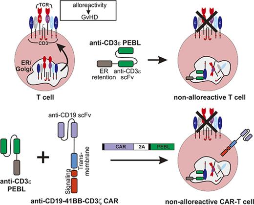
Newly designed PEBLs prevent surface expression of T-cell receptor in T cells without affecting their function.
Combined with chimeric antigen receptors, PEBLs can rapidly generate powerful antileukemic T cells without alloreactivity.
Abstract
Practical methods are needed to increase the applicability and efficacy of chimeric antigen receptor (CAR) T-cell therapies. Using donor-derived CAR-T cells is attractive, but expression of endogenous T-cell receptors (TCRs) carries the risk for graft-versus-host-disease (GVHD). To remove surface TCRαβ, we combined an antibody-derived single-chain variable fragment specific for CD3ε with 21 different amino acid sequences predicted to retain it intracellularly. After transduction in T cells, several of these protein expression blockers (PEBLs) colocalized intracellularly with CD3ε, blocking surface CD3 and TCRαβ expression. In 25 experiments, median TCRαβ expression in T lymphocytes was reduced from 95.7% to 25.0%; CD3/TCRαβ cell depletion yielded virtually pure TCRαβ-negative T cells. Anti-CD3ε PEBLs abrogated TCRαβ-mediated signaling, without affecting immunophenotype or proliferation. In anti-CD3ε PEBL-T cells, expression of an anti-CD19-41BB-CD3ζ CAR induced cytokine secretion, long-term proliferation, and CD19+ leukemia cell killing, at rates meeting or exceeding those of CAR-T cells with normal CD3/TCRαβ expression. In immunodeficient mice, anti-CD3ε PEBL-T cells had markedly reduced GVHD potential; when transduced with anti-CD19 CAR, these T cells killed engrafted leukemic cells. PEBL blockade of surface CD3/TCRαβ expression is an effective tool to prepare allogeneic CAR-T cells. Combined PEBL and CAR expression can be achieved in a single-step procedure, is easily adaptable to current cell manufacturing protocols, and can be used to target other T-cell molecules to further enhance CAR-T-cell therapies.
Introduction
Genetically engineered immune cells are a powerful new treatment of cancer. Recent clinical trials with T lymphocytes expressing chimeric antigen receptors (CARs) have provided a compelling demonstration of their potential. CAR-T cells specific for CD19 induced durable remissions in patients with treatment-refractory CD19-positive leukemia and lymphoma.1-10 Other malignancies can be attacked by T cells redirected against different antigens. Hence, the possible applications for genetically engineered cellular therapy in oncology are wide-ranging.10,11
The initial clinical experience with CAR-T cells has also identified limitations that could diminish therapeutic effect and hamper development. A major issue is the variable fitness of immune cells collected from patients with cancer, resulting in an unpredictable capacity to expand in vivo and exert antitumor effects.10,12 This variability complicates the identification of the most effective cell dosages and might lead to infusion of short-lived and ineffective cells. T lymphocytes from healthy donors should offer better consistency and effectiveness, but pose the risk for graft-versus-host disease (GVHD), a potentially fatal consequence of donor lymphocyte infusion.13,14 In such an allogeneic setting, additional modifications to the infused T cells are required to suppress their capacity to recognize host tissues; namely, downregulation of CD3/TCRαβ.15,16
Contemporary methodologies for gene editing have opened new opportunities relevant to cell therapy of cancer.17 Zinc finger meganucleases, TALEN, and CRISPR-Cas9 can delete genes encoding TCRαβ chains, leading to T cells that lack alloreactivity,15,18,19 whereas other genes can be targeted to delay rejection.15 A report using TALEN deletion of the TCRα and CD52 loci together with anti-CD19 CAR expression indicates that combining CAR expression with gene editing is feasible in a clinical setting,20 although it may still be technically challenging.
To expand the arsenal of tools for enhancing cell-based therapies of cancer, we developed a method that allows simple and effective blockade of surface receptor expression in immune cells. Specific constructs, named protein expression blockers (PEBLs), prevent transport of targeted proteins to the cell membrane. PEBL constructs can be readily combined with other gene modifications and be incorporated into existing clinical-grade protocols for ex vivo cell processing of immune cells. We tested the potential of this approach to downregulate CD3/TCRαβ expression in CAR-T cells.
Materials and methods
Cell lines and T cells
Jurkat, Loucy, Nalm6, RS4;11, and K562 were from the American Type Culture Collection (Rockville, MD); OP-1 was established in our laboratory.21 A murine stem cell virus (MSCV) retroviral vector was used to express the firefly luciferase gene plus green fluorescent protein (GFP) in Nalm6, and CD19 plus DsRed in K562.22
Peripheral blood from healthy donors was obtained from anonymized byproducts of platelet donations at the National University Hospital Blood Bank, Singapore, with Institutional Review Board (National University of Singapore) approval in accordance with the Declaration of Helsinki. Mononucleated cells were separated by centrifugation on Lymphoprep (Axis-Shield, Oslo, Norway). T cells, enriched with Dynabeads Human T-Activator CD3/CD28 (Thermo Fisher Scientific, Waltham, MA), were cultured in RPMI-1640 (Thermo Fisher), 10% fetal bovine serum (GE-Healthcare, Chicago, IL), and antibiotics, with interleukin 2 (IL-2; Proleukin, Novartis, Basel, Switzerland; 100 IU/mL) added every 2 days.
PEBL and CAR constructs
From RNA of the PLU4 murine hybridoma, secreting an anti-human CD3 monoclonal antibody (immunoglobulin G2a [IgG2a] isotype; Creative Diagnostics, Shirley, NY), we synthesized cDNA by Moloney Murine Leukemia Virus reverse transcriptase and Oligo(dT)15 primer (Promega, Madison, WI). We amplified variable regions of heavy and light chain with IgG Library Primer Set Mouse BioGenomics (US Biological, Salem, MA) and assembled them into a single-chain variable fragment (scFv) by a flexible linker sequence encoding (Gly4Ser)4. CD8α signal peptide and transmembrane domains were from human-activated T-cell cDNA.
To generate PEBL constructs, each retention-signaling domain (supplemental Table 1) was added to the 3′ end of the variable heavy chain fragment by PCR. Constructs were subcloned into the MSCV retroviral vector containing an internal ribosome entry site and GFP or mCherry. Preparation of retroviral supernatant and gene transduction were performed as previously described.23 Briefly, retroviral vector-conditioned medium was added to polypropylene tubes coated with RetroNectin (Takara, Otsu, Japan); after removing the supernatant, activated T cells were added and left at 37°C for 12 hours; fresh viral supernatant was added on 2 other successive days. T lymphocytes were maintained in RPMI-1640 with fetal bovine serum, antibiotics, and 200 IU/mL IL-2 until the time of the experiments.
To remove residual CD3/TCRαβ-positive T cells after PEBL transduction, we used allophycocyanin (APC)-conjugated anti-CD3 (BD Biosciences, San Jose, CA; or Miltenyi Biotec, Bergisch Gladbach, Germany) and anti-TCRαβ antibodies (BioLegend, San Diego, CA), with anti-APC MicroBeads and LD column (Miltenyi Biotec).
A CAR constituted by an anti-CD19 scFv, CD8α hinge and transmembrane domains, and cytoplasmic domains of 41BB and CD3ζ (anti-CD19-41BB-CD3ζ)22 was inserted in the MSCV vector, as described for PEBLs. In some experiments, CAR-T cells were expanded by coculture with 100 Gy-irradiated K562 cells transduced with CD19, at a 1:1 E:T ratio. We also transduced T cells with a MSCV vector containing both CAR and PEBL constructs separated by a sequence encoding a self-cleaving 2A peptide.24 Electroporation of anti-CD19-41BB-CD3ζ mRNA was performed as previously described.25,26 Cells electroporated without mRNA were used as control.
Determination of scFv specificity, gene expression, and cell marker profile
To identify the CD3 subunit bound to the antibody derived from the PLU4 hybridoma, the cDNA of each CD3 subunit (Origene, Rockville, MD) was subcloned into MSCV-internal ribosome entry site-GFP and transduced into K562 cells. K562 cells were then permeabilized with 8E reagent (a permeabilization reagent developed in our laboratory), incubated with supernatant from PLU4 hybridoma cells, followed by Alexa Fluor 647 conjugated goat anti-mouse IgG (Southern Biotech, Birmingham, AL).
CAR and PEBL expression was detected by biotin-conjugated goat anti-mouse IgG F(ab’)2 antibody (Jackson ImmunoResearch, West Grove, PA), and phycoerythrin (PE)- or APC-conjugated streptavidin (Jackson ImmunoResearch). For intracellular staining, cells were permeabilized with 8E. To determine whether PEBLs were secreted, supernatant from anti-CD3ε scFv- or PEBL-transduced Jurkat was added to Loucy and incubated in 4°C for 45 minutes; surface-bound scFv and PEBLs were detected with biotin-conjugated goat anti-mouse IgG F(ab’)2 antibody and streptavidin APC.
CD3 expression was detected with anti-CD3 APC (SK7, BD Biosciences). PE- or APC-conjugated anti-TCRαβ (IP26), CD2 APC (RPA-2.10), CD137 APC (4B4-1), CD279 PE (EH12.2H7), and CD366 PE (F38-2E2) were from BioLegend (San Diego, CA). Anti-CD4 PE-Cy7 (SK3), CD8 PE (RPA-T8), CD7 PE (M-T701), CD25 PE-Cy7 or APC (2A3), CD62L APC (DREG-56), and CD69 PE or APC (L78) were from BD Biosciences; CD223 APC (3DS223H) was from Thermo Fisher. Cell staining was analyzed using Fortessa or Accuri C6 flow cytometers (BD Biosciences).
T-cell activation, cytokine production, proliferation, and cytotoxicity
OKT3 (10 μg/mL, Miltenyi Biotech) or isotype-matched control (R&D, Minneapolis, MN) was dispensed into 96-well flat-bottom plates (Corning, Corning, NY) and left at 4°C for 12 hours. After removing soluble antibody, 1 to 2 × 10⁵ Jurkat cells per well were seeded and cultured at 37°C, 5% CO2 for 24 hours. PE- or APC-conjugated anti-CD25 and anti-CD69 antibodies were used to determine T-cell activation, with isotype-matched nonreactive antibodies as control (all from BD Biosciences).
Jurkat cells were transduced with a TCR specific for the hepatitis B virus (HBV) s183 peptide in the context of HLA-A2 (provided by A. Bertoletti, Duke-NUS, Singapore).27 The TCR was inserted into a MSCV vector containing a neomycin-resistant gene, and transduced cells were selected by exposure to neomycin. Expression of TCRβ on the cell surface and intracellularly (after cell permeabilization with 8E) was detected with an anti-TCR Vβ3 antibody conjugated to fluorescein isothiocyanate (Beckman Coulter, Brea, CA). HBV s183-Jurkat cells, transduced with an mCherry vector with or without anti-CD3 PEBL, were cocultured with T2 cells (also from A. Bertoletti) pulsed with 1 µg/mL HBV s183 peptide (Genscript, Piscataway, NJ) at a 1:1 E:T ratio. After 24 hours, cells were stained with anti-CD25 PE and anti-CD69 APC.
To measure interferon γ (IFNγ) production, 1 × 10⁵ T cells and 2 × 10⁵ RS4;11 cells were seeded in a 96-well round bottom plate. After 8 hours in the presence of 0.1% Brefeldin A (GolgiPlug, BD Biosciences), cells were labeled with APC- or PE-conjugated anti-IFNγ (clone 25723.11, BD Biosciences) after cell membrane permeabilization.
To measure cell proliferation, 5 × 10⁴ T cells transduced with CAR or GFP only were placed in 96-well round bottom plate in RPMI-1640 with 10% fetal bovine serum, antibiotics, and 200 IU/mL of IL-2. OP-1 cells were irradiated (100 Gy) and mixed with the T cells at 1:1 E:T. Every 2 days, 200 IU/mL of IL2 was added. GFP+ cells were counted by flow cytometry; a new set of irradiated OP-1 cells was added at 1:1 E:T every 7 days.23
To test cytotoxicity, target cells were labeled with calcein red-orange AM (Thermo Fisher) and plated into a 96-well round bottom plate at a concentration of 5 × 10⁴ cells per 100 μL. T cells were added at various E:T ratios and cultured at 37°C, 5% CO2. After 4 hours, the number of viable target cells was counted by flow cytometry. In some tests, luciferase-labeled cells were used as a target. The assay was performed in a 96-well flat bottom plate, BrightGlo (Promega) was added to the wells after 4 hours, and luminescence was measured using a Flx 800 plate reader (BioTek, Winooski, VT).23
Mouse models
To model GVHD, NOD.Cg-Prkdcscid IL2rgtm1Wjl/SzJ (NSG) mice (Jackson Laboratory, Bar Harbor, ME) received 2.5 Gy total body irradiation. One day later, 1 × 10⁷ T cells transduced with either anti-CD3 PEBL plus GFP or GFP alone were IV infused. All mice received IL-2 (20 000 IU) 3 times per week intraperitoneally (IP). Body weight and GVHD symptoms were monitored 3 times per week; blood was collected once a week by cheek prick. Mice were euthanized when body weight reduction exceeded 20% of the baseline in 2 consecutive measurements. Histopathology evaluation for GVHD was performed at the Advanced Molecular Pathology Laboratory, Institute of Molecular and Cell Biology (Singapore). Anti-human CD3 polyclonal antibody (Agilent Technologies, Santa Clara, CA), anti-human CD4 (EPR6855), and anti-human CD8 (EP1150Y; both from Abcam, Cambridge, United Kingdom) were used for immunohistochemistry.
For the acute lymphoblastic leukemia (ALL) model, Nalm6 cells expressing luciferase (0.5 × 10⁶ cells per mouse) were IV injected, followed 3 days later by T cells transduced with anti-CD19-41BB-CD3ζ and either anti-CD3 PEBL or mCherry (2 × 10⁷ per mouse IV); control mice received RPMI-1640 medium. In a second experiment, mice received 2.5 Gy total body irradiation on day 3 before infusion of T cells or RPMI-1640. All mice received IL-2 (20 000 IU) 3 times per week IP. Leukemia cell load was determined with the Xenogen IVIS-200 System (Perkin Elmer, Waltham, MA; supplemental Methods) after injecting 150 μg/g body weight of aqueous d-luciferin potassium salt (Perkin Elmer) IP. Luminescence was analyzed with Living Image 3.0 software. Mice were euthanized when the luminescence reached 1 × 10¹⁰ photons per second, or earlier if body weight reduction exceeded 20% of their baseline in 2 consecutive measurement or there were other physical signs warranting euthanasia.
Results
Design and functional screening of PEBL constructs
The CD3/TCRαβ complex is assembled in the endoplasmic reticulum (ER); all components are required for its cell surface expression.28-30 To determine the CD3 specificity of the PLU4 antibody, we transduced K562 cells with CD3ε, CD3γ, CD3δ, and CD3ζ alone or in combination and tested PLU4 reactivity by flow cytometry (supplemental Figure 1). The staining pattern indicated reactivity with an epitope of CD3ε most accessible when associated with either CD3γ or CD3δ.
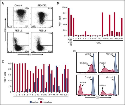
We generated an scFv from the PLU4 hybridoma cDNA and linked it to sequences encoding peptides predicted to anchor it to the ER and/or the Golgi apparatus (supplemental Table 1). We tested 21 constructs for their capacity to suppress CD3 surface expression and compared them with a construct containing SEKDEL, a sequence reported to suppress expression of surface proteins when linked to an scFv.31 In Jurkat cells, retroviral transduction of many of the PEBLs caused a nearly complete elimination of surface CD3 expression (Figure 1A-B), whereas most cells transduced with the SEKDEL construct remained CD3-positive.
Anti-CD3ε PEBLs block surface CD3 expression. (A) Flow cytometric dot-plots illustrate surface CD3 downregulation in Jurkat by anti-CD3ε PEBLs compared with cells transduced with GFP alone (“Control”) or SEKDEL. (B) Surface CD3 expression in Jurkat transduced with the indicated constructs. Bars show a mean of 2 to 3 experiments for KEDL, SEKDEL, and PEBL 2,4,5,8,9,11, or individual results for the remainder. (C) Intracellular or surface expression of PEBL-derived anti-CD3ε scFv in Jurkat. Bars show a mean of 2 to 3 experiments for PEBL 2,4,8,9,11, or individual results for the remainder. (D) Flow cytometric histograms illustrate CD3 expression in peripheral blood T cells transduced with anti-CD3ε SEKDEL or PEBLs, relative to that of lymphocytes transduced with GFP alone, 8 to 13 days posttransduction.
Anti-CD3ε PEBLs block surface CD3 expression. (A) Flow cytometric dot-plots illustrate surface CD3 downregulation in Jurkat by anti-CD3ε PEBLs compared with cells transduced with GFP alone (“Control”) or SEKDEL. (B) Surface CD3 expression in Jurkat transduced with the indicated constructs. Bars show a mean of 2 to 3 experiments for KEDL, SEKDEL, and PEBL 2,4,5,8,9,11, or individual results for the remainder. (C) Intracellular or surface expression of PEBL-derived anti-CD3ε scFv in Jurkat. Bars show a mean of 2 to 3 experiments for PEBL 2,4,8,9,11, or individual results for the remainder. (D) Flow cytometric histograms illustrate CD3 expression in peripheral blood T cells transduced with anti-CD3ε SEKDEL or PEBLs, relative to that of lymphocytes transduced with GFP alone, 8 to 13 days posttransduction.
Expression of PEBLs 1-8, 11, and 19 was confined intracellularly; other PEBLs had varying degrees of surface expression (Figure 1C; supplemental Figure 2). PEBLs colocalized with CD3 intracellularly; no secretion was detected in tests with PEBLs 1-5 (supplemental Figure 2). Importantly, PEBLs also effectively downregulated CD3 expression in activated peripheral blood T lymphocytes (Figure 1D).
CD3 downregulation with PEBLs suppresses TCRαβ expression
In addition to CD3, PEBL transduction also downregulated TCRαβ expression in peripheral blood T lymphocytes. In 25 experiments, median percentage of T cells expressing TCRαβ was reduced from 95.7% (range, 89.4% to 99.0%) to 25.0% (range, 3.5% to 55.2%; Figure 2A). The main factor determining the extent of CD3/TCRαβ downregulation was the efficiency of retroviral transduction, which ranged between 58.5% and 99.8% (median GFP+ cells, 94.2%). Magnetic removal of residual CD3/TCRαβ-positive cells yielded virtually pure populations of CD3/TCRαβ-negative T cells (Figure 2B); in 11 T-cell preparations from 6 donors, T cells expressing normal levels of CD3/TCRαβ were 0.01% (<0.01% to 0.15%) after only 1 round of CD3/TCRαβ depletion.
Anti-CD3ε PEBLs downregulate TCRαβ. (A) TCRαβ expression in GFP-positive T lymphocytes 5 to 9 days after PEBL transduction. Mean (± standard deviation [SD]) is shown for cells transduced with GFP only (“Control”; n = 25), PEBL2 (n = 4), and PEBL5 (n = 18); other data represent results of 1 or mean of 2 experiments. (B) Flow cytometric dot-plots illustrate TCRαβ downregulation in T lymphocytes compared with cells transduced with GFP only. (C) CD3/TCRαβ expression in Jurkat cells transduced with PEBL after long-term culture; Control, cells transduced with GFP alone. (D) Collective results of CD3/TCRαβ expression in long-term cultures of T lymphocytes (with 200 IU/mL IL-2) or Jurkat cells. Symbols indicate persistence of more than 90% reduction of surface CD3/TCRαβ in GFP+ transduced cells.
Anti-CD3ε PEBLs downregulate TCRαβ. (A) TCRαβ expression in GFP-positive T lymphocytes 5 to 9 days after PEBL transduction. Mean (± standard deviation [SD]) is shown for cells transduced with GFP only (“Control”; n = 25), PEBL2 (n = 4), and PEBL5 (n = 18); other data represent results of 1 or mean of 2 experiments. (B) Flow cytometric dot-plots illustrate TCRαβ downregulation in T lymphocytes compared with cells transduced with GFP only. (C) CD3/TCRαβ expression in Jurkat cells transduced with PEBL after long-term culture; Control, cells transduced with GFP alone. (D) Collective results of CD3/TCRαβ expression in long-term cultures of T lymphocytes (with 200 IU/mL IL-2) or Jurkat cells. Symbols indicate persistence of more than 90% reduction of surface CD3/TCRαβ in GFP+ transduced cells.
In peripheral blood T lymphocytes and Jurkat cells maintained in continuous culture, CD3/TCRαβ downregulation was persistent, with a follow-up of up to 2 months for T lymphocytes and 21 months for Jurkat cells (Figure 2C-D; supplemental Figure 3).
Function of T cells with downregulated CD3/TCRαβ by PEBL
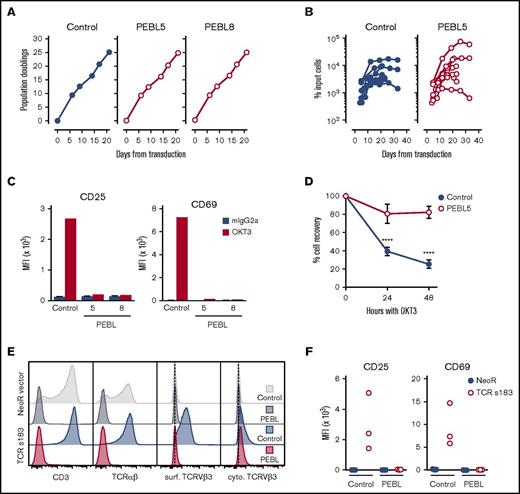
In addition to the lack of surface CD3/TCRαβ, there was no noticeable phenotypic change in lymphocytes transduced with PEBLs; expression of CD4, CD8, CD2, CD7, CD25, CD62L, CD69, CD137 (4-1BB), CD223 (LAG3), CD279 (PD-1), and CD366 (TIM-3) was not significantly altered (supplemental Table 2). T-cell proliferation was also unaffected. In experiments with Jurkat cells, the proliferative rate of PEBL-transduced cells was identical to that of cells transduced with GFP alone (Figure 3A). Likewise, expansion and survival of peripheral blood T cells with IL-2 (200 IU/mL) were not affected by PEBL transduction and CD3/TCRαβ downregulation (Figure 3B).
CD3/TCRαβ downregulation by PEBL does not affect cell proliferation, but abrogates CD3/TCRαβ signaling. (A) Growth rate of Jurkat transduced with anti-CD3 PEBLs or GFP only (“Control”). Symbols indicate mean (± SD) of triplicate measurements. (B) Survival of PEBL-transduced or control T lymphocytes from 5 donors (7 experiments) cultured with IL-2 (200 IU/mL). Symbols indicate mean of triplicate measurements. (C) CD25 and CD69 mean fluorescence intensity (MFI) in Jurkat after 24 hours with OKT3 or nonreactive mouse IgG2a. Bars indicate mean (±SD) of triplicate measurements. (D) Viable PEBL or control T lymphocytes recovered from cultures with OKT3 compared with cultures without OKT3, all containing IL-2 (200 IU/mL). Symbols represent mean (±SD) of 9 measurements with cells from 3 donors. P values by Student t test are shown for significant differences (****P < .0001). (E) Jurkat cells transduced with either a TCR specific for HBV s183 or a vector containing neomycin-resistant gene only (“NeoR”) were transduced with anti-CD3 PEBL or mCherry only (“Control”) after neomycin selection. CD3, TCRαβ, and TCRVβ3 chain (part of the HBV s183 TCR) expression is shown; TCRVβ3 expression was tested on the cell surface, and intracellularly after cell permeabilization. (F) Transduced Jurkat cells shown in panel E were cocultured with T2 cells loaded with HBV s183 peptide for 24 hours. Shown are CD25 and CD69 MFI minus those measured after culture with T2 cells, but without peptide. Symbols represent mean of triplicate measurements.
CD3/TCRαβ downregulation by PEBL does not affect cell proliferation, but abrogates CD3/TCRαβ signaling. (A) Growth rate of Jurkat transduced with anti-CD3 PEBLs or GFP only (“Control”). Symbols indicate mean (± SD) of triplicate measurements. (B) Survival of PEBL-transduced or control T lymphocytes from 5 donors (7 experiments) cultured with IL-2 (200 IU/mL). Symbols indicate mean of triplicate measurements. (C) CD25 and CD69 mean fluorescence intensity (MFI) in Jurkat after 24 hours with OKT3 or nonreactive mouse IgG2a. Bars indicate mean (±SD) of triplicate measurements. (D) Viable PEBL or control T lymphocytes recovered from cultures with OKT3 compared with cultures without OKT3, all containing IL-2 (200 IU/mL). Symbols represent mean (±SD) of 9 measurements with cells from 3 donors. P values by Student t test are shown for significant differences (****P < .0001). (E) Jurkat cells transduced with either a TCR specific for HBV s183 or a vector containing neomycin-resistant gene only (“NeoR”) were transduced with anti-CD3 PEBL or mCherry only (“Control”) after neomycin selection. CD3, TCRαβ, and TCRVβ3 chain (part of the HBV s183 TCR) expression is shown; TCRVβ3 expression was tested on the cell surface, and intracellularly after cell permeabilization. (F) Transduced Jurkat cells shown in panel E were cocultured with T2 cells loaded with HBV s183 peptide for 24 hours. Shown are CD25 and CD69 MFI minus those measured after culture with T2 cells, but without peptide. Symbols represent mean of triplicate measurements.
As expected, knock-down of surface CD3 abrogated CD3 signaling. Thus, in Jurkat cells cultured with the anti-CD3 antibody OKT3, the activation markers CD25 and CD69 were not upregulated if cells had been transduced with anti-CD3 PEBL (Figure 3C). Moreover, if peripheral blood T cells were cultured with OKT3 for 48 hours, viability of cells transduced with GFP alone rapidly decreased, whereas numbers of PEBL-transduced cells remained high (Figure 3D). Finally, we transduced Jurkat cells with a TCR against the HBV s183 peptide expressed in the context of HLA-A2.27 Anti-CD3ε PEBL transduction blocked the surface expression of CD3, of anti-HBV TCRαβ, and of its TCRVβ3 chain (Figure 3E); it abrogated the cells’ capacity to respond to HLA-A2-expressing cells (T2) pulsed with the HBV s183 peptide (Figure 3F).
Function of anti-CD19 CAR in T cells transduced with anti-CD3ε PEBL
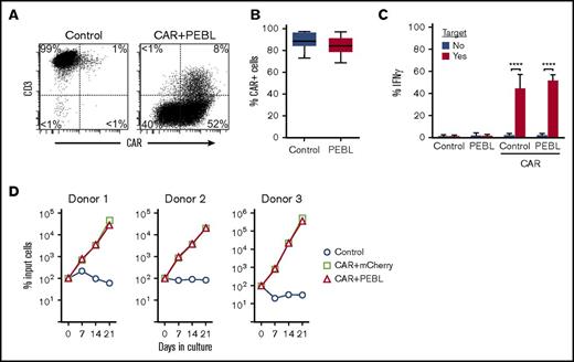
PEBL transduction did not affect T-cell immunophenotype and proliferation, suggesting that expression of a CAR in PEBL-T cells might induce target-specific cytotoxicity, as well as in CD3/TCRαβ-positive T cells. To test this notion, we expressed the anti-CD19-41BB-CD3ζ CAR and anti-CD3 PEBL in T cells and compared their function with that of CAR-T cells without PEBL. In 9 paired experiments, CAR expression by either viral transduction (n = 4) or mRNA electroporation (n = 5) was high, regardless of CD3/TCRαβ expression (Figure 4A-B). Neither PEBL expression nor CD3/TCRαβ downregulation affected CAR function, including CAR-mediated IFNγ secretion and T-cell proliferation (Figure 4C-D).
CAR expression and signaling in T cells with CD3/TCRαβ expression blockade. (A) Flow cytometric dot-plots illustrate CD3 downregulation and anti-CD19-41BB-CD3ζ CAR expression. Cells were transduced with the CAR construct followed by anti-CD3ε PEBL, or with GFP only followed by mCherry only (“Control”). (B) Percentage of T lymphocytes transduced with PEBL or GFP alone (“Control”) expressing anti-CD19-41BB-CD3ζ CAR 24 hours after CAR mRNA electroporation (n = 5), or 5 to 6 days after CAR viral transduction (n = 4); P = .207. (C) IFNγ production by PEBL or control T cells electroporated with CAR mRNA or no mRNA and cultured with CD19+ RS4;11 for 8 hours at E:T 1:2. Bars represent mean (±SD) of 9 measurements with cells from 3 donors; ****P < .0001. (D) T lymphocytes were first transduced with CAR and then transduced with either mCherry alone or anti-CD3 PEBL. Cells were then cultured with irradiated CD19+ OP-1 for 3 weeks. Results were compared with cells transduced with GFP only and then with mCherry only (“Control”). Symbols indicate mean (± SD) percentage cell recovery relative to number of input cells in triplicate cultures.
CAR expression and signaling in T cells with CD3/TCRαβ expression blockade. (A) Flow cytometric dot-plots illustrate CD3 downregulation and anti-CD19-41BB-CD3ζ CAR expression. Cells were transduced with the CAR construct followed by anti-CD3ε PEBL, or with GFP only followed by mCherry only (“Control”). (B) Percentage of T lymphocytes transduced with PEBL or GFP alone (“Control”) expressing anti-CD19-41BB-CD3ζ CAR 24 hours after CAR mRNA electroporation (n = 5), or 5 to 6 days after CAR viral transduction (n = 4); P = .207. (C) IFNγ production by PEBL or control T cells electroporated with CAR mRNA or no mRNA and cultured with CD19+ RS4;11 for 8 hours at E:T 1:2. Bars represent mean (±SD) of 9 measurements with cells from 3 donors; ****P < .0001. (D) T lymphocytes were first transduced with CAR and then transduced with either mCherry alone or anti-CD3 PEBL. Cells were then cultured with irradiated CD19+ OP-1 for 3 weeks. Results were compared with cells transduced with GFP only and then with mCherry only (“Control”). Symbols indicate mean (± SD) percentage cell recovery relative to number of input cells in triplicate cultures.
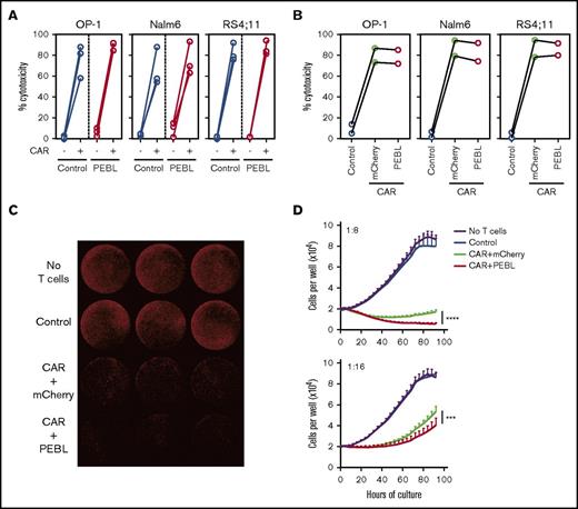
CAR expression in PEBL-transduced T cells induced strong cytotoxicity against CD19+ leukemia cell targets, regardless of whether the CAR was expressed by mRNA electroporation or viral transduction (Figure 5A-B; supplemental Figure 4). We also determined CAR cytotoxicity at low E:T ratios over longer periods, using a live-cell imaging system. CAR+PEBL-T cells were at least as effective as CAR-T cells without PEBL at exerting antileukemic cell killing, with higher cytotoxicities seen at 1:8 and 1:16 E:T (Figure 5C-D).
Cytotoxicity of CAR+PEBL T lymphocytes. (A) Four-hour cytotoxicity assays of PEBL or control (mCherry-transduced) T cells from 3 donors electroporated either with anti-CD19-41BB-CD3ζ CAR mRNA or no mRNA against CD19+ ALL cell lines at 2:1 E:T (see also supplemental Figure 4). Symbols indicate mean of 3 measurements for each donor. (B) Cytotoxicity of CAR-transduced T lymphocytes from 2 donors, sequentially transduced with a retroviral vector containing either mCherry alone or anti-CD3 PEBL was tested against CD19+ cell lines. Control, cells transduced with GFP only followed by mCherry only. Shown are data for 4-hour assays against CD19+ ALL cell lines at 1:1 E:T (full set of data in supplemental Figure 4). Each symbol indicates mean of triplicate experiments for each donor. (C-D) T lymphocytes transduced as in panel B were tested for long-term cytotoxicity against Nalm6 transduced with mCherry. Leukemia cell growth was measured with IncuCyte Zoom System (Essen BioScience). Whole-well imaging of triplicate cultures at 80 hours; E:T 1:8, is shown in panel C; leukemia cell growth measurements at the indicated E:T ratios in panel D. ***P < .001; ****P < .0001.
Cytotoxicity of CAR+PEBL T lymphocytes. (A) Four-hour cytotoxicity assays of PEBL or control (mCherry-transduced) T cells from 3 donors electroporated either with anti-CD19-41BB-CD3ζ CAR mRNA or no mRNA against CD19+ ALL cell lines at 2:1 E:T (see also supplemental Figure 4). Symbols indicate mean of 3 measurements for each donor. (B) Cytotoxicity of CAR-transduced T lymphocytes from 2 donors, sequentially transduced with a retroviral vector containing either mCherry alone or anti-CD3 PEBL was tested against CD19+ cell lines. Control, cells transduced with GFP only followed by mCherry only. Shown are data for 4-hour assays against CD19+ ALL cell lines at 1:1 E:T (full set of data in supplemental Figure 4). Each symbol indicates mean of triplicate experiments for each donor. (C-D) T lymphocytes transduced as in panel B were tested for long-term cytotoxicity against Nalm6 transduced with mCherry. Leukemia cell growth was measured with IncuCyte Zoom System (Essen BioScience). Whole-well imaging of triplicate cultures at 80 hours; E:T 1:8, is shown in panel C; leukemia cell growth measurements at the indicated E:T ratios in panel D. ***P < .001; ****P < .0001.
Downregulation of CD3 with CAR expression and function could also be effectively achieved by using a bicistronic vector containing both CAR and PEBL (supplemental Figure 5).24
Xenoreactivity and antileukemic potency of PEBL-T cells in immunodeficient mice
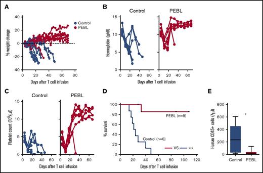
To further test the effectiveness of CD3/TCRαβ knock-out by PEBL, we infused anti-CD3 PEBL T cells in NSG mice that had received 2.5 Gy radiation and evaluated the T cells’ capacity to cause GVHD. All 8 mice injected with human T cells transduced with GFP alone exhibited weight loss, anemia, and thrombocytopenia, whereas these GVHD signs were seen in only 1 of the 8 mice injected with PEBL-transduced T cells (Figure 6A-D; P = .0003 in log-rank test of survival). Human T-cell numbers measured in their peripheral blood were markedly higher overall in mice injected with GFP-transduced T cells (Figure 6E), suggesting that PEBL suppressed T-cell stimulation by xenoantigens. The occurrence of GVHD was confirmed by pathological findings (supplemental Figure 6; supplemental Table 3).
CD3/TCRαβ knock-down by PEBL prevents GVHD. (A) NSG mice were irradiated with 2.5 Gy and IV injected 1 day later with 1 × 10⁷ T lymphocytes transduced with either anti-CD3 PEBL or GFP only (“Control”; n = 8 per group). Body weight is expressed as change relative to weight on day 3 after irradiation. (B) Hemoglobin levels and (C) platelet counts in peripheral blood. (D) Kaplan-Meier overall survival curves and log-rank test. Mice were euthanized when weight reduction exceeded 20% in 2 consecutive measurements (additional data in supplemental Figure 6 and Table 3). (E) Human CD45+ cell counts in blood 18 days after T-cell injection. *P = .0148; ***P < .001.
CD3/TCRαβ knock-down by PEBL prevents GVHD. (A) NSG mice were irradiated with 2.5 Gy and IV injected 1 day later with 1 × 10⁷ T lymphocytes transduced with either anti-CD3 PEBL or GFP only (“Control”; n = 8 per group). Body weight is expressed as change relative to weight on day 3 after irradiation. (B) Hemoglobin levels and (C) platelet counts in peripheral blood. (D) Kaplan-Meier overall survival curves and log-rank test. Mice were euthanized when weight reduction exceeded 20% in 2 consecutive measurements (additional data in supplemental Figure 6 and Table 3). (E) Human CD45+ cell counts in blood 18 days after T-cell injection. *P = .0148; ***P < .001.
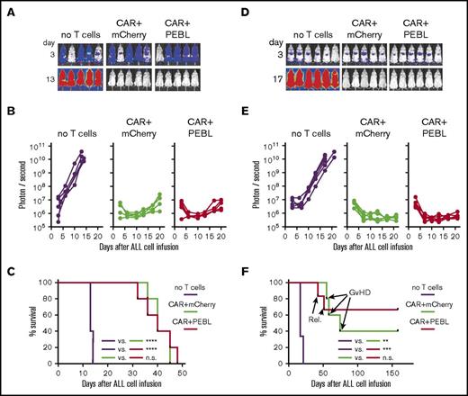
Results of in vitro experiments indicated that PEBL-transduced T cells expressing anti-CD19 CAR retained CAR-mediated cytotoxic capacity. Thus, we tested their antileukemic capacity in a xenograft ALL model. As shown in Figure 7A-C, leukemia cell growth occurred in all untreated control mice, whereas CAR+PEBL-T cells effectively killed Nalm6 leukemic cells at rates that overlapped those of CAR-T cells transduced with mCherry instead of anti-CD3ε PEBL. In a third model, we combined the conditions of the previous 2. After injecting mice with Nalm6 cells and assessing engraftment, mice were irradiated at 2.5 Gy and then treated with CAR-T cells, either transduced with PEBL or with mCherry alone. As shown in Figure 7D-E, all untreated control mice developed leukemia regardless of irradiation, whereas CAR-T cells markedly reduced leukemia burden. Notably, 3 of the 5 mice who received CAR-T cells without PEBL developed GVHD (>20% weight loss, fur loss, reduced mobility), whereas none of the 6 that received CAR+PEBL T cells did (Figure 7F). The remaining 2 mice in the CAR+mCherry group, and 4 of the 6 mice that received CAR+PEBL T cells, remain in remission more than150 days after leukemia cell engraftment (Figure 7F).
T cells with CD3/TCRαβ knock-down by PEBL and CAR expression kill leukemia cells in mice. (A) NSG mice were IV injected with 5 × 10⁵ Nalm6-luciferase cells. Three days later, mice received 2 × 10⁷ T-lymphocytes transduced with anti-CD19-41BB-CD3ζ CAR plus either PEBL or mCherry alone; other mice received tissue culture medium instead (“no T cells”). Bioluminescence images on day 3 are shown with enhanced sensitivity to illustrate Nalm6 engraftment. (B) Symbols correspond to the average bioluminescence signal in ventral and dorsal imaging. (C) Kaplan-Meier curves and log-rank test for overall survival. Mice were euthanized when the ventral and dorsal bioluminescence average signal reached 1 × 10¹⁰ photons per second. ****P < .0001. (D) NSG mice were IV injected with 5 × 10⁵ Nalm6-luciferase cells and with 2 × 10⁷ T lymphocytes on day 3 as described in panel A. Before T lymphocytes injection, mice received 2.5 Gy total body irradiation. Bioluminescence images on day 3 are shown with enhanced sensitivity to illustrate Nalm6 engraftment. (E) Symbols correspond to bioluminescence average by ventral and dorsal imaging. (F) Kaplan-Meier curves and log-rank test for overall survival. Mice were euthanized when the ventral and dorsal bioluminescence average signal reached 1 × 10¹⁰ photons per second, or when signs of GVHD (>20% weight reduction exceeded in 2 consecutive measurements, with reduced mobility and/or fur loss) were evident. GVHD occurred in 3 of the 5 CAR+mCherry mice and 0 of the 6 CAR+PEBL mice; relapse (“Rel.”) rates were 0 of 5 vs 2 of 6, respectively. **P = .0014; ***P = .0006.
T cells with CD3/TCRαβ knock-down by PEBL and CAR expression kill leukemia cells in mice. (A) NSG mice were IV injected with 5 × 10⁵ Nalm6-luciferase cells. Three days later, mice received 2 × 10⁷ T-lymphocytes transduced with anti-CD19-41BB-CD3ζ CAR plus either PEBL or mCherry alone; other mice received tissue culture medium instead (“no T cells”). Bioluminescence images on day 3 are shown with enhanced sensitivity to illustrate Nalm6 engraftment. (B) Symbols correspond to the average bioluminescence signal in ventral and dorsal imaging. (C) Kaplan-Meier curves and log-rank test for overall survival. Mice were euthanized when the ventral and dorsal bioluminescence average signal reached 1 × 10¹⁰ photons per second. ****P < .0001. (D) NSG mice were IV injected with 5 × 10⁵ Nalm6-luciferase cells and with 2 × 10⁷ T lymphocytes on day 3 as described in panel A. Before T lymphocytes injection, mice received 2.5 Gy total body irradiation. Bioluminescence images on day 3 are shown with enhanced sensitivity to illustrate Nalm6 engraftment. (E) Symbols correspond to bioluminescence average by ventral and dorsal imaging. (F) Kaplan-Meier curves and log-rank test for overall survival. Mice were euthanized when the ventral and dorsal bioluminescence average signal reached 1 × 10¹⁰ photons per second, or when signs of GVHD (>20% weight reduction exceeded in 2 consecutive measurements, with reduced mobility and/or fur loss) were evident. GVHD occurred in 3 of the 5 CAR+mCherry mice and 0 of the 6 CAR+PEBL mice; relapse (“Rel.”) rates were 0 of 5 vs 2 of 6, respectively. **P = .0014; ***P = .0006.
Discussion
We developed a method that allows rapid and efficient downregulation of CD3/TCRαβ in T cells. Anti-CD3ε PEBL transduction caused intracellular retention of CD3ε, which, in turn, prevented expression of TCRαβ on the surface of T lymphocytes. We identified PEBL constructs that had minimal or no extracellular leakage and were highly effective at blocking TCRαβ signaling. PEBL-T cells transduced with an antiviral TCR were unable to respond to a cognate viral peptide; PEBL transduction markedly lessened the capacity of human T cells to cause GVHD in mice. PEBL expression and CD3/TCRαβ downregulation was durable; it did not affect expression of other surface molecules, T-cell survival, or proliferation. Importantly, PEBL-T cells responded normally to CAR signaling and killed CAR-targeted ALL cells in vitro and in vivo.
The best PEBLs in our study contained either the KDEL or KKXX (KKMP or KKTN) retention domains, which anchor associated luminal ER proteins, preventing their secretion or membrane expression.32,33 Thus, our anti-CD3ε PEBLs blocked CD3ε assembly with the other components of the CD3/TCRαβ complex and its surface expression. KDEL peptides (such as SEKDEL) have been previously linked to scFv to block protein expression with varying efficiency in experiments performed primarily with cell lines.31,34 A protein trafficking study found that the amino acids in positions −5 and −6 beyond KDEL were important in the ER localization of soluble proteins.35 In the PEBL context, we found that the intervening sequence between scFv and KDEL was critical for its function and identified sequences that improved protein retaining compared with SEKDEL. Protein trafficking studies had also indicated that carboxyl-terminal KKXX motifs direct ER localization and that KKXX positioning in relation to the membrane was critical for its effective function.36 We found that the KKXX motif linked to the CD8α transmembrane domain constituted a robust anchoring platform for PEBLs, and that the spacer between these 2 components affected PEBL function.
Because we did not directly target TCRα or TCRβ chains, a potential concern is that low levels of TCRαβ, undetectable by flow cytometry but sufficient to induce signals, may still persist. We found, however, that T cells transduced with anti-CD3ε PEBL were generally nonresponsive to TCR-mediated signaling. Although it is possible that retention of CD3/TCR and/or PEBL could lead to their accumulation and stress response, we have been unable to detect any deleterious effects. In addition to observing normal growth of PEBL-transduced CD3/TCR-negative Jurkat cells for nearly 2 years, there was no defect in the proliferative and cytotoxic potential of PEBL-transduced T cells. Conceivably, the murine-derived scFv of PEBLs might accelerate rejection of the infused CAR-T cells. This concern, however, could be addressed by using a scFv of human origin, as has been reported for the scFv contained in CARs.37
Contemporary gene editing methods have interesting applications in CAR-T-cell therapy.15,18,20 For example, CRISPR/Cas9 was recently used to insert the anti-CD19 CAR gene into the TCRα-constant (TRAC) locus, eliminating TCRαβ expression.19,38 One of the advantages of the PEBL method is that it does not require major modifications of current protocols for clinical-grade large-scale cell processing. Because the anti-CD3 PEBL gene can be combined with the CAR gene in a single bicistronic construct, an allogeneic CAR-T cell product can be obtained after a single transduction procedure. Manufacturing T cells with PEBL and CAR expression relies on viral vector and gene components that are essentially identical to those used for CAR expression in current clinical trials. Therefore, this approach is unlikely to raise safety concerns beyond those related to standard CAR expression; uncertainties regarding the application of gene editing methodologies do not pertain. That notwithstanding, the PEBL approach can also be combined with gene editing methods to engineer CAR-T cells resistant to rejection and with higher potency.15,39,40 Another application is to block expression of T-cell antigens shared by normal and malignant T cells, thus avoiding CAR-mediated fratricide while targeting T-cell leukemias and lymphomas.41
Clinical results with autologous CAR-T cells have demonstrated their extraordinary potential.1-10 A critical next step for this technology is to improve its consistency and manufacturing, so that patients can have access to uniformly robust and timely products. To this end, methods to reliably generate allogeneic CAR-T cells are an important advance. Allogeneic cells can be available regardless of the patient immune cell status and his/her fitness to undergo apheresis. CAR-T cells could be prepared with the optimal cellular composition, high CAR expression, and maximum functional potency. Clinical observations and experimental data suggest that the risk for GVHD with allogeneic CAR-T cells may be lower than expected if CARs rely on CD28 costimulation and are infused in HLA-matched recipients.42-45 This, however, may not extend to other CARs and/or different transplant settings. Thus, grade II GVHD was reported in 2 of 3 patients who received infusion of CD137 costimulated donor CAR-T cells,46 and grade II/III GVHD in 3 of 6 patients who received infusion of haploidentical CAR-T cells costimulated with CD28, CD137, and CD27.47 In these studies, GVHD required administration of corticosteroids, which are likely to eliminate the CAR-T cells. Regardless of the relative merits of different costimulatory molecules in terms of clinical efficacy and toxicity,48 lack of TCRαβ expression reportedly can enhance antitumor activity of CAR-T cells.19 Interestingly, in our tests of long-term cytotoxicity in vitro, T cells transduced with PEBL plus CAR performed better than those with CAR alone, in agreement with this observation. Overall, removing CD3/TCRαβ from allogeneic CAR-T cells products is likely to be advantageous, particularly if it can be accomplished with minimal disruption of established manufacturing protocols.
The full-text version of this article contains a data supplement.
Acknowledgments
The authors thank Antonio Bertoletti, Duke-NUS, Singapore, for valuable material.
This work was supported by grant NMRC/STaR/0025/2015a from the National Medical Research Council of Singapore.
This study was part of T.K.’s PhD program (Director: Tomohiro Morio, Department of Pediatrics and Developmental Biology, Tokyo Medical and Dental University, Tokyo, Japan).
Authorship
Contribution: T.K. developed PEBLs, performed experiments, and analyzed data; D.W. and Y.T.P. performed experiments and analyzed data; and D.C. designed the study, analyzed data, and wrote the manuscript with T.K. and the input of the other authors.
Conflict-of-interest disclosure: T.K., Y.T.P., and D.C. are coinventors in patent applications describing the technologies used. D.W. declares no competing financial interests.
Correspondence: Dario Campana, Department of Pediatrics, National University of Singapore, Centre for Translational Medicine, 14 Medical Dr, Singapore 117599; e-mail: paedc@nus.edu.sg.

![Figure 2. Anti-CD3ε PEBLs downregulate TCRαβ. (A) TCRαβ expression in GFP-positive T lymphocytes 5 to 9 days after PEBL transduction. Mean (± standard deviation [SD]) is shown for cells transduced with GFP only (“Control”; n = 25), PEBL2 (n = 4), and PEBL5 (n = 18); other data represent results of 1 or mean of 2 experiments. (B) Flow cytometric dot-plots illustrate TCRαβ downregulation in T lymphocytes compared with cells transduced with GFP only. (C) CD3/TCRαβ expression in Jurkat cells transduced with PEBL after long-term culture; Control, cells transduced with GFP alone. (D) Collective results of CD3/TCRαβ expression in long-term cultures of T lymphocytes (with 200 IU/mL IL-2) or Jurkat cells. Symbols indicate persistence of more than 90% reduction of surface CD3/TCRαβ in GFP+ transduced cells.](https://ash.silverchair-cdn.com/ash/content_public/journal/bloodadvances/2/5/10.1182_bloodadvances.2017012823/3/m_advances012823f2.jpeg?Expires=1769094040&Signature=RDSeMbNkxOQhCWkWna5MotX6SaFGVdceYfWqPdZ~spu1~2J47Uit9R5YiKdFmgKuzvsUZPMJmTauAGId8416P8OPQuSfaCOIqnQrldWobjO0HSJsFR7OC8KAZxUyh4z9o6-wK9z2arHL6Msi5Y3KWAmqpafCY0vvTKXxFsio1dDqV4ZTyopHc-QGCeTgg98mMDiBoKc55s83OwQg2mcyTVMpmn26INqHOgKyi8~BwSC8wXo8qrqOR0YoDeLmxsmGR0Sm8d3uXaQnIWpQ0M2eOTs0jGtlYlMcxHy10Gh4WuTu9KpViawU0OX7jv0ng4qycw4a5Y6rtEzLLUiKKOLBRg__&Key-Pair-Id=APKAIE5G5CRDK6RD3PGA)
