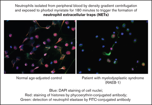
Key points
The formation of NETs, an important bactericidal mechanism, is impaired in patients with MDS.
This finding supports the notion that neutrophil dysfunction, in addition to neutropenia, contributes to the risk of infection in MDS.
Abstract
Neutrophil extracellular traps (NETs) are networks of extracellular fibers primarily composed of DNA and histone proteins, which bind pathogens. We investigated NET formation in 12 patients with myelodysplastic syndrome (MDS) and 15 age-adjusted normal controls after stimulation with phorbol-12-myristate-13-acetate (PMA). Histones and neutrophil elastase were visualized by immunostaining. Since NET formation is triggered by reactive oxygen species (ROS), mainly produced by reduced NADP-oxidase and myeloperoxidase (MPO), ROS were analyzed by flow cytometry using hydroethidine, 3’-(p-aminophenyl) fluorescein, and 3’-(hydroxyphenyl) fluorescein. On fluorescence microscopy, PMA-stimulated MDS neutrophils generated fewer NETs than controls (stimulated increase from 17% to 67% vs 17% to 85%) (P = .02) and showed less cellular swelling (P = .04). The decrease in mean fluorescence intensity (MFI) of 4’,6-diamidino-2-phenylindole, indicating chromatin decondensation, was significantly less in MDS neutrophils than controls (ΔMFI 3467 vs ΔMFI 4687, P = .03). In addition, the decrease in MFI for fluorescein isothiocyanate, indicating release of neutrophil elastase from cytoplasmic granules, was diminished in patients with MDS (P = .00002). On flow cytometry, less cell swelling after PMA (P = .02) and a smaller decrease in granularity after H2O2 stimulation (P = .002) were confirmed. PMA-stimulated ROS production and oxidative burst activity did not reveal significant differences between MDS and controls. However, inhibition of MPO activity was more easily achieved in patients with MDS (P = .01), corroborating the notion of a partial MPO defect. We conclude that NET formation is significantly impaired in MDS neutrophils. Although we found abnormalities of MPO-dependent generation of hypochloride, impaired ROS production may not be the only cause of deficient NETosis in MDS.
Introduction
Myelodysplastic syndromes (MDS) are age-related clonal bone marrow (BM) disorders characterized by a proliferative advantage at the hematopoietic stem cell level, leading to clonal dominance.1 Impaired fitness at the level of more differentiated clonal progeny causes disturbed cellular maturation and premature cell death. Therefore, hematopoiesis is ineffective, as reflected by peripheral blood cytopenias. Precursor cells in the BM show various dysplastic features, cytogenetic and molecular genetic alterations, and functional defects.2-4 The latter also affects clonal cells that manage to survive and appear in the peripheral blood. The prognosis of patients with MDS is limited by the degree of BM failure and the likelihood of clonal evolution to acute myeloid leukemia (AML).5
The clinical impact of BM failure is determined not only by the severity of cytopenia but also by the functional impairment of blood cells. For instance, patients with lower-risk MDS usually have higher platelet counts than patients with higher-risk MDS but have a similar risk of dying from severe bleeding, indicating that platelet dysfunction, in addition to thrombocytopenia, plays an important role.6 Similarly, severe infections, constituting the most common cause of death in MDS,7 are due to neutropenia as well as neutrophil dysfunction.8,9 Historically, defects in neutrophil granulocyte function were first detected in patients with MDS when these disorders were still called refractory megaloblastic anemia10 or preleukemia.11,12 A few years later, impairment of granulocyte function was described in patients with primary MDS.8,13 However, to the best of our knowledge, the formation of neutrophil extracellular traps (NETs), an important bactericidal mechanism,14 has not been studied in MDS as yet.
NETs are networks of extracellular fibers, primarily composed of DNA and histone proteins, which bind pathogens. This component of the immune system's first line of defense was first described in 2004.15 NETosis is a unique form of cell death that is characterized by the release of decondensed chromatin and granular contents to the extracellular space. The extracellular chromatin acts to immobilize microbes and prevent their dispersal in the host.14 Formation of NETs is triggered by reactive oxygen species (ROS), which in neutrophils are mainly produced by reduced NADP (NADPH) oxidase and myeloperoxidase (MPO). After activation, neutrophils undergo morphological changes, including decondensation of chromatin and disintegration of the nuclear membrane, leading to cell swelling. Prior to release into the extracellular space, chromatin is decorated with antimicrobial components from the granula, such as neutrophil elastase. The clinical importance of NETs was first recognized in the context of chronic granulomatous disease, where congenital deficiency of NADPH oxidase impairs ROS production and NET formation,16 resulting in severe, particularly fungal, infections.17 We used fluorescence microscopy to examine NET formation in patients with MDS in comparison with an age-adjusted control group and flow cytometry to further characterize NET formation and assess the production of ROS by NADPH oxidase and MPO.
Materials and methods
Isolation of peripheral blood neutrophils
Blood samples were obtained after written informed consent from MDS patients seen at the Department of Hematology, Oncology and Clinical Immunology and from age-matched individuals who underwent vascular surgery. The investigation was approved by the institutional review board, ie, the ethics committee of the Medical Faculty of Heinrich Heine University Düsseldorf (reference number 4638). Table 1 summarizes patient characteristics. Patients were selected to provide a roughly balanced representation of lower-risk and higher-risk MDS. There was no selection for distinct genetic characteristics.
MDS patient characteristics
| Case no. . | Age . | Sex . | MDS type . | Karyotype . | WBC (×1000/µl) . | ANC (×1000/µl) . |
|---|---|---|---|---|---|---|
| 1-15 | Age-adjusted controls | |||||
| 16 | 79 | m | MDS del(5q) | 47,XY, del(5)(q11q33),+21 | 9,9 | 7,7 |
| 17 | 77 | f | RCMD | 46,XX | 1,3 | 0,5 |
| 18 | 72 | f | RAEB I | 46,X,idic(X)(q13)[16]/ 46, XX[13] | 4,3 | 1,9 |
| 19 | 69 | m | RCMD RS | 46, XY | 3,5 | 2,2 |
| 20 | 79 | m | RCMD | 46,XY,del(11)(q21)[6]/ 46,XY,del(9)(q22)[4]| 46,XY[18] | 3,3 | 2,1 |
| 21 | 71 | f | RARS | 46,XX | 5,2 | 4,3 |
| 22 | 74 | m | RCMD | 46,XY | 6,2 | 3,3 |
| 23 | 71 | f | RAEB II | 46,XX | 2,7 | 0,3 |
| 24 | 79 | m | RAEB I | 46,XY | 3,4 | 1,5 |
| 25 | 75 | m | RCMD | 46,XY | 13,7 | 7,8 |
| 26 | 76 | m | RAEB II | 46,XY | 2,3 | 0,4 |
| 27 | 73 | f | RCMD | 46,XX,del(5)(q14q33)[4]/ 46,XX[13] | 2,3 | 0,9 |
| Mean | 75 | m/f (7/5) | 4,8 | 2,7 |
f, female; M, male; MDS del 5q, MDS with deletion of chromosome 5q as sole cytogenetic anomaly; RAEB I, refractory anemia with excess of blasts <10%; RAEB II, RAEB with ≥10% blasts; RARS, refractory anemia with ring sideroblasts; RCMD, refractory cytopenia with multilineage dysplasia; RCMD-RS, RCMD with ring siderolblasts.
Neutrophil granulocytes were isolated from freshly drawn EDTA-anticoagulated blood samples through density gradient centrifugation with Histopaque 1119 (Sigma Aldrich). The neutrophil fraction was washed with PBS, pelleted, and resuspended in 5 mL 0.83% ammonium chloride to lyse residual red cells. After further washing with phosphate-buffered saline (PBS), neutrophils were counted and resuspended at a concentration of 106 cells per 100 µL in RPMI culture medium plus 1% human serum albumin (HSA). In all, we analyzed 12 samples from MDS patients and 15 samples from an age-adjusted control group. Not all of the tests could be performed on all of the samples because it was not always possible to isolate sufficient numbers of neutrophils, especially in the MDS group. Density gradient centrifugation yielded substantially lower numbers of neutrophils for fluorescence microscopy in MDS patients vs age-adjusted controls, which may be attributable not only to neutropenia but also to lower robustness and altered sedimentation behavior of the MDS neutrophils.
Stimulation of NET formation
After placing coverslips (13 mm) in 24-well culture plates, 2 × 10⁵ cells (in 500 µL RPMI) were pipetted on each coverslip. After allowing the cells to sediment for 30 minutes at 37°C, they were incubated with 1.62 nM phorbol-12-myristat-13-acetat (PMA) in 500 µl RPMI + 1% HSA. PMA stimulation was stopped after 30 minutes and 180 minutes, respectively, by adding 500 µL 4% paraformaldehyde for cell fixation. PMA-treated samples and untreated controls were then kept at 4°C.
Detection of NETs by fluorescence microscopy
NET formation can be visualized through the staining of DNA with 4’,6-diamidino-2-phenylindole (DAPI), and histones and elastase with fluorescent antibodies (see Figure 3). Cells in suspension are exposed to Triton X-100 for 1 minute, washed in PBS, and incubated with primary antibodies against histones and elastase, respectively, for 1 hour. After blocking unspecific binding using 10% goat serum, samples were incubated with fluorescent secondary antibodies (goat anti-rabbit Alexa 488, goat anti-mouse Alexa 568) at room temperature, transferred to coverslips, and embedded in a mounting medium containing DAPI.
Assessment of NET formation
The proportion of granulocytes forming NETS was assessed according to a protocol established by Brinkmann and Zychlinsky.18,19 Mean fluorescence intensity (MFI) of cells was determined from 5 representative images per coverslip before and after stimulation with PMA for 30 minutes and 180 minutes. Image data were loaded into Image J/FIJI Software (https://imagej.nih.gov/ij/) and analyzed using 3 fluorescence detection channels. The proportion of NET-forming neutrophils was defined as the number of cells detected by histone staining divided by the number of cells detected by DAPI. The evaluation included 9 MDS samples and 14 age-adjusted controls. In addition, images derived from digital overlay of all 3 fluorescence channels were visually examined for the activity of NET formation using a visual analog score ranging from 0 to 3 (Figure 3). This semiquantitative visual assessment included 7 MDS samples and 11 controls.
Flow cytometric assessment of cell size and granularity
Forward scatter was used to assess cell size, and side scatter was used to assess granularity before and after stimulation with PMA. After defining cutoffs, the proportion of cells exceeding the cutoff was determined.
Flow cytometric analyses of ROS generation
In order to assess inadequate NADPH-dependent and/or MPO-dependent generation of ROS as a potential cause of decreased NET formation, flow cytometric detection of ROS was carried out in leukocytes from freshly drawn blood samples subjected to red cell lysis in ammonium chloride solution (0.83%). 1 × 10⁶ cells per aliquot in 100 µl PBS were incubated with and without ABAH (4-aminobenzoic hydrazide) and AP (4-dimethylamino-antipyrine), each at 200 µM for 20 minutes at 37°C, to inhibit MPO-dependent ROS production20,21 and with and without DPI (diphenyl-iodoniumchloride), at 20 µM for 20 minutes at 37°C, to inhibit NADPH oxidase-dependent ROS production22 (Figure 1). Subsequently, fluorochromes 3'(p-aminophenyl) fluorescein (APF) and 3'(p-hydroxyphenyl) fluorescein (HPF), each at 5 µM, were used to detect hypochloride (OCL-) (Figure 2). Superoxide anion was detected by using dihydroethidium (HE) at 25 µM.23 ROS production was stimulated with PMI, or H2O2, for assessment of MPO activity.
Diagram of ROS production, enzyme inhibitors, and fluorochromes used to measure ROS.
Diagram of ROS production, enzyme inhibitors, and fluorochromes used to measure ROS.
HPF signal is subtracted from APF signal to determine hypochloride production by MPO.
HPF signal is subtracted from APF signal to determine hypochloride production by MPO.
Data obtained by flow cytometry were captured using CellQuest Software (Becton Dickinson). Raw data were further analyzed using FCS Express Reader (De Novo Software) and Excel (Microsoft).
Results
Fluorescence microscopy
Semiquantitative visual assessment of NET formation.
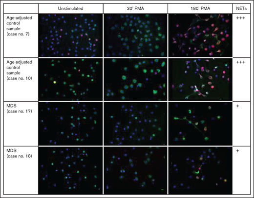
Images derived from digital overlay of all 3 fluorescence channels were visually examined for the activity of NET formation (Figure 3) (Figure 5). Particular attention was paid to the morphological appearance of cell nuclei. Characteristically, there is an early change in the nuclear-cytoplasmic ratio, followed by chromatin decondensation and dissolution of the nucleus. Neutrophil elastase (NE) escapes from azurophilic granules and translocates to the nucleus, where it partially degrades specific histones, promoting chromatin decondensation.24 Finally, chromatin and neutrophil elastase are released to the extracellular space.
Visual assessment of NET formation. NET formation was assessed microscopically in unstimulated neutrophils and after 30 minutes and 180 minutes of stimulation with PMA, in MDS patients and age-adjusted normal controls (representative examples).
Visual assessment of NET formation. NET formation was assessed microscopically in unstimulated neutrophils and after 30 minutes and 180 minutes of stimulation with PMA, in MDS patients and age-adjusted normal controls (representative examples).
Using a visual analog score ranging from 0 to 3, the assessment yielded a mean score of 2.92 in a healthy standard control that was always run in parallel with the study samples, 2.2 in the age-adjusted control samples, and 1.4 in the MDS group.
Automated assessment of NET formation.
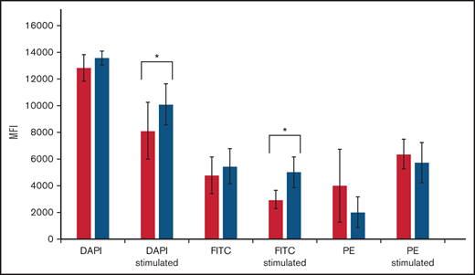
MFI for DAPI prior to PMI stimulation was slightly higher in MDS (13566 ± 530) than controls (12821 ± 1009) (statistically not significant; P = .06). After stimulation with PMI for 180 minutes, DAPI MFI decreased substantially in MDS (to 10099 ± 1530) and controls (to 8134 ± 2121). The decrease was smaller in MDS (ΔMFI 3467) vs controls (ΔMFI 4687), leading to a significantly higher DAPI MFI in MDS vs controls after stimulation (P = .03) (Figure 4). This indicates that dilution of DAPI fluorescence intensity through nuclear swelling occurred to a lesser extent in MDS samples vs controls.
Automated microscopic assessment of NET formation. MFIs were determined for DAPI (detection of nuclear swelling), PE (detection of histones), and fluorescein isothiocyanate (FITC) (detection of neutrophil elastase). Comparison of MFI pre and poststimulation with PMA for 180 minutes between age-adjusted controls (black columns) and MDS samples (gray columns). *Statistically significant difference.
Automated microscopic assessment of NET formation. MFIs were determined for DAPI (detection of nuclear swelling), PE (detection of histones), and fluorescein isothiocyanate (FITC) (detection of neutrophil elastase). Comparison of MFI pre and poststimulation with PMA for 180 minutes between age-adjusted controls (black columns) and MDS samples (gray columns). *Statistically significant difference.
MFI for PE (detection of histones) prior to PMI stimulation was significantly lower in MDS (2033 ± 1128) than controls (4024 ± 2727) (P = .05). After stimulation with PMI for 180 minutes, PE MFI increased in MDS (to 5765 ± 1486) as well as controls (to 6398 ± 1105). The increase was greater in MDS (ΔMFI 1991) vs controls (ΔMFI 633), but the difference did not reach statistical significance (P = .25) (Figure 4). The significantly lower PE MFI in MDS samples prior to stimulation suggests a denser chromatin structure of cell nuclei.
MFI for fluorescein isothiocyanate (FITC) (detecting neutrophil elastase) prior to stimulation was 5484 ± 1299 in MDS and 4804 ± 1365 in controls. After stimulation with PMI for 180 minutes, FITC MFI decreased nonsignificantly in MDS (to 5032 ± 1142) (P = .44) and significantly (to 2992 ± 671) (P = .0001) in age-adjusted controls. The difference in FITC MFI after stimulation was statistically highly significant (P = .00002) (Figure 4). This finding suggests that there was substantially less release (and thus dilution) of neutrophil elastase from cytoplasmic granules in MDS vs controls.
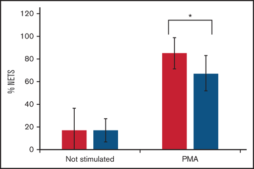
Automated image analysis prior to stimulation yielded a mean of 17% NETs in MDS samples as well as in age-adjusted controls (Figure 5). After stimulation with PMA for 180 minutes, the mean proportion of neutrophils identified as NET forming (PE positive) was lower in MDS samples (67%) than controls (85%) (P = .02).
Proportion of NETs pre and poststimulation with PMA in age-adjusted controls (black columns) and MDS samples (gray columns). *P = .02.
Proportion of NETs pre and poststimulation with PMA in age-adjusted controls (black columns) and MDS samples (gray columns). *P = .02.
Flow cytometric analyses
Flow cytometric assessment of cell size and granularity.
By flow cytometry, the reaction of neutrophils to stimulation with PMA was confirmed to differ between MDS and age-adjusted controls. Cellular swelling after stimulation with PMA was significantly less pronounced in MDS neutrophils (mean Δ28 ± 16% above the cutoff) than in control neutrophils (mean Δ47 ± 17.3% above cutoff) (P = .02). In contrast to PMA, stimulation with H2O2 did not induce significant changes in cell size in both groups.
The change in granularity after H2O2 stimulation also differed between MDS and control neutrophils. Granularity decreased in both groups due to the release of granule contents. The decrease in MDS (Δ18 ± 8% above cutoff) was significantly smaller than the decrease in controls (Δ32 ± 11% above cutoff) (P = .002).
Assessment of hypochloride (HOCl) production.
Neutrophils were exposed to H2O2 in order to provide the substrate for MPO to produce HOCl, which is known to trigger NET formation. The production of HOCl was assessed using 2 fluorochromes, APF and HPF. MFIs before and after stimulation with H2O2 were measured to determine ΔAPF and ΔHPF. The difference ΔAPF-ΔHPF was then calculated to estimate HOCl production (HPF must be subtracted to exclude OH· and ONOO- from the equation; Figure 2).25 Experiments were performed with and without MPO inhibitors ABAH and AP (Figure 6).
Flow cytometric assessment of APF and HPF fluorescence intensity for calculating HOCl production in neutrophils. Granulocytes were gated according to their forward and side scatter (dot blots). The gated cells were then presented according to side scatter and APF/HPF fluorescence intensity (fluorescein isothiocyanate [FITC]). (A) APF stimulation with H2O2 plus inhibition of MPO using ABAH/AP. (B) HPF stimulation with H2O2 plus inhibition of MPO using ABAH/AP. (C) Histograms representing the findings in (A). (D) Histograms representing the findings in (B). (A-D) A representative example of age-related controls and MDS, respectively. (E) Upon stimulation with H2O2, ΔAPF-ΔHPF is a measure of HOCl production. The difference in HOCl production between age-adjusted controls (black columns) and MDS samples (gray columns) only reached statistical significance when MPO activity was diminished by inhibitors. *P = .01.
Flow cytometric assessment of APF and HPF fluorescence intensity for calculating HOCl production in neutrophils. Granulocytes were gated according to their forward and side scatter (dot blots). The gated cells were then presented according to side scatter and APF/HPF fluorescence intensity (fluorescein isothiocyanate [FITC]). (A) APF stimulation with H2O2 plus inhibition of MPO using ABAH/AP. (B) HPF stimulation with H2O2 plus inhibition of MPO using ABAH/AP. (C) Histograms representing the findings in (A). (D) Histograms representing the findings in (B). (A-D) A representative example of age-related controls and MDS, respectively. (E) Upon stimulation with H2O2, ΔAPF-ΔHPF is a measure of HOCl production. The difference in HOCl production between age-adjusted controls (black columns) and MDS samples (gray columns) only reached statistical significance when MPO activity was diminished by inhibitors. *P = .01.
In age-adjusted controls (n = 13), mean ΔAPF with inhibitors (2057 ± 788) was significantly smaller than ΔAPF without inhibition (2932 ± 826) (P = .003). In the MDS group (n = 12), mean ΔAPF with inhibitors (1407 ± 615) was also significantly smaller than ΔAPF without inhibition (2549 ± 1027) (P = .002).
In age-adjusted controls, mean ΔHPF with inhibitors (151 ± 68) was significantly smaller than ΔHPF without inhibition (959 ± 322) (P = 4.4 × 10⁻¹⁰). In the MDS group, mean ΔHPF with inhibitors (152 ± 99) was also significantly smaller than ΔHPF without inhibition (983 ± 376) (P = 1.0 × 10⁻⁷). As shown in Figure 6D, the blue curves generally displayed 2 peaks, in MDS as well as controls, suggesting that there are 2 subpopulations of granulocytes differing in their response to ABAH/AP.
Without MPO inhibition, the calculated H2O2-induced HOCl production was not significantly different between MDS and controls (P = .31). However, when MPO activity was reduced through inhibition with ABAH and AP, this intervention caused a greater decrease in HOCl production in MDS neutrophils vs controls (P = .01).
Assessment of O2-production.
The production of O2- was measured using the fluorochrome dihydroethidium (HE). Neutrophils were stimulated with PMA, with or without DPI present (an inhibitor of NADPH oxidase).
In the age-adjusted control group, MFI for HE after PMA stimulation was 95 ± 30, corrected for autofluorescence. In the presence of DPI, MFI was only 15 ± 6 (P = 2.9 × 10⁻¹¹).
In the MDS group, MFI for HE after PMS stimulation was 124 ± 44. In the presence of DPI, MFI was only 21 ± 12 (P = 1.2 × 10⁻⁸). The results were not significantly different between MDS and controls.
Discussion
In patients with myelodysplastic syndrome, neutrophils may be deficient not only in number but also in microbicidal capacity.8,12,13 Such functional impairment of granulocytes is thought to increase the risk of severe infections, which are the major cause of death in MDS.7 However, to the best of our knowledge, the capacity to form NETs, an important component of the immune system's first line of defense,26 has never been investigated in granulocytes from patients with MDS.
Using fluorescence microscopy, we examined NET formation in MDS neutrophils in comparison with neutrophils from age-matched controls. Automated image analysis according to a protocol established by Brinkmann and Zychlinsky18,19 determined the proportion of NET forming neutrophils as the number of cells detected by histone staining divided by the number of cells detected by DAPI staining of nuclear DNA. This approach is based on the observation that NET formation is associated with nuclear swelling and chromatin decondensation, thus exposing histone proteins and making them accessible to histone-targeted antibodies. We found that prior to stimulation with PMA, the proportion of NETS was 17% in MDS samples as well as in age-adjusted controls. After stimulation with PMA for 180 minutes, the mean proportion of NETs was lower in MDS (67%) than controls (85%). The difference was statistically significant (P = .02) but did not fully reflect our visual perception of markedly diminished NET forming capacity in MDS samples.
We therefore performed a semiquantitative assessment of images derived from a digital overlay of 3 fluorescence channels, reflecting cell nuclei (DAPI), histones (FITC), and neutrophil elastase (HE). Considering nuclear swelling, changes in the nuclear-cytoplasmic ratio, appearance of histone staining, nuclear membrane dissolution, release of neutrophil elastase from cytoplasmic granules, and release of chromatin and elastase to the extracellular space, a visual analog score of NET formation was applied, ranging from 0 to 3. Our assessment yielded a mean score of 2.92 in young healthy controls, 2.2 in the elderly age-adjusted control group, and 1.4 in the MDS group. In addition, flow cytometric assessment of forward and side scatter, respectively, showed less cellular swelling after PMA stimulation and less cytoplasmic degranulation after H2O2 stimulation in MDS vs controls. We feel confident that NET formation in MDS patients is significantly impaired in comparison with age-adjusted controls.
We feel less confident about the cause of impaired NET formation. Starting from the well-described phenomenon of partial MPO deficiency that is detectable by cytochemical staining or flow cytometric analysis27 of neutrophils in many MDS patients, we hypothesized that impaired NET formation may be caused by impaired ROS production, which is at least partly dependent on MPO activity.28-30 We therefore used flow cytometry to examine the production of ROS by NADPH oxidase and MPO. Stimulated production of superoxide anion (O2-) by NADPH oxidase, tested with dihydroethidium, was not significantly different between MDS and age-adjusted controls. Regarding MPO, the calculated H2O2-induced production of HOCl was not significantly different between MDS and controls (P = .31). However, when we partially impeded MPO activity by adding ABAH/AP,31 we observed a greater impairment in HOCl production in MDS neutrophils vs controls (P = .01). This would appear to be consistent with a preexisting partial MPO deficiency.
However, we do not seek a monocausal explanation for impaired NET formation in MDS. MDS are a heterogeneous group of BM disorders characterized by a wide spectrum of acquired somatic mutations.4,32,33 The most common mutations affect components of the RNA splicing apparatus, causing a multitude of aberrantly spliced messenger RNAs. The resulting dysregulation of cellular processes34 provides ample opportunity beyond impaired ROS production to impede NETosis of neutrophils. It would be interesting to investigate possible correlations between NET formation and distinct genetic MDS subtypes. Considering the variety of cytogenetic aberrations and somatic mutations detectable in MDS, such an analysis would require the examination of a large number of patients.
NET formation may play a role in other myeloid malignancies, too. While AML would not appear to be the preferred object of study, since AML blasts are not expected to form NETs, and residual granulocytes may not represent the neoplastic clone, myeloproliferative neoplasms (MPNs) have indeed been looked at. Interestingly, in contrast to MDS, Wolach et al discovered that neutrophils from patients with MPNs are primed for NET formation, an effect blunted by pharmacological inhibition of JAK signaling with ruxolitinib, which abrogated NET formation and reduced thrombosis in a mouse model.35
While the mutational landscape of MDS is now well described, the functional consequences of mutations are not fully understood. Our observation of impaired NET formation adds to the overall picture of functional cellular deficits and helps to explain why MDS patients are susceptible to life-threatening infections. Unfortunately, to the best of our knowledge, there are no drugs that could be used for improving the capability of neutrophils to form NETs. While granulocyte colony-stimulating factor comes to mind, clinical experience has taught us that granulocyte colony-stimulating factor does not prevent infection in patients with MDS.36,37
Authorship
Contributions: C.B. and N.G. designed the research and wrote the paper; C.B., J.F., and P.C. performed the experiments and analyzed the data; U.G. collected the clinical data; and R.H. helped to write the paper.
Conflict-of-interest disclosure: The authors declare no competing financial interests.
Correspondence: Carolin Brings, Department of Vascular and Endovascular Surgery, Heinrich Heine University, Moorenstr. 5, 40225 Düsseldorf, Germany, carolin.brings@med.uni-duesseldorf.de; and Norbert Gattermann, Department of Hematology, Oncology and Clinical Immunology, Heinrich Heine University, Moorenstr. 5, 40225 Düsseldorf, Germany, gattermann@med.uni-duesseldorf.de
References
Author notes
For data sharing, please contact the corresponding authors at carolin.brings@med.uni-duesseldorf.de or gattermann@med.uni-duesseldorf.de.


![Flow cytometric assessment of APF and HPF fluorescence intensity for calculating HOCl production in neutrophils. Granulocytes were gated according to their forward and side scatter (dot blots). The gated cells were then presented according to side scatter and APF/HPF fluorescence intensity (fluorescein isothiocyanate [FITC]). (A) APF stimulation with H2O2 plus inhibition of MPO using ABAH/AP. (B) HPF stimulation with H2O2 plus inhibition of MPO using ABAH/AP. (C) Histograms representing the findings in (A). (D) Histograms representing the findings in (B). (A-D) A representative example of age-related controls and MDS, respectively. (E) Upon stimulation with H2O2, ΔAPF-ΔHPF is a measure of HOCl production. The difference in HOCl production between age-adjusted controls (black columns) and MDS samples (gray columns) only reached statistical significance when MPO activity was diminished by inhibitors. *P = .01.](https://ash.silverchair-cdn.com/ash/content_public/journal/bloodadvances/6/1/10.1182_bloodadvances.2021005721/5/m_advancesadv2021005721f6.png?Expires=1769163378&Signature=H58UNsi52W3WEEZ-CUUY7xl6dCXx7bpjb3unsG4251~skLQB08LDBUPU-L~h1gGu4IuT2xNP~Zn7iC8~a0IVCv-d-kdiuptA89CUbUgq1C2KMolL2OoId-SR7kbTkOfIY0H9nsdQHi88~lZHc2Xih~bf9KQQGL4qgSmvVthapZxA~5cxWR~Lx38W9NBMAPjeRBjwyg7TJGkfBZ45iwqkYGDmkoKcVRzhaQ22esq3Cj6KNwBs7rKI1HfgsWP2OZA0VeQpcq1SL66w4KC6a9u~~iGv-v3IvUcsvGsHc4xXHuS3kUMVxQfGfpEIyCPg-YheCDmdmYOsc0LZlAP7vKIU6w__&Key-Pair-Id=APKAIE5G5CRDK6RD3PGA)