TO THE EDITOR:
Transient abnormal myelopoiesis (TAM) is a myeloid proliferative condition with leukemic potential, almost exclusively seen in infants with trisomy 21 (Down syndrome [DS]). We report here an unusual case of a neonate without DS who presented with a petechial rash and a highly elevated white blood cell (WBC) count with myeloid blasts, initially concerning for acute myeloid leukemia (AML). Trisomy 21 and a GATA1 frameshift mutation were present in the myeloid blast-predominant peripheral blood, leading to the diagnosis of TAM. Trisomy 21 was absent in the lymphocytes and skin fibroblasts, supporting the diagnosis of mosaic DS in this infant. This case highlights the importance of rapid cytogenetic and molecular diagnostic techniques in the evaluation of neonatal leukemia.
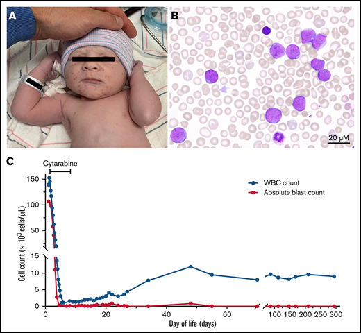
The patient, a phenotypically normal male infant, was born at 37 weeks via cesarean delivery after an uneventful pregnancy with full prenatal care, including normal prenatal cell-free DNA screening. A petechial rash on face, scalp, and torso was present at birth (Figure 1A). He did not have hepatosplenomegaly, and results of a physical examination were otherwise normal. A complete blood count showed a WBC count of 112 × 103 cells/µL with 76% blasts (Figure 1B), a hemoglobin of 10 g/dL, and a platelet count of 65 × 103 cells/µL. Over the next 24 hours, the WBC count increased to 153 × 103 cells/µL, and signs of tumor lysis syndrome developed, with peak uric acid of 7.1 mg/dL and lactate dehydrogenase of 1733 U/L. There was no coagulopathy. Peripheral blood flow cytometry showed 84% myeloid blasts expressing: CD45 (dim), CD34, CD117, CD33, CD7, CD4 (subset dim), CD56 (small subset), CD123 (dim), CD36, CD38, CD41 (small subset with weak expression), CD61 (small subset with weak expression), and CD71 (Table 1). The patient was diagnosed with acute myeloid leukemia (AML) with partial megakaryoblastic differentiation and started on 100 mg/m2 cytarabine every 12 hours for cytoreduction, given the rapidly rising WBC counts. Given the newborn status, TAM was considered in the differential diagnosis.
Patient presentation and acute leukemia evaluation. (A) Petechial rash on face, scalp, and torso was present at birth and prompted evaluation with a complete blood count. (B) Peripheral blood smear shows presence of myeloblasts. (C) Graph shows white blood cell and absolute blast count over time, highlighting response to cytarabine therapy.
Patient presentation and acute leukemia evaluation. (A) Petechial rash on face, scalp, and torso was present at birth and prompted evaluation with a complete blood count. (B) Peripheral blood smear shows presence of myeloblasts. (C) Graph shows white blood cell and absolute blast count over time, highlighting response to cytarabine therapy.
Test results at time of diagnosis
| Diagnostic test . | Tissue type . | Findings . |
|---|---|---|
| Flow cytometry | Peripheral blood | Population of immature cells (comprising ∼84% of viable cells): Positive for CD45 (dim), CD34, CD117, CD33, CD7, CD4 (subset dim), CD56 (small subset), CD123 (dim), CD36, CD38, CD41 (small subset with weak expression), CD61 (small subset with weak expression), CD71. Negative for myeloperoxidase, HLA-DR, CD13, CD15, CD16, CD14, CD64, CD65, CD11b, sCD3, CD2, CD5, CD8, CD10, CD19, and CD20. |
| FISH | Peripheral blood | Positive for 3 copies of RUNX1 in 88% of examined cells. Negative for RUNX1T1/RUNX1, KMT2A, PML/RARA or CBFB rearrangements, monosomy 5, monosomy 7, and trisomy 8. |
| Cytogenetics | Peripheral blood | 47,XY, + 21[20] |
| Lymphocytes | 46,XY[8] | |
| Skin fibroblasts | 46,XY[50] | |
| NGS panel | Peripheral blood | GATA1 (NM_002049) c.136_137insCCTCCACTGCCCCGAG (p.T52Lfs*21): 80.1% variant allele frequency. Copy number analysis: gain of RUNX1, ERG, U2AF1 (on 21q). |
On day-of-life (DOL) 4, fluorescence in-situ hybridization (FISH) of cells from peripheral blood obtained on DOL1 demonstrated 3 copies of RUNX1, raising suspicion of trisomy 21. On DOL6, a targeted next-generation sequencing (NGS) panel revealed a GATA1 frameshift mutation and confirmed a gain of chromosome 21 (Table 1). This constellation of findings was diagnostic of TAM. With improvement in WBC, cytarabine was discontinued on DOL6. The patient developed chemotherapy-induced pancytopenia and was treated with supportive care. Peripheral myeloid blasts were detected through 2 months of age and then resolved (Figure 1C). A metaphase karyotyping of peripheral blood at the time of diagnosis showed trisomy 21, whereas phytohemagglutinin-stimulated lymphocytes and skin fibroblasts had a normal male karyotype (Table 1). GATA1 mutation testing was not performed at the time of remission. Given the risk of developing AML after TAM, the patient has been observed closely by an oncologist and remains healthy at 14 months of age.
For leukemia diagnosis, flow cytometry (Boston Children’s Hospital, Boston, MA), FISH (Integrated Oncology, New York, NY), cytogenetics (Integrated Oncology, New York, NY), and rapid heme panel (Brigham and Women’s Hospital, Boston, MA) were performed. The rapid heme panel assessed selected exons in 88 genes recurrently mutated in hematologic neoplasms. Extracted DNA is submitted to NEB next direct chemistry (New England Biolabs, Inc, Ipswich, MA) with dual indices and unique molecular identifiers. Sequencing was performed on the NextSeq 550Dx system (150-bp paired-end sequencing; Illumina, San Diego, CA). Our patient’s family gave written consent for this manuscript and provided the photo in Figure 1A.
TAM is caused by the interaction of trisomy 21 (T21) and acquired mutations of the zinc finger hematopoietic transcription factor GATA1. During the fetal period, hematopoiesis primarily occurs in the liver rather than the bone marrow. T21 in fetal liver hematopoietic stem cells impairs lymphocyte differentiation and causes increased proliferation of megakaryocyte-erythroid progenitors.1 These progenitor cells can subsequently acquire N-terminal mutations (typically, frameshift or splice variants) in the transcription factor GATA1.2,3 Truncation of the GATA1 N-terminal domain, which is necessary for terminal erythroid and megakaryocytic differentiation, results in TAM, a myeloproliferative disorder resembling AML with a megakaryoblastic immunophenotype.4-6
TAM occurs in up to 10% of neonates with DS.1 Although most of the individuals with DS (90% to 95%) have constitutional T21, 2% to 4% have chromosome 21 translocations, and another 1.3% to 5% have somatic T21 mosaicism.7 T21 mosaicism is rare, with an estimated frequency of 1 in 16 670 to 1 in 41 670 live births; however, low-level mosaicism may have a subtle or undetectable phenotype, and thus may not be diagnosed.9 Neonates with T21 mosaicism are at risk for T21-associated complications, such as TAM, in affected tissues, although recognition of this entity can be challenging in the absence of the phenotypic features of DS.8,9
In most cases, there is extinction of the abnormal myeloid clone early in infancy, and TAM self-resolves within months.10 Cytoreductive treatment with leukapheresis or chemotherapy may be necessary in cases of life-threatening complications, such as cardiorespiratory compromise, hyperleukocytosis, renal or liver dysfunction, disseminated intravascular coagulopathy, hydrops fetalis, or hyperviscosity.10 In about 10 to 20% of TAM cases, the TAM clone acquires additional mutations in genes involved in transcription regulation (RAD21, STAG1/2, SMC1/3 or CTCF), epigenetic modification (EZH2, KANSL1 or SUZ12), or cellular signaling (JAK1/2/3, RAS, MPL or KIT), and TAM progresses to AML.2,6,11,12 The megakaryoblasts of DS-associated AML (DS-AML) and TAM have similar morphology and immunophenotype.13 Patients who develop AML have the same GATA1 mutation as detected during TAM, highlighting the clonal evolution of this disease.13 GATA1 mutations are not present during remission of TAM or DS-AML, suggesting that they are disease specific.13
Though TAM is often self-resolving, follow-up is crucial. As noted, up to 20% of patients with TAM develop AML within the first 5 years of life; the risk is most likely similar for a patient with TAM and mosaic DS, although infrequent occurrence and variable levels of mosaicism limit investigation.10,14 Thus, patients with TAM should be followed closely with physical examinations and serial complete blood counts for development of DS-AML. Outcomes for DS-AML are favorable as compared with non–DS-AML, with long-term survival of 74% to 91%.15
The clinical suspicion of TAM in a phenotypically normal infant and integration of rapid NGS testing were critical in the diagnosis of our patient. Two key findings led to the diagnosis of TAM: the presence of T21 on FISH, cytogenetics, and NGS, and GATA1 mutation on NGS. T21 is a rare finding in non-DS myeloid leukemia, present in ∼5% of overall pediatric cases of AML,4,16 and its presence should raise suspicion for TAM or DS-AML, even in the absence of DS.17 GATA1 mutations are nearly pathognomonic for TAM and DS-AML.18-22
In summary, TAM should be included in the differential diagnosis for neonatal leukemia, even in the absence of DS phenotypic features. The presence of T21 within myeloid blasts is uncommon and should prompt consideration of TAM or DS-AML in an older child. Access to a rapid NGS platform for assessment of GATA1 facilitates the diagnosis of TAM. Early identification of TAM allows for proper therapy and ensures appropriate screening for future development of leukemia.
Acknowledgments: This work was supported by National Institutes of Health (NIH), National Cancer Institute grant K08 CA222684 (Y.P.) and NCI research training grant T32-CA136432-12 (R.A.-B.) and by a Hood Foundation grant (Y.P.).
Authorship: Y.P. and R.A.-B. conceptualized the manuscript; R.A.-B. performed the literature search and drafted the original manuscript, figure, and table; F.W. and Y.P. critically revised the original manuscript; R.C. provided the diagnostic pathology evaluation; N.I.L. and A.S.K. performed the NGS testing; B.A.D. provided disease-specific expertise and recommendations; K.D. provided ongoing clinical data; and all authors were involved in the writing of the manuscript and approved of the final version.
Conflict-of-interest disclosure: The authors declare no competing financial interests.
Correspondence: Yana Pikman, Dana-Farber Cancer Institute, 450 Brookline Ave, Boston, MA 02215; e-mail: yana_pikman@dfci.harvard.edu.
References
Author notes
Requests for data sharing may be submitted to Yana Pikman (yana_pikman@dfci.harvard.edu).

