Key Points
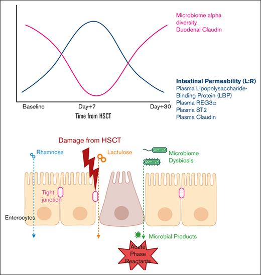
L:R ratios rise after conditioning, independent of regimen or diagnosis, and return to baseline by day +30 in most patients receiving allogeneic HSCT.
Intestinal permeability correlates directly with LBP levels and inversely with dysbiosis.
Abstract
Intestinal permeability may correlate with adverse outcomes during hematopoietic stem cell transplantation (HSCT), but longitudinal quantification with traditional oral mannitol and lactulose is not feasible in HSCT recipients because of mucositis and diarrhea. A modified lactulose:rhamnose (LR) assay is validated in children with environmental enteritis. Our study objective was to quantify peri-HSCT intestinal permeability changes using the modified LR assay. The LR assay was administered before transplant, at day +7 and +30 to 80 pediatric and young adult patients who received allogeneic HSCT. Lactulose and rhamnose were detected using urine mass spectrometry and expressed as an L:R ratio. Metagenomic shotgun sequencing of stool for microbiome analyses and enzyme-linked immunosorbent assay analyses of plasma lipopolysaccharide binding protein (LBP), ST2, REG3α, claudin1, occludin, and intestinal alkaline phosphatase were performed at the same timepoints. L:R ratios were increased at day +7 but returned to baseline at day +30 in most patients (P = .014). Conditioning regimen intensity did not affect the trajectory of L:R (P = .39). Baseline L:R ratios did not vary with diagnosis. L:R correlated with LBP levels (r2 = 0.208; P = .0014). High L:R ratios were associated with lower microbiome diversity (P = .035), loss of anaerobic organisms (P = .020), and higher plasma LBP (P = .0014). No adverse gastrointestinal effects occurred because of LR. Intestinal permeability as measured through L:R ratios after allogeneic HSCT correlates with intestinal dysbiosis and elevated plasma LBP. The LR assay is well-tolerated and may identify transplant recipients who are more likely to experience adverse outcomes.
Introduction
Intestinal permeability may be associated with posttransplant outcomes
There is growing evidence that dysregulated intestinal physiology contributes to adverse outcomes during hematopoietic stem cell transplantation (HSCT). Cytotoxic conditioning for HSCT causes gut mucosal barrier injury and dysregulation of the intestinal microbiome.1,2 Dysbiosis is associated with morbidity and nonrelapse mortality after transplant,3-5 particularly related to graft versus host disease (GVHD)6-9 and bloodstream infections (BSIs).10-12 REG3α (regenerating family member–3 alpha), a marker of intestinal damage from gastrointestinal GVHD (GI-GVHD), and ST2 (suppression of tumorigenicity–2 protein) are biomarkers predicting GVHD risk and treatment response.13-16 The inflammatory response to bacterial lipopolysaccharide (LPS) is mediated by LPS binding protein (LBP), and circulating LBP levels are high in patients with bacterial or fungal sepsis, including oncology patients having febrile neutropenia.17-19 Impaired intestinal barrier function may be a mechanistic link between altered microbial composition, intestinal damage, and elevated levels of circulating LBP, allowing translocation of pathogenic microbes or microbial products. However, the association of impaired intestinal barrier function with outcomes after HSCT is not well-delineated because measurement of intestinal barrier function is rare, likely because of lack of a convenient clinical assay. Typical assays measure fractional excretion of orally ingested probes, most commonly saccharides such as lactulose, mannitol, and rhamnose. These decades-old techniques require urine collection from 5 to 24 hours and ingestion of 50 to 100 mL solution of 51Cr-EDTA, lactulose-mannitol, or lactulose-rhamnose (including 5g lactulose),20-23 which is not feasible in peritransplant patients experiencing significant mucositis, vomiting, or diarrhea. These assays are even less feasible in children.
Interaction between intestinal barrier and microbiome
Epithelial tight junctions of the intestinal tract closely regulate the passage of small molecules. Intestinal barrier injury can be generated by inflammation from inflammatory bowel disease,24-26 microbial toxins,27,28 or chemotherapy21,29-32 but can be ameliorated by intestinal flora. p40 and p75 proteins produced by Lactobacillus rhamnosus regulate intestinal epithelial cell survival and stabilize tight junction proteins.33,34 Furthermore, microbial metabolites butyrate and tryptophan also regulate intestinal epithelial function.35,36 Studies conducted in patients with autoimmune diseases, such as multiple sclerosis and type 1 diabetes, and acute pathologies such as pancreatitis report both increased intestinal permeability and dysbiosis; however, the causal and temporal relationships remain unclear.37-39
Challenges of direct intestinal permeability quantification
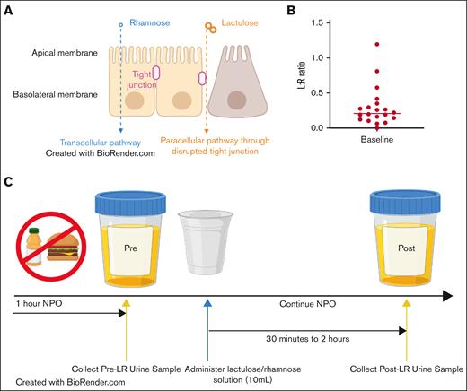
Clinical measurement of intestinal permeability requires active participation and is challenging in patients undergoing HSCT, especially during the period of mucositis when barrier function is likely altered maximally. Traditional saccharide-based permeability assays rely on the different molecular weights and volumes of the disaccharide, lactulose and monosaccharides, mannitol and L-rhamnose, which are absorbed by the gut but are poorly metabolized.40 The smaller size of monosaccharides enables transcellular transport through the intestinal epithelium, whereas lactulose can only permeate when tight junctions between enterocytes are disrupted (Figure 1A). Using a ratio of disaccharide:monosaccharide, therefore, distinguishes intestinal absorption pathways. Compared with mannitol, L-rhamnose is less often added to food and thereby less likely to contaminate human samples.41
Study background and schema. (A) Rationale of saccharide ratios to measure intestinal permeability. (B) Urine lactulose:rhamnose (L:R) ratios of healthy control children after administration of lactulose/rhamnose solution. Horizontal line represents median. An abnormal L:R ratio is >1. (C) Study schema and timeline.
Study background and schema. (A) Rationale of saccharide ratios to measure intestinal permeability. (B) Urine lactulose:rhamnose (L:R) ratios of healthy control children after administration of lactulose/rhamnose solution. Horizontal line represents median. An abnormal L:R ratio is >1. (C) Study schema and timeline.
Novel validated intestinal permeability protocol in children
Faubion et al refined the traditional saccharide permeability assay and validated it in infants in the United States, Zambia, and Peru to study environmental enteric dysfunction.42 The accuracy of high-performance liquid chromatography mass spectrometry for detection of urinary lactulose:rhamnose (L:R) levels allowed for a 5-fold dose and volume reduction of LR solution. Urine collection was also shortened to 2 hours after LR ingestion to focus on small intestinal permeability, and evaluable samples were obtained from 87% of participants. This modified LR protocol was well-tolerated by young children and applicable in both resource-abundant and resource-limited settings, and its results correlated with environmental enteritis markers.
Surrogates of direct intestinal permeability measurements
Others have addressed the difficulty of clinical intestinal permeability measurements by investigating small molecule regulators, correlative markers, and tight junction components. Plasma levels of zonulin, a modulator of intercellular tight junctions, have been used as a measure of intestinal permeability in several studies.43,44 There are 3 genotypes of zonulin, also known as prehaptoglobin2, of which HP1-1 homozygotes do not produce active zonulin, whereas HP1-2 and HP2-2 individuals do.45 HP2 genotypes are more frequent in celiac disease and inflammatory bowel disease cohorts.46,47 Loss of expression of another molecule, intestinal alkaline phosphatase (IAP), is associated with decreased expression of intestinal junctional proteins and barrier function in mice, and in humans biallelic mutations in IAP cause inflammatory bowel disease.48-51 Tissue expression of tight junction proteins, claudin and occludin, are known to change with inflammation,52-56 and circulating levels are being explored in inflammatory and infectious states.57-60 In contrast to the above surrogate markers, LR excretion is a direct measure of intestinal permeability.
Study objectives
We take advantage of the refined LR protocol from the study by Faubion et al to test the hypothesis that intestinal permeability in pediatric and young adult allogeneic HSCT recipients increases during transplant and that subjects with increased intestinal permeability are more likely to have dysbiosis. We also wanted to investigate zonulin genotypes as a potential risk factor for outcomes after HSCT and to compare L:R ratios with LBP, IAP, claudin, occludin, REG3α, and ST2 to understand how altered gut permeability correlates with existing markers of adverse outcomes.
Methods
Lactulose/rhamnose assay
Patients scheduled to undergo allogeneic HSCT at Cincinnati Children’s Hospital Medical Center (CCHMC) were recruited between August 2019 and November 2021. Patients were excluded if they were unable to take anything via mouth or nasogastric, gastric or jejunal tube. Participants were separately enrolled on a biobank repository study, from which blood and stool samples were obtained. LR solution consisted of 10 mL water, 1.0 g lactulose, and 0.2 g L-rhamnose (TCI America). Participants gave 1 urine sample before LR ingestion and were nil per os for a minimum of 1 hour before ingestion of the LR solution until the urine sample after LR ingestion was obtained. The test was administered at evaluation before transplantation, day +7 (±7 days) and day +30 (±7 days) (Figure 1B). Urine samples were collected between 30 minutes and 2 hours after ingestion of LR solution. The L:R ratio was calculated by dividing urinary lactulose with rhamnose levels, which were determined using high-performance liquid chromatography mass spectrometry at the Mayo Clinic Immunochemical Core Lab. Twenty additional healthy children were recruited as control participants for evaluation of L:R assay performance at our institution. Demographic and transplant data were collected prospectively, and outcomes were followed until day +180. All HSCT recipients were prospectively monitored for transplant-associated thrombotic microangiopathy (TA-TMA) according to our standard risk stratification classification, and participants without high-risk features did not receive targeted TA-TMA therapy.61
Microbiome data analysis
Shotgun metagenome sequencing methods may be found in supplemental Materials and Methods.62-64 Species that accounted for <0.01% of the study-wide microbial population were excluded. Three samples with 250 000 reads aligned at the species level were likewise excluded. Principal component analysis was performed on a Bray-Curtis distance matrix calculated from normalized species abundance data using the ade4, Vegan and FactoExtra packages in R.
ELISA
Plasma levels of ST2 and REG3α (R&D Systems), claudin1 and occludin (MyBioSource), LBP (Abcam), and IAP (MyBioSource) at the same timepoints as the L:R assays were measured using enzyme-linked immunosorbent assay (ELISA).
In a distinct cohort of 43 recipients after HSCT retrospectively identified to have developed BSIs or mucosal barrier injury–related laboratory-confirmed bloodstream infections (MBI-LCBI), plasma LBP levels were measured via ELISA and ferritin levels were measured via chemiluminescence (Siemens Atellica) from samples serially collected and stored in our transplant biorepository at baseline before start of transplant conditioning, 14 and 7 days before the infection, on the day of infection, and 7 days after.
Mice
C57BL/6 (JAX stock #380051) congenic mice were purchased from the Jackson laboratory. C57BL/6 congenic mice (12-week-old) were used as recipients for HSCT experiments and were lethally irradiated (11.75Gy delivered in split doses 3 hours apart) and retro-orbitally injected with 300 000 whole bone marrow cells from C57BLl/6 wild-type donors. Fourteen days after transplantation, mice were killed. Control C57BL/6 mice who were neither exposed to radiation nor transplanted were killed at 14 weeks, and organs from both groups were harvested for immunohistochemistry (supplemental Materials and Methods).
Zonulin genotyping
To test if zonulin genotypes influenced intestinal permeability and whether we needed to incorporate testing in our prospective cohort, we obtained available samples taken before transplantation from 116 consecutive patients who underwent allogeneic HSCT from our biorepository. These patients were distinct from our prospective cohort of patients. For more details refer to the supplemental Materials and Methods.65
Statistics
Primary outcomes included changes in peritransplant intestinal permeability, correlation of L:R values with LBP and known biomarkers REG3α and ST2, and correlation of L:R values with changes in microbiome. Secondary outcomes included correlation of L:R values with D+100 incidence of AGVHD, BSI, TA-TMA, and correlation of L:R with other markers of interest. The Mann-Whitney U test was used to calculate the differences between medians. Fisher exact test was used to compare rates of adverse outcomes. For immunohistochemistry analysis, differences between means were compared using 2-tailed unpaired t-test. Statistical significance of differences in overall microbiome composition were determined using multiresponse permutations procedures, a form of PERMANOVA.66 Fold-change and log2 fold-change were calculated with the gtools package in R. Stool samples at all time points between patients with normal and abnormal intestinal permeability were grouped for comparison. Difference in species abundance between individuals with normal vs elevated L:R were identified with shrinkage linear discriminant analysis.67 Significance of the association was tested using Wilcoxon rank sum. Examination of clinical covariates with microbial abundance was performed using zero inflated negative binomial generalized linear mixed models (ZINB-GLMM68) using the “glmer” command from the lme4 package in R and performed nonparametric tests (Mann-Whitney U test or Kruskal-Wallis test), followed by Bonferroni correction for multiple testing. Each microbial species was subjected to ZINB-GLMM with L:R ratio (as continuous variable), underlying diagnosis, conditioning regimen intensity, antibiotic exposure, BSI, GVHD, and TMA as fixed variables and subject as random variable. Estimates are reported for each species and fixed variable. P-values were subjected to false-discovery rate correction for multiple testing.
Study approval
Human studies research was approved by CCHMC institutional review board. All participants provided written informed consent, and pediatric participants aged ≥11 provided assent. All animal experiments were approved by the CCHMC Institutional Animal Care and Use Committee.
Results
Intestinal permeability in healthy children
We sought to confirm the threshold of a L:R ratio ≥1 for abnormal intestinal permeability established by Faubion et al42 and determine the baseline environmental contamination of lactulose and L-rhamnose in 20 healthy pediatric participants (Figure 1B-C; supplemental Figure 1A). In our study, urine samples after LR ingestion collected from patients with urine levels before LR ingestion of lactulose >5 μg/mL and rhamnose >10 μg/mL were considered unevaluable, and an L:R ratio ≥1 was considered abnormal.
Participant demographics and outcomes
Eighty patients who received allogeneic HSCT were enrolled prospectively on the permeability study (Table 1). The median age of participants was 9.4 years (range 0.5-37.9). Eleven young adults (13.8%) aged >18 participated. No participants experienced adverse gastrointestinal effects after ingesting LR. All but 1 provided a urine specimen after LR ingestion before initiation of the preparative regimen. Fifty-six participants (70%) provided urine specimens at all 3 timepoints, including 43 who also provided stool samples. No bladder catheterizations were performed to obtain samples. Sixteen participants (20.0%) developed acute GVHD by day +100. Skin GVHD was most common (81.3% of participants with GVHD). Only 6 participants (31.6%) had GI-GVHD. BSI occurred in 21 participants by day +30 (26.3%). Fourteen participants (17.5%) developed MBI-LCBI. Thirty-two participants (40.0%) met criteria for moderate or high-risk TA-TMA.
Demographic and HSCT data of study participants
| Characteristic . | Number (n = 80) . |
|---|---|
| Median age (range), y | 9.4 (0.5-37.9) |
| Female | 28 |
| Underlying diagnosis | |
| Malignancy | 32 |
| Primary immune deficiency | 17 |
| Hemoglobinopathy | 4 |
| Metabolic disorder | 4 |
| Bone marrow failure | 22 |
| Platelet function disorder | 1 |
| Conditioning regimen | |
| Myeloablative | 57 |
| Reduced intensity | 23 |
| HLA match | |
| 5/10 | 3 |
| 6/10 | 2 |
| 7/10 | 1 |
| 8/10 | 5 |
| 9/10 | 13 |
| 10/10 | 56 |
| Donor relation | |
| Related | 18 |
| Unrelated | 62 |
| Stem cell source | |
| Bone marrow | 42 |
| Peripheral blood stem cells | 33 |
| Umbilical cord blood | 5 |
| Acute GVHD prophylaxis | |
| CNI + MMF | 20 |
| CNI + MMF + abatacept | 27 |
| T-cell depletion∗ | 25 |
| CNI + methotrexate + abatacept | 2 |
| CNI + MMF + posttransplant cyclophosphamide | 2 |
| CNI + methylprednisolone | 3 |
| Sirolimus + abatacept | 1 |
| Clinical outcomes | |
| Acute GVHD | 16 |
| Skin GVHD | 13 |
| GI GVHD | 6 |
| BSI by day +30 | 21 |
| Mucosal barrier injury–related BSI by day +30 | 14 |
| Moderate/high-risk TA-TMA | 32 |
CNI, calcineurin inhibitor; MMF, mycophenolate mofetil.
In vitro T-cell depletion included either CD34+ selection or T -cell receptor alpha/beta/CD19+ depletion.
L:R ratios in allogeneic HSCT recipients
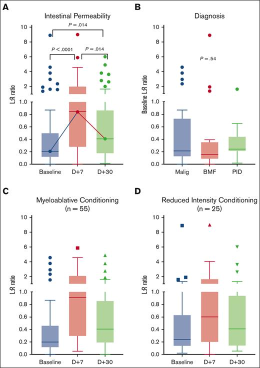
Intestinal permeability as measured using the L:R ratio followed a consistent trajectory in most allogeneic HSCT recipients. Permeability at baseline showed a median L:R ratio of 0.19 (range 0.00-8.90), which increased to 0.86 (range 0.00-9.00) at day +7, but returned to median levels of 0.40 (range 0.02-6.00) at day +30, which did not differ statistically from baseline (Figure 2A).
Intestinal permeability measured by urine L:R ratio. (A) Longitudinal intestinal permeability of 80 allogeneic BMT recipients. (B) Baseline L:R ratio by diagnosis group. Trajectory of L:R ratio in patients who received a (C) myeloablative conditioning and (D) reduced intensity conditioning regimen. There were no significant differences between the L:R ratios of (C) and (D) at any of the timepoints. BMF, bone marrow failure; BMT, bone marrow transplant; Malig, malignancy; PID, primary immune deficiency.
Intestinal permeability measured by urine L:R ratio. (A) Longitudinal intestinal permeability of 80 allogeneic BMT recipients. (B) Baseline L:R ratio by diagnosis group. Trajectory of L:R ratio in patients who received a (C) myeloablative conditioning and (D) reduced intensity conditioning regimen. There were no significant differences between the L:R ratios of (C) and (D) at any of the timepoints. BMF, bone marrow failure; BMT, bone marrow transplant; Malig, malignancy; PID, primary immune deficiency.
Eight participants (10.0%) had high baseline intestinal permeability. Baseline L:R ratios neither varied with underlying diagnoses (Figure 2B), nor did they vary significantly with age (supplemental Figure 1C) or sex (P = .67). There were no differences in the trajectory of L:R ratios between patients who received myeloablative or reduced intensity conditioning (RIC) (P = .39, Figure 2C-D), total body irradiation (supplemental Figure 1D), or who had baseline elevated L:R ratio (supplemental Figure 1E).
Intestinal permeability and HSCT outcomes
L:R ratios at any of the 3 timepoints did not predict development of TA-TMA, BSI, GVHD, or GI-GVHD by day +100 (Figure 3A-D). Patients who developed a BSI within 7 days of day +7 L:R measurement appeared to have higher day +7 L:R ratio (P = .0513, supplemental Figure 1F-G). In contrast to the standard trajectory of L:R ratios through HSCT, 12 participants had an elevated L:R ratio at day +30 compared with baseline, of whom 8 had adverse clinical outcomes by day+100, including severe adenovirus colitis, Clostridium difficile colitis, Enterococcus bloodstream infection, GI-GVHD, and TA-TMA (P = .51).
Associations of intestinal permeability with HSCT outcomes. L:R ratios at baseline (BL), day +7 (D7), and day +30 (D30) in subjects who did or did not develop (A) TMA, (B) BSI, (C) GVHD, and (D) intestinal involvement of GVHD.
Associations of intestinal permeability with HSCT outcomes. L:R ratios at baseline (BL), day +7 (D7), and day +30 (D30) in subjects who did or did not develop (A) TMA, (B) BSI, (C) GVHD, and (D) intestinal involvement of GVHD.
Microbiome alterations
Consistent with previous studies, the microbial composition of baseline stool samples was significantly different from day +7 and day +30 (P = .001; Figure 4A). Similarly, the Shannon Diversity Index of baseline stool samples was significantly higher than day +7 or day +30 (P = .0008; Figure 4B). At individual timepoints, the fecal microbiome of participants with elevated L:R ratios was distinct from patients with normal L:R ratios. Participants with increased day +7 L:R ratios had significantly lower Shannon Diversity Index scores than those with normal day +7 L:R ratios (P = .044; Figure 4C). Similarly, participants with increased day +30 L:R ratios had significantly lower Shannon Diversity Index scores than those with normal day +30 L:R ratios (P = .047; Figure 4D). The downward trajectory of intestinal microbiome diversity through transplant in participants with abnormal L:R ratios at day +30 was also different from the stable plateau in alpha diversity in participants with normal L:R ratios at day +30 (Figure 4E-F). Across all samples and timepoints, L:R values had a significantly negative association with Shannon Diversity Index scores (P = .035; Figure 4G).
Microbiome diversity in intestinal permeability cohort. (A) Principal component analysis of intestinal microbiome composition from stool samples at baseline ([BL], blue), day +7 ([D+7], red), and day +30 ([D+30], green). Colored ellipses represent the 95th percentile for the mean composition of the samples grouped by timepoint. (B) Alpha diversity of study cohort before, during, and after transplant. Alpha diversity at (C) day +7 and (D) day +30 grouped by intestinal permeability as elevated (red) or not elevated (blue). Trajectory of alpha diversity during transplant in (E) participants with normal day +30 L:R ratio and (F) participants with abnormally elevated day +30 L:R ratio. (G) Linear regression model of log (L:R ratio) and alpha diversity.
Microbiome diversity in intestinal permeability cohort. (A) Principal component analysis of intestinal microbiome composition from stool samples at baseline ([BL], blue), day +7 ([D+7], red), and day +30 ([D+30], green). Colored ellipses represent the 95th percentile for the mean composition of the samples grouped by timepoint. (B) Alpha diversity of study cohort before, during, and after transplant. Alpha diversity at (C) day +7 and (D) day +30 grouped by intestinal permeability as elevated (red) or not elevated (blue). Trajectory of alpha diversity during transplant in (E) participants with normal day +30 L:R ratio and (F) participants with abnormally elevated day +30 L:R ratio. (G) Linear regression model of log (L:R ratio) and alpha diversity.
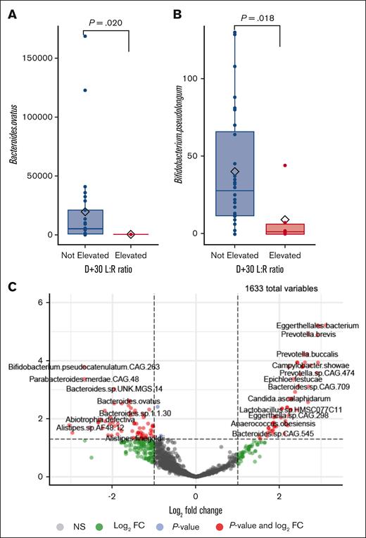
Analyses of stool species abundance also reported correlations with intestinal permeability. Comparison of species abundance revealed a number of species that were significantly differentially abundant between individuals with normal and elevated L:R as assessed by shrinkage linear discriminant analysis and Wilcoxon rank sum test, respectively (supplemental Figure 1A-B). Species associated with normal L:R ratios at D+7 and D+30 predominated, including normally commensal gut inhabitants (supplemental Figure 1A-B), whereas participants with abnormally elevated L:R ratios had significantly lower abundance of presumed beneficial organisms, such as Bacteroides ovatus (Figure 5A) and Bifidobacterium pseudolongum species (Figure 5B). To assess the association between L:R ratio and microbiome composition in the context of other important clinical variables, we used generalized linear mixed modeling as described previously in “Statistics.” This analysis identified many species that were differentially abundant at day +7 (920 of 1635 total species) or day +30 (919 species) relative to other timepoints (Figure 5C; supplemental Table 1). Elevation of L:R at day +7 was associated with differential abundance of 143 species whereas elevation of L:R ratio at day +30 was associated with differential abundance of 25 species. Several Prevotella species, which are oral and intestinal microbiome inhabitants, were associated with increased intestinal permeability. Lower abundance of Lactobacillus, Ruminococcus, Clostridium, and Blautia species were also associated with high intestinal permeability at day +7 (supplemental Tables 2 and 3). Elevated L:R ratio at baseline was only correlated with 3 species.
Microbiome composition in intestinal permeability cohort. Normalized count abundance of (A) Bacteroides species and (B) Bifidobacterium species in subjects with elevated (red) and not elevated (blue) day +30 L:R ratios. (C) Volcano plot of species abundance associated with high day +7 L:R ratios by GLLM-ZINB.
Microbiome composition in intestinal permeability cohort. Normalized count abundance of (A) Bacteroides species and (B) Bifidobacterium species in subjects with elevated (red) and not elevated (blue) day +30 L:R ratios. (C) Volcano plot of species abundance associated with high day +7 L:R ratios by GLLM-ZINB.
LBP, tight junction proteins, and other biomarkers in allogeneic HSCT recipients
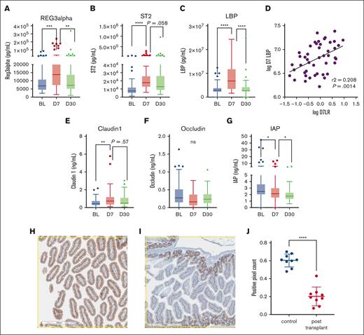
Plasma levels of intestinal epithelial tight junction proteins claudin1 and occludin and other associated markers including Reg3α, ST2, LBP, and IAP were quantified in our cohort using ELISA to further investigate mechanisms of altered permeability. The trajectories of plasma Reg3α and ST2 mirrored those of measured intestinal permeability (Figure 6A-B), with significant increase from baseline at day +7 and overall return to near-baseline by day +30. Similarly, LBP increased significantly from baseline to day +7, which resolved by day +30 (Figure 6C). Notably, there was a significant correlation (r2 = 0.208; P = .0014) between day +7 L:R ratio and LBP values (Figure 6D). Claudin1 levels increased from baseline to day +7, although there were no differences in occludin levels (Figure 6E-F). Plasma IAP, another previously reported marker of intestinal permeability, decreased from baseline to day +30 (Figure 6G). In a separate cohort of patients with BSIs and MBI-LCBIs, ferritin (a nonspecific acute-phase reactant) and LBP levels at day-14 and day-7 before the day of BSI were higher than those at baseline. However, the overall trajectories were not similar, suggesting that LBP was more than a nonspecific acute-phase reactant (supplemental Figure 2C-D), although there was a modest correlation between individual values of LBP and ferritin (P = .026; supplemental Figure 2E).
Human plasma and murine tissue biomarkers in HSCT recipients. Plasma (A) Reg3α, (B) ST2, and (C) LBP levels. (D) day +7 LBP and L:R ratios. Plasma (E) claudin1, (F) occludin, and (G) IAP levels. Representative 8× section (H) of control mouse duodenum stained for claudin1 (brown) and (I) from mouse 14 days after lethal irradiation and HSCT. (J) Positive pixel count of 3 sections from 3 mice in each group. ∗P < .05; ∗∗P < .01; ∗∗∗P < .001; ∗∗∗∗P < .0001.
Human plasma and murine tissue biomarkers in HSCT recipients. Plasma (A) Reg3α, (B) ST2, and (C) LBP levels. (D) day +7 LBP and L:R ratios. Plasma (E) claudin1, (F) occludin, and (G) IAP levels. Representative 8× section (H) of control mouse duodenum stained for claudin1 (brown) and (I) from mouse 14 days after lethal irradiation and HSCT. (J) Positive pixel count of 3 sections from 3 mice in each group. ∗P < .05; ∗∗P < .01; ∗∗∗P < .001; ∗∗∗∗P < .0001.
After the change seen in plasma claudin levels, duodenal claudin expression was compared in mice who underwent lethal irradiation and allogeneic HSCT with age-matched controls with no transplant. Transplanted mice had significantly lower expression of claudin in the duodenum (Figure 6H-J). There were no differences in occludin expression between transplanted and nontransplanted mice (data not shown).
Zonulin genotypes
Zonulin genotypes were studied as a potential genetic modifier of risk for adverse outcomes after HSCT. The distribution of genotypes of zonulin in 116 consecutive HSCT recipients was consistent with population data, with 24 (20%) genotype HP1-1, 46 (40%) 1 to 2, and 46 (40%) 2 to 2. No associations were found between zonulin genotypes and the development of GVHD, GI-GIVHD, BSI, or mortality (not shown).
Discussion
To our knowledge, our study is the first directly measuring intestinal permeability in pediatric and young adult patients receiving HSCT. More than two-thirds of participants tolerated the LR solution and provided timed urine samples at all timepoints. The modified protocol requires ingestion of less lactulose, rhamnose, and water than previous methods, is easy to administer, cheap (<$90/sample), and well tolerated by participants as young as 6 months old, even during the peritransplant period. This method is feasible for use in any participants with limited oral tolerance, paving the way for future studies of intestinal permeability in all HSCT recipients.
We observed a consistent pattern of intestinal permeability changes during transplant. Earlier work used urine samples collected between 5 and 24 hours and oral lactulose-rhamnose, lactulose-mannitol, or 51Cr-EDTA, comprised mostly autologous HSCT recipients and reported a similar peak of intestinal permeability from 1 to 2 weeks after conditioning, followed by a return to normal within 3 to 4 weeks.20-22 Mechanistically, the increase in plasma claudin1 and decrease in duodenal claudin after transplant confirms the dysregulation of tight junctions after HSCT. The uniform return of the L:R ratio to baseline by day +30 after HSCT likely reflects age and regenerative capacity of the gut despite stressors.
The loss of diversity during HSCT is well-studied,1,3,4,5,6,7,69,70,71 but an inverse correlation between intestinal permeability and microbiome composition has only been previously demonstrated in 1 cohort of patients with type 1 diabetes.38 This correlation has not previously been reported in allogeneic HSCT recipients. Notably, the effect size analyses show that most species associated with permeability are higher in samples from participants with normal permeability and generally comprise commensal anaerobic organisms, including Bacteroides ovatus and Bifidobacterium pseudolongum, suggesting that the intestinal microbial balance has been shifted to favor dysbiosis in the elevated permeability samples. Moreover, ZINB-GLMM analysis of the microbiome in participants with high L:R at day +7 revealed the reduced abundance of recognized beneficial organisms such as Lactobacillus, Clostridium, and Blautia, as well as increased abundance of Prevotella species. Although most patients return to normal permeability, participants with abnormally elevated permeability at day +30 have further losses of microbial diversity from day +7, identifying a small group of participants who are at risk for adverse outcomes and deserve additional study. If the loss of gut barrier function is the initiating event, early interventions such as oral IAP supplement,48 vitamin D,72-74 vitamin A,75,76 or zonulin inhibitors77,78 before, or at the initiation of conditioning chemotherapy could be pursued. In contrast, if altered intestinal permeability only propagates pathologic processes set into motion by separate mechanisms,79,80 this would inform a targeted intervention toward the initiating event, supplemented by permeability-directed interventions.
Ten percent of the participants in our study before transplant and 13 of 35 participants (37%) in the study by Keefe et al, had elevated baseline intestinal permeability levels.21 The 5-hour urine collection from the participants of Keefe study may have captured lower colonic sites of permeability not measured using our 2-hour protocol. Baseline intestinal permeability in our participants did not vary with age or underlying diagnosis. Antibiotic treatments before HSCT could differentially affect intestinal permeability at baseline. We were however unable to comment on the impact of antibiotics on intestinal permeability before HSCT, because 90% of our patients had antibiotic exposures before HSCT and believe that future studies with comparable numbers of patients with and without antibiotic exposures before HSCT may be able to study differences in intestinal permeability reliably.
The trajectory of intestinal permeability was also independent of conditioning intensity. Data from study by Johansson et al suggested that the gut barrier is preserved during allogeneic HSCT using RIC regimens, with no detectable changes in intestinal permeability throughout transplant.23 However, their cohort included only patients with leukemia or renal carcinoma, and the RIC regimens used were different from our institutional practice, which may be reasons for the difference.
We observed a similar trajectory of L:R ratios and Reg3 α, ST2, claudin1, and LBP after HSCT. As LBP has been previously described as an acute-phase reactant,81 we investigated its relationship to ferritin, another well-described acute-phase reactant, and found a correlation between individual values but distinct trajectories. Based on this, we postulate that LBP and L:R values, although affected by general inflammation, reflect specific changes in the gut epithelium, corroborating animal studies.82-85 In conjunction with our findings of dysbiosis, we propose that HSCT results in altered gut microbial composition and increased permeability, which together allow leakage of microbial products into the blood, triggering systemic response and LBP elevation. Plasma REG3α correlates inversely with intestinal Paneth cell numbers and REG3α expression levels.86 Our data suggest a similar relationship for the tight junction protein claudin1, because our mice after transplant had significantly lower duodenal expression than controls, whereas human participants after transplant had higher plasma levels from baseline. Unlike claudin1, plasma occludin levels did not vary significantly at our timepoints, corroborating our immunohistochemistry findings in mice and suggesting that occludin expression and regulation are independent of claudin. Plasma IAP also consistently decreased after transplant, not following L:R values. At least 2 possible theories may explain these differences: (1) IAP expression may be more influenced by enteral nutritional intake than direct cytotoxicity from chemotherapy and radiation,87 and enteral intake at days +7 and +30 is typically reduced from baseline, or (2) ability of IAP to detoxify LPS88 and the peak in LBP at day +7 may enhance IAP depletion.
We had expected to observe a correlation between intestinal permeability and adverse transplant outcomes, such as GVHD and BSI similar to previously published work.20 Our sample timepoints were predetermined and not time-of-events, and only 4 patients developed GVHD, including 1 with GI-GVHD, within the timeframe of permeability measurements (up to day +37). Johansson et al also reported a brief difference in intestinal permeability at day +4 between patients who went onto develop grades 2 to 4 GVHD, which was not present at day-1 or day +7.89 We were unable to capture differences of such short duration and recognize that intensifying tracking after HSCT could have identified relationships between alterations in intestinal permeability and acute GVHD. Moreover, only 16 participants developed acute GVHD, including 6 patients with GI-GVHD, by day +100. This may be related to our early adoption of abatacept as standard of care because of its success in preventing acute GVHD in HSCT recipients90,91 or the lower incidence of acute GVHD in pediatric HSCT recipients. An adult cohort, with possibly higher rates of GVHD and BSIs,92 may have different results. We also did not see associations between zonulin genotypes and these outcomes, which may be because of low incidence of these events or could suggest a zonulin-independent regulatory mechanism of intestinal permeability after HSCT.
A limitation of our study was the variability of timing of the test at day+7 (±7 days) which was proposed because of inability of patients to undergo this test during mucositis during the initial phase of this study. We felt it was important to introduce this variability to obtain crucial data which has otherwise been significantly lacking in pediatric patients who received HSCT. In this pilot study, our primary aim was to describe longitudinal changes in intestinal permeability. We did not have prior data to help guide what sample sizes would be needed to observe meaningful changes in intestinal permeability based on outcomes, but we believe that future studies will be helpfully guided by these data to have sample size estimates. We have missing data and dropouts similar to most human studies, and these patients were excluded from our analyses. Our results demonstrate that changes in intestinal permeability during HSCT can be easily quantified, even in infants. Our data suggest that patients with abnormally high intestinal permeability also have dysbiosis and a systemic inflammatory response, thereby identifying a population of patients who could benefit from early interventions such as prebiotics, to restore intestinal microbiome health in the future. Future studies would ideally schedule more frequent L:R and microbiome sampling, as we are unable to discern the temporal relationship between permeability and dysbiosis, which would inform the development and timing of potential interventions to decrease systemic inflammation and improve outcomes after HSCT. Future murine models could be developed to correlate small and large intestinal permeability after HSCT.
Acknowledgments
The authors thank the nurses, parents, and patients who made this study possible. The authors also thank Damien Reynaud for his efforts spearheading the animal work, Betsy DiPasquale for immunohistochemistry work, and Verzhiniya Markova for excellent administrative assistance.
This study was funded by a Cincinnati Children’s Hospital Medical Center Research Innovation Pilot grant (Y.M.W.).
Authorship
Contribution: Y.M.W., S.A., N.L., and L.L. collected and processed samples and performed experiments; A.D., K.L., B.H., O.V.S., and M.T. collected samples and clinical data; J.K. and K.N. conceptualized and performed animal studies; D.B.H. performed microbiome analysis; Y.M.W. and P.K. wrote the manuscript; P.K. conceptualized the study with S.M.D. and W.A.F.; and all authors critically reviewed the manuscript.
Conflict-of-interest disclosure: The authors declare no competing financial interests.
Correspondence: YunZu Michele Wang, Cincinnati Children’s Hospital Medical Center, Cancer and Blood Diseases Institute, 3333 Burnet Ave MLC 11027, Cincinnati, OH 45229; e-mail: yunzu.wang@cchmc.org.
References
Author notes
Data are available on request from the corresponding author, YunZu Michele Wang (yunzu.wang@cchmc.org). Sequence Read Archive (SRA) submission: SUB13114228.
The full-text version of this article contains a data supplement.


![Microbiome diversity in intestinal permeability cohort. (A) Principal component analysis of intestinal microbiome composition from stool samples at baseline ([BL], blue), day +7 ([D+7], red), and day +30 ([D+30], green). Colored ellipses represent the 95th percentile for the mean composition of the samples grouped by timepoint. (B) Alpha diversity of study cohort before, during, and after transplant. Alpha diversity at (C) day +7 and (D) day +30 grouped by intestinal permeability as elevated (red) or not elevated (blue). Trajectory of alpha diversity during transplant in (E) participants with normal day +30 L:R ratio and (F) participants with abnormally elevated day +30 L:R ratio. (G) Linear regression model of log (L:R ratio) and alpha diversity.](https://ash.silverchair-cdn.com/ash/content_public/journal/bloodadvances/7/17/10.1182_bloodadvances.2023009960/2/m_blooda_adv-2023-009960-gr4.jpeg?Expires=1765883022&Signature=r8sS3vWye55whgcePzMyQzW8ILQeN3~oQubHlhfPNaFO2lirDzsb9Q~RTL1DOZ37IIjOQN5FxLIr5Zig4cbqtcgk-caUDrV4kuPms5CXgHqmNX4~dGEQo-ZAE-X69VYyGhw8K7LTzwiRS11uW9sltgIIZTJDD3Vcp1fglwEMcJkgeN7UUNrCwP7pCVE63gAsG6jeSAmmeDXKsspr~1A24XyC1sSfcpngMhCcn4fV6x2s85SqWOcFlezARGSes2VjFg6Z7plAFJhzT8IvHsxE2dYsSkhaj3BgsT41wbel0BBd~VbeKjKwCUXR93CQoV1-QASHSuhya6RUfohz5dsgNQ__&Key-Pair-Id=APKAIE5G5CRDK6RD3PGA)
