Key Points
Expression analysis of >1500 pediatric AML samples demonstrates MNX1 expression as a universal feature of t(7;12)(q36;p13) AML.
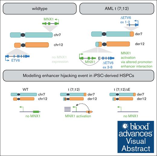
MNX1 is activated by an enhancer-hijacking event in t(7;12)(q36;p13) AML and not, as previously postulated, by an MNX1::ETV6 oncofusion.
Visual Abstract
Acute myeloid leukemia (AML) with the t(7;12)(q36;p13) translocation occurs only in very young children and has a poor clinical outcome. The expected oncofusion between break point partners (motor neuron and pancreas homeobox 1 [MNX1] and ETS variant transcription factor 6 [ETV6]) has only been reported in a subset of cases. However, a universal feature is the strong transcript and protein expression of MNX1, a homeobox transcription factor that is normally not expressed in hematopoietic cells. Here, we map the translocation break points on chromosomes 7 and 12 in affected patients to a region proximal to MNX1 and either introns 1 or 2 of ETV6. The frequency of MNX1 overexpression in pediatric AML is 2.4% and occurs predominantly in t(7;12)(q36;p13) AML. Chromatin interaction assays in a t(7;12)(q36;p13) induced pluripotent stem cell line model unravel an enhancer-hijacking event that explains MNX1 overexpression in hematopoietic cells. Our data suggest that enhancer hijacking may be a more widespread consequence of translocations in which no oncofusion product was identified, including t(1;3) or t(4;12) AML.
Introduction
Acute myeloid leukemia (AML) has been successfully investigated in the past using cytogenetic analysis. This led to the discovery of numerous recurrent chromosomal translocations (eg, t(8;21)(q22; q22) or t(15;17)(q22;q12)) generating oncofusion proteins (eg, RUNX1::RUNX1T1 or PML::RARA, respectively) that drive leukemogenesis. For many years, these translocations have served as diagnostic and prognostic markers and affected patients can now be treated with specific targeted therapies (eg, retinoic acid and arsenic trioxide in t(15;17) cases).1 In 1998, 2 publications reported the translocation t(7;12)(q36;p13) in AML of infants2,3 occurring predominantly in children aged <18 months and not in adult AML. A recent meta-analysis of the Nordic Society for Pediatric Hematology and Oncology (NOPHO-AML) determined that t(7;12)(q36;p13) AML constituted 4.3% of all children with AML aged <2 years and found a 3-year event-free survival of 24% (literature-based data) and 43% (NOPHO-AML data).4 Cytogenetically, t(7;12)(q36;p13) AML is often associated with the occurrence of trisomy 19,4,5 but no other recurrent aberrations have been described.
Reported break points in t(7;12)(q36;p13) AML have mainly been evaluated by fluorescent in situ hybridization analysis. The breakpoints on chromosome 12 (chr12) are located within intron 1 or 2 of ETS variant transcription factor 6 (ETV6) and proximal to motor neuron and pancreas homeobox 1 (MNX1) and within the 3’ end of nucleolar protein with MIF4G domain 1 (NOM1) on chr7.6 A MNX1::ETV6 fusion transcript was described only in a subset of t(7;12)(q36;p13) AML cases.4-7 However, all AML cases with t(7;12)(q36;p13) have high expression of MNX1,5 suggesting a yet unknown mechanism of MNX1 activation. Consistent with the activation of a silenced gene locus, a translocation of the MNX1 locus from the nuclear periphery to the internal nucleus was seen, an observation that is in line with the idea that condensed and silent chromatin is located in the nuclear periphery.6 Furthermore, interactions of ETV6 downstream elements with the MNX1 locus have been postulated as possible mechanisms for MNX1 activation.6,8 The first clue for the existence of possible aberrant promoter-enhancer interactions leading to MNX1 activation came from our investigations of the GDM-1 AML cell line, which harbors a t(6;7)(q23;q36) translocation. In GDM-1, the MNX1 promoter interacts with an enhancer element from the MYB locus on chr6q23.9
Recently, Nilsson et al. reported the introduction of a translocation between chr7q36 and chr12p13, modeling the one found in t(7;12)(q36;p13) AML, into the human induced pluripotent stem cell (iPSC) line, ChiPSC22WT.8 The derivative line, ChiPSC22t(7;12), can be differentiated into hematopoietic stem and progenitor cells (HSPCs) and, as such, expresses MNX1, suggesting that hematopoietic enhancers play a role in MNX1 activation. Enhancer hijacking has initially been described as a mechanism for oncogene activation in AML with inv(3)/t(3;3)(q21q26) AML, in which activation of EVI1, an isoform encoded from the MDS and EVI1 complex locus (MECOM), results from the repositioning of a GATA2 enhancer.10-12 Enhancer hijacking is also implicated in acute leukemia of ambiguous lineage in which translocated hematopoietic enhancers from different chromosomes are involved in activating BCL11B.13
Here, we provide a detailed description of the molecular alterations found in 6 patients with t(7;12)(q36;p13) AML and dissect the molecular mechanism leading to MNX1 activation through the use of CRISPR-engineered ChiPSC22t(7;12) iPSCs and HSPCs. We identified that a previously proposed8 enhancer-hijacking event activates the MNX1 promoter via hematopoietic enhancers from the ETV6 locus and validated this event in the iPSC/HSPC system. Our data suggest that enhancer hijacking may be a more widespread, but so far largely unappreciated, mechanism for gene activation in AML with cytogenetic abnormalities.
Methods
Samples and cell lines
Pediatric leukemia samples T1, T2, and T3 (supplemental Table 1) were obtained at diagnosis after informed consent of patients’ legal guardians in accordance with the institution’s ethical review board (University Essen and Medical University Hannover, MHH, no. 2899). Sample T4 (supplemental Table 1) came from a pediatric AML cohort in Gothenburg, Sweden, and informed consent was obtained from the legal guardians in accordance with the local ethical review board. Human iPSC line ChiPSC22 (Cellartis/Takara Bio Europe AB) was cultivated in the feeder-free DEF-CS system (Cellartis/Takara Bio Europe) under standard conditions. Before differentiation, cells were transferred to Matrigel (Corning) and mTeSR1 medium (STEMCELL Technologies Inc) for 2 to 3 passages. ChiPSC22 was authenticated; this line and its derivatives were regularly tested for mycoplasma contamination using a commercial test kit (VenorGeM Classic, Minerva Biolabs).
Differentiation to hematopoietic cells
Differentiation of ChiPSC22 was done as previously described.8
Whole-genome sequencing (WGS)
Genomic DNA was isolated using the Quick DNA Miniprep kit (Zymo Research), and libraries were sequenced in an Illumina HiSeq X Ten sequencer. FASTQ files were aligned with the Burrows-Wheeler Aligner (maximal exact match option) to the hg19 reference genome. Single nucleotide variants (SNVs) were called using mutect2. Because of the lack of matched germ line sequences, only 52 known AML driver genes (supplemental Table 2) were screened for mutations. Structural variants (SVs) and somatic copy number alterations (SCNAs) were called using the Hartwig Medical Foundation (HMF) pipeline (https://github.com/hartwigmedical/hmftools). HMF tools were used in tumor-only mode, and putative germ line SVs were filtered out using a large panel of HMF-provided normals. SVs <20 kb were filtered out. Processed data of 2 samples of the TARGET-AML data set (supplemental Table 1; database of Genotypes and Phenotypes (dbGaP) accession: phs000465.v22.p8) was downloaded using the Globus platform.14
RNA isolation, sequencing, and quantitative Reverse Transcriptase-Polymerase Chain Reaction (qRT-PCR)
Total RNA was isolated using the RNeasy Plus Mini kit (Qiagen). After library preparation, RNA was sequenced on NOVASEQ 6000 with 100-bp paired end. The FASTQ files were processed using the nf-core15 RNA-sequencing (RNAseq) v3.9 pipeline, with alignment performed using Spliced Transcripts Alignment to a Reference (STAR)16 and quantification performed with Salmon.17 Allele-specific expression was examined by first detecting heterozygous single-nucleotide polymorphisms in exons using the genomic analysis toolkit (GATK) HaplotypeCaller and then counting the allelic expression in RNA using GATK ASEReadCounter.18 Differential expression analysis was performed using limma for Affymetrix array data and pydeseq2 for RNAseq data.
qRT-PCR was performed as previously described using TaqMan Universal Master Mix II with uracil-N-glycosylase (ThermoFisher Scientific, Applied Biosystems) and TaqMan gene expression assays (ThermoFisher Scientific, Applied Biosystems; supplemental Table 3).8
Detection of fusion transcripts
For our own samples, fusion transcripts were detected using STAR-Fusion v1.10.1.19 For the TARGET-AML cohort, we downloaded the processed STAR-Fusion results from the Genomic Data Commons data portal.
Protein extraction, western blotting, and protein detection
Protein extraction, western blotting, and protein detection with antibodies (supplemental Table 4) was done as described previously.9
Expression screens
RNAseq expression data were downloaded from the TARGET cohort20 (both TARGET-NCI (Therapeutically Applicable Research to Generate Effective Treatments, National Cancer Institute) and TARGET-FHCRC (Fred Hutchinson Cancer Research Center); https://target-data.nci.nih.gov/Public/AML/mRNA-seq/L3/expression/BCCA/). MNX1 is not expressed in normal hematopoietic cells; hence, in RNAseq, a MNX1 expression >0.5 transcripts per million was considered overexpression. Gene expression data files of the Balgobind cohort21 were downloaded from Gene Expression Omnibus (GSE17855) and normalized using the affy R package (https://bioconductor.org/packages/release/bioc/html/affy.html). Log-expression values of microarray data were assumed to be normally distributed. We computed the mean and standard deviation for MNX1 expression across all samples; those whose MNX1 expression was higher than the mean plus 3 standard deviations were considered to express MNX1 (3-sigma rule).
4C
Circular chromosome conformation capture (4C) with 2 million cells was done and analyzed as described9 using HindIII in combination with DpnII (supplemental Table 5).
High-throughput chromosome conformation capture (Hi-C)
Hi-C libraries were prepared and analyzed as previously described22 with minor modifications. One million cells were fixed at a final concentration of 1% formaldehyde in RPMI 1640 medium. Digestion was performed using DpnII. Two to 3 Hi-C library replicates per sample were sequenced with 240 million reads per replicate. The FASTQ files were processed using the nf-core/hic v.2.1.0 pipeline. Hi-C figures were generated using figeno (https://github.com/CompEpigen/figeno)23.
Two-color fluorescence in situ hybridization (FISH)
Two NOVA-probe sets targeting MNX1 (chr7:156802250-156807250) and ETV6 (chr12:11949500-11954500) carrying multiple ATTO594 or ATTO647N dyes were synthesized as described previously24 (supplemental Table 6). Two-color FISH was conducted as previously described with minor adaptations.25,26 ChiPSC22t(7;12) iPSCs were seeded on DEF-CS COAT-1–coated coverslips (Cellartis, Takara BioSciences) and ChiPSC22t(7;12) HSPCs on poly-L-lysine–coated coverslips. After washing and fixation steps, coverslips were mounted on microscopic slides with Mowiol (2.5% 1,4-Diazabicyclo[2.2.2]octane and pH 7.0; Carl Roth), dried for 30 minutes, and sealed with nail polish.27 Automated Stimulated Emission Depletion (STED) microscopy was performed according to Brandstetter et al.25 FISH signals within confocal scans were detected using a Laplacian-of-Gaussian blob detector and subsequently imaged using 3-dimensional (3D) STED settings. Subpixel localization of FISH spots in both channels was performed by fitting a multidimensional Gaussian function plus a constant background using the Levenberg-Marquardt algorithm. The peak height of the fitted Gaussians was used to determine spot intensity. Only distances <600 nm were considered.
ACT-seq and ATAC-seq
Genome-wide targeting and mapping of histone modifications (supplemental Table 4) and mapping of open chromatin were done by antibody-guided chromatin tagmentation sequencing (ACT-seq) and assay for transposase-accessible chromatin by sequencing (ATAC-seq), respectively, as described previously.9 For read normalization using spiked-in yeast DNA in ACT-seq, trimmed reads were additionally aligned against the Saccharomyces cerevisiae R64 reference genome followed by postalignment filtering. An ACT-seq library–specific scaling factor was obtained by calculating the multiplicative inverse of the number of filtered alignments against the yeast genome.9 ACT-seq peak calling was done applying MACS v.2.2.6 (https://pypi.org/project/MACS2/) with a q-value cutoff of 0.05 and default parameters using a wrapper script with settings narrowPeak and broadPeak for acetylated lysine 27 of histone 3 (H3K27ac) and (monomethylated lysine 4 of histone 3) H3K4me1, respectively. To facilitate visualization of hematopoietic-specific enhancers in the Integrative Genomics Viewer (version 2.11.7),28 we generated HSPC-specific H3K27ac and H3K4me1 bw-tracks with callpeaks using corresponding iPSC data as internal reference.
Deletion of the enhancer region
A region of 213.5 kb (chr12:11951022-12164578, GRCh37/hg19) covering the 4 enhancers located closest to the break point in ChiPSC22t(7;12) was deleted by CRISPR/Cas9 editing as described previously8 using CRISPR RNAs designed with the Alt-R Custom Cas9 crRNA Design Tool (Integrated DNA Technologies). To join the 2 ends by homology-directed repair, a 150 single-stranded deoxynucleotide was designed with 75 bases sequence homology on each side. Deletion was done in ChiPSC22WT and ChiPSC22t(7;12) sublines 14D7 and 24C7.8 The presence of the deletion on the translocated and the wild-type allele was validated by PCR using the Terra PCR Direct Polymerase Mix (Takara Bio Europe; supplemental Table 7). From line 14D7, cell line 2304B4 was generated, and from 24C7, lines 2305B10 and 2305C9 were generated.
Phenotypic characterization of enhancer deletion clones
For flow cytometry analysis, cells were resuspended in phosphate-buffered saline plus EDTA and incubated with the mix of antibodies for 15 to 20 minutes in the dark at room temperature. Cells were washed once and resuspended in phosphate-buffered saline plus EDTA. Data were collected on BD FACS Aria (BD Biosciences) and analyzed using BD FACSDiva. The colony-forming unit assay was done using MethoCult H4034 Optimum (STEMCELL Technologies) following the manufacturer’s protocol. Proliferation was analyzed by continuous culture of the HSPCs in StemSpan SFEM II + CC100 (STEMCELL Technologies) for 14 days. Cells were counted every 48 hours, centrifuged, and 75% fresh medium was added to 25% old medium. Cell division was calculated as follows: ln(B/A)/(ln2), in which A = number of seeded cells and B = number of cells after 48 hours.
Results
WGS of t(7;12)(q36;p13) AML
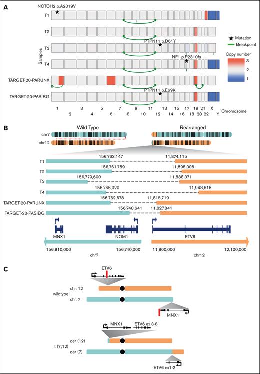
To precisely map structural rearrangements, SCNAs, and genetic mutations in t(7;12)(q36;p13) AML, we performed WGS of 4 t(7;12)(q36;p13) AML cases, T1, T2, T3, and T4 (Figure 1A; supplemental Table 1). We additionally used published WGS data from 2 samples with t(7;12)(q36;p13) from the TARGET cohort29 (supplemental Table 1). The presence of t(7;12)(q36;p13) as a reciprocal balanced translocation was verified in all 6 samples (Figure 1A). The break point on chr12 is located in 5 samples in intron 1 and in 1 sample in intron 2 of ETV6. On chr7, all break points are located proximal to MNX1; in 4 cases within NOM1, located next to MNX1; and in 2 cases, between MNX1 and NOM1 (Figure 1B). In none of these cases, an oncofusion gene between MNX1 and ETV6 is supported by the observed translocation break points, leaving the main MNX1 variant (RefSeq: NM_005515) unaffected by the genomic rearrangements (Figure 1C). Accompanying cytogenetic data (supplemental Table 1) revealed trisomy 19 in all cases, a result confirmed by SCNA analysis for all cases except for T1, for which SCNA analysis identified a trisomy 22 but no trisomy 19 (Figure 1A). We found mutations in common leukemia genes (supplemental Table 2), namely in NOTCH2 (p.A2319V), NF1 (p.P2310fs), and PTPN11 (p.D61Y and p.E69K; Figure 1A).
WGS analysis of t(7;12)(q36;p13) AML. (A) Copy numbers (blue, loss; red, gain), structural rearrangements (green bow connecting 2 chromosomes), and mutations in known AML driver genes for 6 t(7;12)(q36;p13) AML samples based on WGS. Samples T1, T2, T3, and T4 were profiled in this study, whereas TARGET-20-PARUNX and TARGET-20-PASIBG are from the TARGET-AML cohort 15. (B) Sketch of the rearranged chr7 and chr12 and zoom-in on the region around the break points. (C) Schematic overview of chr7 (turquoise), chr12 (orange), and derivative chromosomes der(12) and der(7) resulting from the reciprocal t(7;12) translocation involving MNX1 on chr7 and ETV6 on chr12. Red lines indicate positions of break/fusion points.
WGS analysis of t(7;12)(q36;p13) AML. (A) Copy numbers (blue, loss; red, gain), structural rearrangements (green bow connecting 2 chromosomes), and mutations in known AML driver genes for 6 t(7;12)(q36;p13) AML samples based on WGS. Samples T1, T2, T3, and T4 were profiled in this study, whereas TARGET-20-PARUNX and TARGET-20-PASIBG are from the TARGET-AML cohort 15. (B) Sketch of the rearranged chr7 and chr12 and zoom-in on the region around the break points. (C) Schematic overview of chr7 (turquoise), chr12 (orange), and derivative chromosomes der(12) and der(7) resulting from the reciprocal t(7;12) translocation involving MNX1 on chr7 and ETV6 on chr12. Red lines indicate positions of break/fusion points.
T1, T2, and T3 were profiled with RNAseq, but no fusion transcript MNX1::ETV6 could be identified. The TARGET-AML cohort contains 14 t(7;12) samples, and of these, only 1 has an MNX1::ETV6 fusion detected by STAR-Fusion (TARGET-20-PAWNHH), and 1 has an ETV6::LMBR1 fusion (TARGET-20-PAWNYK). Therefore, fusion transcripts do not appear to be the driving factor behind t(7;12)(q36;p13).
MNX1 is highly expressed in all t(7;12)(q36;p13) AML and is associated with a characteristic gene expression signature
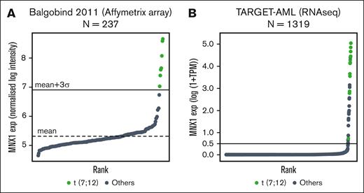
Although normally not expressed in the hematopoietic lineage, MNX1 is highly expressed in all analyzed t(7;12)(q36;p13) AML.5,6,30 In line with this, AML cases T1 to T4 showed high MNX1 expression (supplemental Table 1). We additionally evaluated MNX1 expression in 2 pediatric AML cohorts with available expression data: Balgobind et al (237 samples profiled with Affymetrix array31; Figure 2A) and TARGET-AML (1319 samples profiled with RNAseq29; Figure 2B). MNX1 was expressed in 7 of 237 samples (2.9%) of the Balgobind cohort and in 31 of 1319 (2.3%; including resample for samples TARGET-20-PARUNX and TARGET-21-PASVJS) samples of the TARGET-AML cohort. All t(7;12) samples showed MNX1 expression but also some samples without 7q36-rearrangements. Accordingly, there might be alternative mechanisms leading to MNX1 activation. Most t(7;12)(q36;p13) samples were diagnosed at younger than 2 years; however, most MNX1-overexpressing samples without t(7;12) were diagnosed at an older age (supplemental Table 1). A characteristic gene expression signature for t(7;12)(q36;p13) AML compared with other cytogenetic subgroups in pediatric AML has been described.31 The majority of these genes are either consistently downregulated in t(7;12)(q36;p13) AML (eg, TP53BP2) or upregulated together with MNX1 (EDIL3, LIN28B, BAMBI, MAF, FAM171B, AGR2, CRISP3, KRT72, and MMP9). These do not lie on the translocated piece of chr7; and, hence, their expression change might be a secondary effect of the translocation. We performed differential expression analysis between the t(7;12)(q36;p13) and the other cases from each the Balgobind and the TARGET-AML cohort (supplemental Table 8; supplemental Figure 1). Our lists of upregulated and downregulated genes include the genes identified by Balgobind et al,31 which are indeed consistently deregulated in t(7;12)(q36;p13) across several cohorts, as well as other genes not reported before. The samples with MNX1 expression but without genomic rearrangement close to MNX1 did not exhibit this typical gene signature (supplemental Figures 1 and 2). Several experimental systems have recently been developed to model t(7;12): an HSPC system with a t(7;12) translocation8,32 and a mouse model of leukemia induced by MNX1 overexpression.33 The HSPC system partially recapitulated the patients’ gene expression signature, whereas in the mouse model, most genes of the t(7;12) signature were not differentially expressed (supplemental Figure 2).
MNX1 expression in pediatric AML with t(7;12)(q36;p13) translocation. (A-B) MNX1 expression in 2 different pediatric AML cohorts. (A) Balgobind et al31; 237 samples profiled with Affymetrix arrays. The mean expression level is shown with a dashed line, and the mean plus 3 standard deviations is shown with a horizontal line. (B) TARGET-AML29; 1319 samples profiled with RNAseq (cutoff, 0.5 TPM). Samples with cytogenetically detected t(7;12)(q36;p13) translocation are shown in green and other samples in gray. exp, expression; TPM, transcripts per million.
MNX1 expression in pediatric AML with t(7;12)(q36;p13) translocation. (A-B) MNX1 expression in 2 different pediatric AML cohorts. (A) Balgobind et al31; 237 samples profiled with Affymetrix arrays. The mean expression level is shown with a dashed line, and the mean plus 3 standard deviations is shown with a horizontal line. (B) TARGET-AML29; 1319 samples profiled with RNAseq (cutoff, 0.5 TPM). Samples with cytogenetically detected t(7;12)(q36;p13) translocation are shown in green and other samples in gray. exp, expression; TPM, transcripts per million.
The break points on chr12 in the t(7;12)(q36;p13) samples led to a corrupted ETV6 allele and reduced ETV6 expression compared with other samples (supplemental Figure 3), but this was not significant due to the low number of t(7;12) cases and the high ETV6 expression variability even in the absence of rearrangements. Heterozygous single-nucleotide polymorphisms in exons of ETV6 were found in T1 and T3, and the allele frequency in RNA suggested monoallelic expression of ETV6 in the leukemic cells (supplemental Figure 3).
A t(7;12)(q36;p13) cell line model exhibits MNX1 protein expression and chromatin interactions between the MNX1 and ETV6 regions
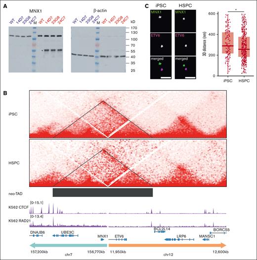
Previously, Nilsson et al engineered an iPSC line harboring a balanced translocation t(7;12)(q36;p13) (ChiPSC22t(7;12)) with a break point in ETV6 intron 2 and a second one ∼21 kb proximal to MNX1 in the common break point region.8,32 Upon differentiation of ChiPSC22t(7;12) cells to HSPCs, MNX1 became activated. We confirmed this result at the protein level in 3 ChiPSC22t(7;12) sublines, 14D7, 23G8, and 24C7; the MNX1 protein was only expressed in the ChiPSC22t(7;12) HSPCs but not in the ChiPSC22t(7;12) iPSCs, ChiPSC22WT iPSCs or ChiPSC22WT HSPCs (Figure 3A). WGS of the 3 sublines did not reveal any relevant changes to the original line ChiPSC22, except for the presence of the heterozygous t(7;12). To examine whether the t(7;12)(q36;p13) translocation juxtaposes enhancers from the ETV6 region with the MNX1 promoter, we profiled the iPSCs and HSPCs of both ChiPSC22WT and ChiPSC22t(7;12) lines with Hi-C. No interactions were seen between chr7 and chr12 in the ChiPSC22WT (supplemental Figure 4), but we observed a new topologically associating domain (neo-TAD) around the break point in both iPSC and HSPC ChiPSC22t(7;12) (Figure 3B). The neo-TAD extends up to chr12:12200000, meaning that enhancers located between the break point and the end of the neo-TAD could interact with the MNX1 promoter. Public ChIPseq data from K562 cells revealed binding of CCCTC-Binding Factor (CTCF) and Radiation Gene 21 (RAD21) at the extremities of the neo-TAD, which would explain its formation (Figure 3B). For an independent proof of interaction, we performed 4C using an MNX1 viewpoint (chr7:156805780-156806574) and a viewpoint located in the neo-TAD, close to the ETV6 break point (chr12:11953871-11954315; supplemental Table 5). Reciprocal 4C confirmed the interaction between the MNX1 and ETV6 regions in HSPCs and, less strongly, in 1 iPSC sample (supplemental Figure 5).
MNX1 protein expression and chromatin interaction of the MNX1 gene with the ETV6 region in ChiPSC22t(7;12) cells. (A) Western blot with an MNX1 antibody (left) and iPSC (blue) and HSPC (red) protein extracts from ChiPSC22WT and ChiPSC22t(7;12) sublines 14D7, 23G8, and 24C7. The MNX1 protein (asterisk) is only detected in HSPCs of ChiPSC22t(7;12) sublines 14D7, 23G8, and 24C7. The common band at ∼120 kD results from an unknown protein cross-reacting with the MNX1 antibody. To demonstrate loading of equal protein amounts, the unstripped blot was reincubated with an antibody against β-actin (right). (B) Chromatin interactions analyzed by Hi-C seq in the genomic region flanking the translocation break point in the ChiPSC22t(7;12) subline 24C7, either as iPSCs (top) or HSPCs (below). The neo-TAD is indicated by a black bar. ChIPseq data for CTCF and RAD21 in K562 were retrieved from the encode project (IDs ENCFF468HJA and ENCFF000YXZ). (C) Increased proximity between MNX1 and ETV6 in ChiPSC22t(7;12) subline 14D7–derived HSPCs compared with iPSCs. Representative STED images of FISH spots in 2 colors targeting MNX1 and ETV6 in iPSCs and HSPCs (left). Scale bars, 500 nm. 3D distances between the MNX1 and ETV6 signals (right). Red horizontal lines within boxes indicate medians; box limits indicate upper and lower quartiles. iPSCs, n = 154; HSPCs, n = 409, across 3 independent replicates. ∗P < .05, Wilcoxon rank-sum test.
MNX1 protein expression and chromatin interaction of the MNX1 gene with the ETV6 region in ChiPSC22t(7;12) cells. (A) Western blot with an MNX1 antibody (left) and iPSC (blue) and HSPC (red) protein extracts from ChiPSC22WT and ChiPSC22t(7;12) sublines 14D7, 23G8, and 24C7. The MNX1 protein (asterisk) is only detected in HSPCs of ChiPSC22t(7;12) sublines 14D7, 23G8, and 24C7. The common band at ∼120 kD results from an unknown protein cross-reacting with the MNX1 antibody. To demonstrate loading of equal protein amounts, the unstripped blot was reincubated with an antibody against β-actin (right). (B) Chromatin interactions analyzed by Hi-C seq in the genomic region flanking the translocation break point in the ChiPSC22t(7;12) subline 24C7, either as iPSCs (top) or HSPCs (below). The neo-TAD is indicated by a black bar. ChIPseq data for CTCF and RAD21 in K562 were retrieved from the encode project (IDs ENCFF468HJA and ENCFF000YXZ). (C) Increased proximity between MNX1 and ETV6 in ChiPSC22t(7;12) subline 14D7–derived HSPCs compared with iPSCs. Representative STED images of FISH spots in 2 colors targeting MNX1 and ETV6 in iPSCs and HSPCs (left). Scale bars, 500 nm. 3D distances between the MNX1 and ETV6 signals (right). Red horizontal lines within boxes indicate medians; box limits indicate upper and lower quartiles. iPSCs, n = 154; HSPCs, n = 409, across 3 independent replicates. ∗P < .05, Wilcoxon rank-sum test.
We further performed 2-color FISH targeting the MNX1 promoter and a neo-TAD region located close to the ETV6 break point and observed a significantly decreased 3D distance between the targeted regions in ChiPSC22t(7;12) HSPCs compared with the corresponding iPSCs (Figure 3C). This suggests a reinforced contact between an enhancer from the ETV6 neo-TAD region and the MNX1 promoter upon differentiation.
Identification of hematopoietic enhancers in ETV6 and its vicinity
We next searched for hematopoietic enhancers located in the chr12 part of the neo-TAD. Active enhancers reside in open chromatin; hence, we profiled accessible chromatin by ATAC in AML-T1 and -T2 and in HSPCs of our cell line model. The patient samples clustered with the HSPCs, separately from the iPSCs, and had similar peaks as HSPCs (supplemental Figure 6A-B). We found 4 consistent open chromatin sites common to the patient samples and to the ChiPSC22t(7;12) and ChiPSC22WT cells within the ETV6 neo-TAD (Figure 4). Additionally, we mapped peaks of enhancer marks H3K27ac and H3K4me1 in both iPSCs and HSPCs. To facilitate the identification of hematopoietic enhancers in ChiPSC22t(7;12) and ChiPSC22WT HSPCs, we applied MACS2 peak calling using the corresponding iPSC data as internal reference. The 4 open chromatin regions were also marked by HSPC-specific H3K27ac and H3K4me1 peaks. In addition, we used public chromatin immunoprecipitation followed by sequencing (ChIPseq) data sets for the same enhancer marks (Gene Expression Omnibus: GSM772885 and GSM621451) and the histone acetyltransferase P300, generated from CD34+ cells and the chronic myeloid leukemia–derived cell line MOLM-1.10 Again, the 4 ATAC/H3K27ac/H3K4me1 peaks were found as well in MOLM-1 and CD34+ cells (Figure 4). Two of these peaks coincide in addition with p300 peaks (Figure 4). In conclusion, we identified 4 strong enhancer candidates in the ETV6 neo-TAD region, 2 of which coincide with p300 peaks and may drive MNX1 activation in t(7;12)(q36;p13) AML.
Open chromatin and enhancer mark profiles in the ETV6 neo-TAD region of patient and cell line samples. Open chromatin profiles (ATAC) of patients with AML , T1 and T2, and of HSPCs from ChiPSC22WT and ChiPSC22t(7;12) sublines 14D7, 23G8, and 24C7 in the ETV6 neo-TAD region. HSPC-specific enhancer mark H3K27ac and H3K4me1 profiles and publicly available10 p300, H3K27ac, and H3K4me1 profiles from MOLM-1 and CD34+. Relevant common peak positions are highlighted by a gray shading. The chr12 break point (BP) position in T1 and T2 and in the ChiPSC22t(7;12) sublines are indicated.
Open chromatin and enhancer mark profiles in the ETV6 neo-TAD region of patient and cell line samples. Open chromatin profiles (ATAC) of patients with AML , T1 and T2, and of HSPCs from ChiPSC22WT and ChiPSC22t(7;12) sublines 14D7, 23G8, and 24C7 in the ETV6 neo-TAD region. HSPC-specific enhancer mark H3K27ac and H3K4me1 profiles and publicly available10 p300, H3K27ac, and H3K4me1 profiles from MOLM-1 and CD34+. Relevant common peak positions are highlighted by a gray shading. The chr12 break point (BP) position in T1 and T2 and in the ChiPSC22t(7;12) sublines are indicated.
Deletion of enhancers in the ETV6 region abrogates MNX1 expression in ChiPSC22t(7;12) HSPCs
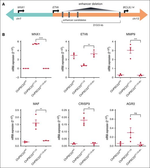
As a further layer of experimental evidence for de novo promoter-enhancer interactions in t(7;12)(q36;p13) AML, we examined MNX1 expression levels in 3 ChiPSC22t(7;12) sublines carrying a deletion of 213.5 kb, which removed the 4 enhancer candidates (ChiPSC22t(7;12)ΔEn; Figure 5A; supplemental Figure 7A). We performed WGS of the ChiPSC22t(7;12)ΔEn derivatives 2304B4 and 2305C9, could confirm the enhancer deletion in both, and found no other rearrangements in 2304B4 but an inversion in the nontranslocated ETV6 allele of 2305B9 (supplemental Figure 8). Consequently, clone 2305B9 was omitted from further analyses. In the other 2 ChiPSC22t(7;12)ΔEn lines, HSPC-specific MNX1 expression was abrogated (Figure 5B), whereas the expression of LRP6 and BCL2L14 near the deletion was not changed (supplemental Figure 7B). This supports the hypothesis that MNX1 activation in ChiPSC22t(7;12) is the result of interactions between the MNX1 promoter and 1 or multiple enhancers located in or close to ETV6. We also observed downregulation of genes that are upregulated together with MNX1 in t(7;12)(q36;p13), such as AGR2, MMP9, MAF, and CRISP3 (Figure 5B), suggesting that they are regulated by MNX1. Similar to differentiated ChiPSC22WT, ETV6 was upregulated, which might be explained by MNX1 regulation as well. Further phenotypic characterization of the ChiPSC22t(7;12)ΔEn sublines 2304B4 and 2305B10 revealed that there is no difference in differentiation capacity compared with the parental ChiPSC22t(7;12) (supplemental Figure 7C). The HSPCs derived from both groups also show similar proliferation rates and give rise to similar colony-forming units (supplemental Figure 7D-E).
Molecular validation of enhancer-promoter interaction in ChiPSC22t(7;12) upon differentiation. (A) Scheme of the enhancer deletion experiment performed to validate the interaction between the MNX1 promoter and enhancers distal to the BP. (B) Gene expression in HSPCs derived from ChiPSC22WT (n = 3), ChiPSC22t(7;12) (n = 4, from 2 independent cell lines), and ChiPSC22t(7;12)ΔEn (n = 3, from 2 independent cell lines) measured via qRT-PCR and shown as 2−ΔCt vs GUSB as endogenous reference. ∗∗∗∗P < .0001; ∗∗∗P < .001; ∗∗P < .01; ∗P < .05. ns, not significant.
Molecular validation of enhancer-promoter interaction in ChiPSC22t(7;12) upon differentiation. (A) Scheme of the enhancer deletion experiment performed to validate the interaction between the MNX1 promoter and enhancers distal to the BP. (B) Gene expression in HSPCs derived from ChiPSC22WT (n = 3), ChiPSC22t(7;12) (n = 4, from 2 independent cell lines), and ChiPSC22t(7;12)ΔEn (n = 3, from 2 independent cell lines) measured via qRT-PCR and shown as 2−ΔCt vs GUSB as endogenous reference. ∗∗∗∗P < .0001; ∗∗∗P < .001; ∗∗P < .01; ∗P < .05. ns, not significant.
In conclusion, we provide experimental evidence for a previously proposed enhancer-hijacking event8 rather than the creation of an oncofusion protein in pediatric AML with t(7;12)(q36;p13). Using an in vitro iPSC/HSPC cell system, we demonstrate that 1 or several enhancers in the ETV6 region interact with MNX1 to regulate its expression.
Discussion
In this manuscript, we describe enhancer hijacking and activation of MNX1 as a novel molecular mechanism resulting from a translocation between chr7 and chr12 [t(7;12)(q36;p13)] in pediatric AML. Our study shifts the focus from a putative MNX1::ETV6 oncofusion transcript42 to the activation of MNX1 as the unifying putative leukemia-driving event. Overexpression of MNX1 is accompanied by monoallelic inactivation of ETV6 on the translocated chromosome, putatively resulting in haploinsufficiency. Our observation has important implications for the diagnosis of this subgroup of patients, as well as novel therapeutic approaches. As shown in the expression reanalysis, all t(7;12)(q36;p13) AML demonstrate overexpression of MNX1. Only few AML cases without t(7;12)(q36;p13) show MNX1 overexpression; but although t(7;12)(q36;p13) AML is diagnosed at a very young age (<20 months), MNX1 overexpression in the absence of this translocation occurs predominantly at a later age, suggesting different, yet unexplained, molecular pathways converging in MNX1 expression. Considering that MNX1 is not expressed in the normal hematopoietic system, quantitative MNX1 expression analysis could be used as a diagnostic marker for this subgroup of pediatric AML.
Enhancer-hijacking events resulting in the activation of proto-oncogenes have been described also in other human malignancies including translocations resulting in the activation of oncogenes MYC, BCL2, or CCND1 in B-cell lymphoma34-36 or rearrangements in medulloblastoma.37 Subtype-specific 3D genomic alterations were recently discovered in AML leading to enhancer-promoter or enhancer-silencer loops.38 We demonstrated that the t(7;12)(q36;p13) translocation results in a neo-TAD, in which the MNX1 promoter is able to interact with the ETV6 region.
Initial evidence for an oncogenic role of MNX1 in leukemogenesis comes from a study by Nagel et al characterizing the MNX1-overexpressing cell line GDM-1.20 Knockdown of MNX1 led to a reduction of cell viability and cell adhesion. In vitro overexpression of MNX1 in HT1080 and NIH3T3 cells leads to premature, oncogene-induced senescence mediated by the induction of p53 signaling.39 In vivo, ectopic MNX1 expression in murine HSPCs resulted in strong differentiation arrest and accumulation at the megakaryocyte/erythrocyte progenitor stage.39 Overall, this phenotype is in line with reports on t(7;12)(q36;p13) AML blast cells that are less differentiated (French-American-British subtype M0 or M2) and demonstrate expression of the stem cell markers CD34 and CD117.5,40 Waraky et al used retroviral transduction of MNX1-expressing constructs into murine fetal HSPCs and were able to induce AML.33 A possible link to leukemogenesis was described with the observation that MNX1 activation resulted in reduced H3K4me1/2/3 and H3K27me3 levels providing increased chromatin accessibility.33
Our study challenges the concept in AML that all reciprocal translocations lead to oncofusion proteins as an overestimated molecular mechanism in AML for gene activation. Future studies unraveling the molecular defects of t(7;12)(q36;p13) AML should focus on the targets of homeobox transcription factor MNX1 rather than the oncofusion, as already initiated in some reports.21,30,39 Furthermore, copy number alterations of genes such as DNA methyltransferase 1 (DNMT1) or RNA polymerase II transcriptional elongation factor (ELL) on chr19, coupregulation of genes such as EDIL3 and LIN28B, or haploinsufficiency of ETV6 might contribute to the leukemogenic process. ETV6 is a strong transcriptional repressor, and haploinsufficiency could result in reactivation of its target genes.41 Moreover, the study of a potential therapeutic benefit by epigenetic drug treatment targeting MNX1 promoter-ETV6 enhancer interaction in the ChiPSC22t(7;12) iPSC/HSPC model is warranted.
We recognize that this study has several limitations that could be overcome in future studies. Due to the rarity of the disease and the limitations in obtaining primary leukemic samples, multiple (epi)genomic studies on a single patient sample are currently not possible but may become possible with improved biobanking, international collaboration, and the development of low-input profiling assays. A step in this direction is the survival analysis in pediatric AML by the NOPHO-AML.4 Another limitation is the mapping of responsible enhancers in the 213 kb ETV6 region, including at least 4 potential hematopoietic enhancers that could drive MNX1 expression. Individual enhancer knockout experiments will determine whether a single enhancer or multiple enhancers are required to activate MNX1.
Acknowledgments
The authors thank the Genomics and Proteomics Core Facility, the Omics IT and Data Management Core Facility of the German Cancer Research Center, Heidelberg, Germany, and the Center for Advanced Light Microscopy at the Ludwig-Maximilians-University Munich for their excellent support.
This work was, in part, supported by funds from the Helderleigh Foundation (Enhance Program) and German Research Foundation, SFB1074 subproject B11N (SFB1074/3 2020 Project Number:217328187 C.P. and A.R.), Carreras Foundation (DJCLS 03 R/2022 C.P. and E.S.) and FOR2674 subprojects A1 (PL 202/7-2), A6 (LI 2492/3-1), and A9 (PL 202/8-2 C.P. and D.B.L.), the Swedish Cancer Society (200925 PjF, CAN2017/461), the Swedish Childhood Cancer Foundation (PR2021-0025 and TJ2022-0017) and Västra Götalandsregionen (ALFGBG-431881 [L.P.]), and the SFB1064 subproject A17 (H.L.). Further support comes from the Helmholtz International Graduate School (A.R. and E.S.).
Authorship
Contribution: Y.L.B., G.G., B.S., D.R., and L.B. provided the clinical specimens; A.R., A.T., M.B., A.Ö., T.N., S.J., M.E., A.W., A.D., C.S., H.H., and H.L. performed the experimental procedures; E.S., U.H.T., J.H., J.A.W., K.B., J.A.W., P.L., and D.W. performed the bioinformatics and statistical analyses; E.S. was responsible for the sequence data upload to the public databases; D.B.L., L.P., D.W., and C.P. designed the study and supervised the experimental and bioinformatics work; D.W., A.R., E.S., and C.P. wrote the paper; and all authors provided feedback on the report.
Conflict-of-interest disclosure: L.B. has received honoraria from AbbVie, Amgen, Astellas, Bristol Myers Squibb, Celgene, Daiichi Sankyo, Gilead, Hexal, Janssen, Jazz Pharmaceuticals, Menarini, Novartis, Pfizer, Roche, and Sanofi; and research support from Bayer and Jazz Pharmaceuticals. D.B.L. receives honoraria from Infectopharm GmbH. The remaining authors declare no competing financial interests.
Correspondence: Christoph Plass, Division of Cancer Epigenomics, German Cancer Research Center, INF 280, 69120 Heidelberg, Germany; email: c.plass@dkfz.de; and Daniel B. Lipka, Section of Translational Cancer Epigenomics, Division of Translational Medical Oncology, German Cancer Research Center, Im Neuenheimer Feld 581, 69120 Heidelberg, Germany; email: d.lipka@dkfz.de.
References
Author notes
D.W., A.R., E.S., and U.H.T. are joint first authors.
D.B.L. and C.P. are joint senior authors.
All cell line sequencing data generated here are based on human reference GRCh37/hg19 and were deposited in the National Center for Biotechnology Information Gene Expression Omnibus database under accession GSE244379. The patient data were deposited in the European Genome-Phenome Archive (accession number EGAS50000000130).
The full-text version of this article contains a data supplement.

