Hematopoietic stem cells (HSCs) have a remarkable capacity for lifelong production of blood cells; however, they are not immune to aging. Surprisingly, as a mouse ages, there is actually an accumulation of phenotypically defined HSCs, with up to five times as many found in old mice compared to young mice.1-3 Despite the increase in number, hematopoietic output shifts toward a myeloid bias, and the repopulating ability of old HSCs is only about one-quarter as efficient as young HSCs. While most of the published work has used mouse models, a retrospective analysis by the National Marrow Donor Program assessing donor characteristics on recipient outcome found that age was the only donor trait significantly associated with overall and disease-free survival. Recipients who received a graft from a donor older than 45 years had significant decreases in five-year survival,4 demonstrating clear deficits in HSC function as a result of aging.
Recently, work from Dr. Jeffrey M. Bernitz and colleagues suggests that not only is quiescence an important feature of HSC maintenance during aging, but that HSCs have the capacity to count the number of self-renewal divisions they have undergone and have a maximal limit of four.5
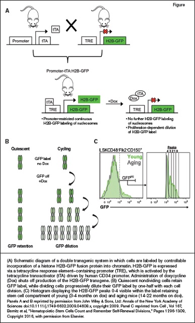
(A) Schematic diagram of a double transgenic system in which cells are labeled by controllable incorporation of a histone H2B-GFP fusion protein into chromatin. H2B-GFP is expressed via a tetracycline response element–containing promoter (TRE), which is activated by the tetracycline transactivator (tTA) driven by human CD34 promoter. Administration of doxycycline (Dox) shuts off production of the H2B-GFP transgene. (B) Quiescent nondividing cells retain GFP label, while dividing cells progressively dilute their GFP label by one-half with each cell division. (C) Histogram displaying the H2B-GFP peaks 0-4 visible within the label retaining stem cell compartment of young (3-4 months on dox) and aging mice (14-22 months on dox). Panels A and B reprinted by permission from John Wiley & Sons, Ltd: Annals of the New York Academy of Sciences doi:10.1111/j.1749-6632.2009.04608.x, copyright 2009. Panel C reprinted from Cell , Vol 167, Bernitz et al, "Hematopoietic Stem Cells Count and Remember Self-Renewal Divisions," Pages 1296-1309, Copyright 2016, with permission from Elsevier.
(A) Schematic diagram of a double transgenic system in which cells are labeled by controllable incorporation of a histone H2B-GFP fusion protein into chromatin. H2B-GFP is expressed via a tetracycline response element–containing promoter (TRE), which is activated by the tetracycline transactivator (tTA) driven by human CD34 promoter. Administration of doxycycline (Dox) shuts off production of the H2B-GFP transgene. (B) Quiescent nondividing cells retain GFP label, while dividing cells progressively dilute their GFP label by one-half with each cell division. (C) Histogram displaying the H2B-GFP peaks 0-4 visible within the label retaining stem cell compartment of young (3-4 months on dox) and aging mice (14-22 months on dox). Panels A and B reprinted by permission from John Wiley & Sons, Ltd: Annals of the New York Academy of Sciences doi:10.1111/j.1749-6632.2009.04608.x, copyright 2009. Panel C reprinted from Cell , Vol 167, Bernitz et al, "Hematopoietic Stem Cells Count and Remember Self-Renewal Divisions," Pages 1296-1309, Copyright 2016, with permission from Elsevier.
The authors hypothesized that proliferation of stem cells was the cause of the deficits in aging and used a mouse model6 to track HSC divisions. This mouse model utilizes a histone H2B-green fluorescent protein (GFP) fusion protein that is capable of incorporating GFP into nucleosomes (Figure, panel A). The H2B-GFP is under the control of a tetracycline-responsive element, and the activator is expressed in HSCs under the control of the human CD34 promoter. Therefore, HSCs incorporate GFP into their nucleosomes until the mice are given doxycycline, where the H2B-GFP expression is then repressed, and no additional incorporation of GFP into the nucleosome occurs. Quiescent stem cells that never divide will maintain high levels of GFP, while each successive division will dilute the GFP, resulting in progressively dimmer signals (Figure, panel B).
After 10 to 22 months of doxycycline, about 3 percent of the HSCs in aged mice still contained GFP, demonstrating long-term quiescence. Transplantation studies revealed that all of the long-term repopulating capacity was contained within those expressing high levels of GFP, indicating that in the aged mouse, only HSCs that had not proliferated extensively were capable of reconstitution.
Further analysis of the HSCs expressing high levels of GFP showed the presence of five distinct peaks of GFP expression (Figure, Panel C). Each of these peaks corresponded to a twofold dilution of GFP signal, and thus represented the divisional history of the stem cell. In the aged mice, twice as many label-retaining HSCs were found; however, 97.4 percent of them had the dimmest level of GFP expression, representing four divisional events. Mathematical modeling demonstrated that all of these HSCs in the aged mice contained in the fourth peak of GFP expression were the result of four rounds of self-renewal divisions. The accumulation of HSCs at this level of GFP expression suggests that HSCs maintain a history of their self-renewal divisions. After they have reached a count of four, they either remain dormant, or perhaps divide a fifth time that ultimately results in the complete loss of long-term stem cell function.
In Brief
Evidence of the ability of stem cells to count their divisions creates many additional questions for further discovery. 1) How do stem cells maintain a record of their divisions? There have been case reports of telomere shortening being associated with poor graft outcomes7 ; however, analysis of mouse stem cells forced to proliferate has shown no differences in telomere length.8 Epigenetic changes, or perhaps the dilution of a factor may govern the ability to track cell division. Determining the mechanisms of this counting function may allow for alterations that can teach stem cells to count beyond four, extending the long-term repopulating ability. 2) What implications does this counting feature have on human stem cell expansion and gene therapy efforts? If HSCs have an internal clock that only allows for a set number of self-renewal divisions, that clock could be a large hindrance to successfully expand stem cells for transplantation. A recent clinical protocol for stem cell expansion9 results in about nine to ten divisions over the course of the ex vivo protocol. So far, engraftment does not appear to be compromised and may suggest that human cells have a larger counter, or that ex vivo systems bypass the normal control mechanisms. 3) What does seem clear is that transplantation, at least transiently, allows HSCs to bypass the normal homeostatic barrier of four self-renewal divisions. Understanding how transplantation creates the workaround may help aid in developing further therapeutic strategies. Intriguingly, the normal maximal amount of serial transplantations in the mouse model is about four to five serial transplantations,10,11 suggesting that the workaround may even have its own counting mechanism. Answers to these questions are likely to significantly advance the field and our understanding of self-renewal.
References
Competing Interests
Dr. Hoggatt indicated no relevant conflicts of interest.

