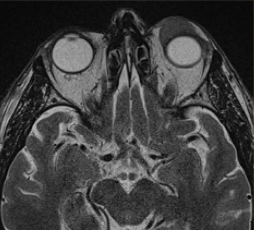
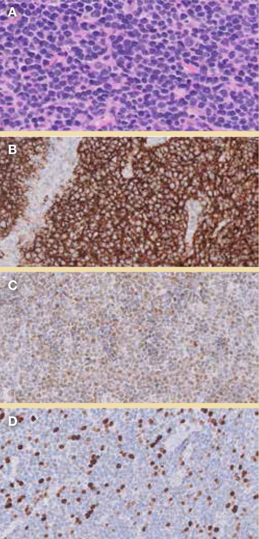
A 62-year-old man presented with progressive ptosis of the left eyelid, which he had been experiencing for the past year. He had no other symptoms or significant medical history. Physical examination revealed a firm mass below the left supraorbital rim, with secondary mechanical ptosis (Figure 1). Magnetic resonance imaging (MRI) revealed an extraconal, expansile lesion with well-defined edges at the level of the left upper eyelid, along with predominantly hypointense homogeneous signal intensity in T1 and T2 sequences showing restricted diffusion (Figure 2). Analysis of the biopsy specimen revealed a diffuse pattern of small lymphocyte proliferation and monomorphic cytology consisting predominantly of centrocyte-like cells of B-cell lineage (CD20+). Cyclin D1 and SOX11 expression were detected, although the specimen was negative for BCL6, CD5, CD10, and CD23 expression (Figure 3). The neoplastic cells exhibited restricted expression of kappa light chain, an unremarkable pattern of p53 expression (wild-type pattern), and a low Ki67 proliferation index. Fluorescence in situ hybridization detected chromosomal rearrangement involving CCND1 at 11q13.
Mechanical ptosis at the level of the left orbit. Patient consent obtained.
Axial MRI showing a lesion in the extraconal compartment of the left orbit.
Biopsy findings. A: Magnified image of hematoxylin and eosin (H&E) staining; B: CD20+ staining; C: Reaction of the tumor cells with anticyclin D1; D: Low Ki67 index.
Biopsy findings. A: Magnified image of hematoxylin and eosin (H&E) staining; B: CD20+ staining; C: Reaction of the tumor cells with anticyclin D1; D: Low Ki67 index.
What is your diagnosis?
Mucosa-associated lymphoid tissue (MALT) lymphoma
Mantle cell lymphoma (MCL)
Small lymphocytic lymphoma (SLL)
Follicular lymphoma (FL)
For the solution to the quiz, visit The Hematologist online, https://ashpublications.org/thehematologist/pages/orbital_tumor_in_a_62_year_old_patient.
Competing Interests
Drs. Suárez, Morillo, and Piris indicated no relevant conflicts of interest.


