Case Description
An 83-year-old woman presented with progressive, bilateral neck swelling. She did not report weight loss, loss of appetite, fever, night sweats, abdominal pain, back/bone pain, or jaundice. Twenty years ago, she was diagnosed with left-sided breast cancer, which was treated with surgery and chemotherapy, and she underwent chemotherapy and immunotherapy for chronic lymphocytic leukemia (CLL) five years ago. Physical examination revealed bilateral discrete mobile cervical lymph nodes, with the largest lymph node in the right supraclavicular region reaching up to 6 cm. A complete blood count with differential revealed macrocytic anemia (hemoglobin: 9.4 g/dL; mean corpuscular volume: 102 fL), normal white blood cell count (4.3×10⁹/L; neutrophils: 3.579/L, lymphocytes: 10.09/L), and thrombocytopenia (platelets: 36 K/uL). Bone marrow aspiration with biopsy and PET-CT were performed.
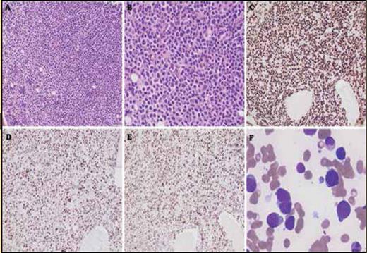
Bone marrow core biopsy showing infiltration by large lymphoid cells, extensively replacing normal hematopoietic elements. (A, hematoxylin and eosin, 200x). The lymphoid cells are large and have irregular nuclear contours with an increased nuclear-to-cytoplasmic ratio (B, hematoxylin and eosin, 400x) The lymphoid infiltrate is positive for PAX5 (C), showing aberrant overexpression of p53 (D) and a high Ki-67 proliferation index (E). (C-E; immunohistochemistry with hematoxylin counterstain, 200x). The bone marrow aspirate smear shows increased numbers of large, atypical lymphoid cells with prolymphocytoid morphology (F, Giemsa, 1,000x).
Bone marrow core biopsy showing infiltration by large lymphoid cells, extensively replacing normal hematopoietic elements. (A, hematoxylin and eosin, 200x). The lymphoid cells are large and have irregular nuclear contours with an increased nuclear-to-cytoplasmic ratio (B, hematoxylin and eosin, 400x) The lymphoid infiltrate is positive for PAX5 (C), showing aberrant overexpression of p53 (D) and a high Ki-67 proliferation index (E). (C-E; immunohistochemistry with hematoxylin counterstain, 200x). The bone marrow aspirate smear shows increased numbers of large, atypical lymphoid cells with prolymphocytoid morphology (F, Giemsa, 1,000x).
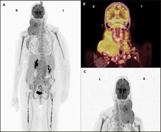
(A-C). PET/CT scan with contrast showing conglomerates of hypermetabolic lymph nodes in the right neck displacing the right oropharynx and extension to the right axilla, with an index lesion of 6.2 x 5.5 cm and standardized uptake value (SUV) of 8.2, and hypermetabolic lymph nodes in the left supraclavicular stations, with an index lesion of 1.7 x 1 cm and an SUV of 3.7.
(A-C). PET/CT scan with contrast showing conglomerates of hypermetabolic lymph nodes in the right neck displacing the right oropharynx and extension to the right axilla, with an index lesion of 6.2 x 5.5 cm and standardized uptake value (SUV) of 8.2, and hypermetabolic lymph nodes in the left supraclavicular stations, with an index lesion of 1.7 x 1 cm and an SUV of 3.7.
What is the diagnosis?
Metastatic carcinoma
Richter transformation
Myelodysplastic syndrome
Megaloblastic anemia
For the solution to the quiz, visit The Hematologist online, www.hematology.org/TheHematologist/Image-Challenge.
Competing Interests
Drs. Jain, Loghavi, and Alvarado indicated no relevant conflicts of interest.


