Abstract
T cells are produced through 2 mechanisms, thymopoiesis and proliferative expansion of postthymic T cells. Thymic output generates diversity of the pool, and proliferation achieves optimal clonal size of each individual T cell. To determine the contribution of these 2 mechanisms to the formation of the initial T-cell repertoire, we examined neonates of 30 to 40 weeks' gestation. Peripheral T cells were in a state of high proliferative turnover. In premature infants, 10% of T cells were dividing; the proliferation rates then declined but were still elevated in mature newborns. Throughout the third trimester, concentrations of T-cell–receptor excision circles (TRECs) were 10 per 100 T cells. Stability of TREC frequencies throughout the period of repertoire generation suggested strict regulation of clonal size to approximately 10 to 20 cells. Neonatal naive CD4+ and CD8+ T cells were explicitly responsive to IL-7; growth-promoting properties of IL-15 were selective for newborn CD8+ T cells. Neonatal T cells expressed telomerase and, in spite of the high turnover, built up a telomeric reserve. Thus, proliferative expansion, facilitated by increased cytokine responsiveness, and thymopoiesis complement each other as mechanisms of T-cell production in neonates. Maintaining optimal clonal size instead of filling the space in a lymphopenic host appears to regulate homeostatic T-cell proliferation during fetal development.
Introduction
De novo T-cell generation requires a functional thymus.1 The current paradigm is that the human T-cell repertoire is established during late fetal development and that, by the time of birth, thymectomy does not cause immediate immunodeficiency.2,3 Thymic epithelial space—the component of the thymus relevant for T-cell production and selection— involutes rapidly after birth. Thymic involution starts at the age of 1 year and continues with a yearly loss of 3%.4,5 The peripheral T-cell compartment is under homeostatic control6 ; with the reduction in thymopoietic capacity, alternative mechanisms of T-cell generation are necessary to prevent lymphopenia. Autoproliferation of postthymic T cells, particularly those in the memory compartment, is now accepted as a mechanism of T-cell production.7,8 With advancing age, a shift occurs from efficient thymic lymphopoiesis to T-cell generation through peripheral replication; however, both mechanisms likely coexist during adult life. Age-related loss of thymic function is not the only condition that necessitates alternative means of repopulating the T-cell compartment. A number of clinical conditions, including chemotherapy, bone marrow transplantation, and infection with human immunodeficiency virus, are associated with T-cell depletion and the subsequent need to regenerate T cells despite limited thymopoietic capability. Evidence shows that recovery of peripheral T-cell numbers after chemotherapy is from thymic sources in children younger than 18 years of age.7 In adults, thymopoiesis may still contribute to some degree, but replication of peripheral T cells becomes the dominant mechanism of filling the pool. It is obvious that the proliferation of postthymic T cells, especially when not entirely random, can have profound consequences on the composition of the T-cell repertoire.9,10 What precisely these consequences are for immunocompetence is not understood.
Functionality of the immune system is tightly linked to the receptor diversity of the T-cell pool.11,12 Obviously, a large spectrum of T-cell receptors (TCRs) increases the chances of recognizing invading microorganisms. Thymopoiesis, the single mechanism of creating novel T cells, should be the key factor in determining immunocompetence. Young adults carry approximately 2 × 10¹¹ CD4+ T cells and approximately 1 × 10¹¹ CD8+ T cells.13,14 The highest degree of diversity would be obtained if each naive T cell in the pool had a unique TCR. However, such a high degree of diversity might not be advantageous because it would decrease the probability of antigen recognition at a given tissue site. Instead, a minimal clonal size for each naive T-cell specificity is required to increase the chance for a T cell to encounter antigen and to mount a defensive response. Generation of a minimal clonal size requires that T cells selected in the thymus undergo intrathymic or postthymic proliferation.
Arstila et al15 have estimated the clonal size of naive T cells in adult humans. They measured an average diversity of 9 × 10⁵ different TCR β-chains and 4.5 × 10⁵ different α-chains and estimated a maximum of 1 × 10⁸ different TCR α-β combinations in individual donors, resulting in an average clonal size of 1000 for naive T cells. Using a different approach, we have determined the median TCR β-chain frequency as 1in2 × 107 naive CD4+ T cells.16
To investigate how the need for diversity and for achieving minimal clonal size of each T cell are balanced during establishment of the T-cell repertoire in late fetal development, we studied parameters of T-cell homeostasis in healthy newborns between the gestational ages of 30 and 40 weeks. We found that the frequency of T cells carrying signal joint TCR excision circles (sjTRECs), which represent recent thymic emigrants, is high but remarkably stable during this critical period of TCR repertoire formation. High thymopoietic capacity is accompanied by high proliferative turnover of postthymic T cells, with up to 10% of the pool dividing. Neonatal T cells are exquisitely sensitive to growth-promoting cytokines, and CD4+ and CD8+ T cells display distinct response patterns. Peripheral expansion of T cells is achieved without telomeric loss because of the spontaneous expression of telomerase. Despite undergoing numerous rounds of replication, naive T cells are equipped with an elongated telomeric reserve, enabling them to respond with prolonged cellular replication later in life. We conclude that lymphopoiesis in the initial phase of repertoire generation is not geared toward maximal diversity but involves the expansion of postthymic T cells to a preset clonal size.
Patients, materials, and methods
Patients and samples
Seventy-five cord blood samples were collected immediately after live birth (median gestational age, 38.5 weeks). Samples from mothers with an infection or an inflammatory disease were excluded. Peripheral blood was collected from 39 healthy donors aged 19 to 30 years (median age, 25 years). This study was approved by the Mayo Clinic Institutional Review Board, and informed consent was obtained as appropriate.
Cell isolation
Peripheral blood mononuclear cells (PBMCs) were isolated using Ficoll-Hypaque (Amersham Biosciences, Arlington Heights, IL) by density centrifugation. CD4+ and CD8+ T cells were positively selected by MACS magnetic microbead separation (Miltenyi Biotec, Auburn, CA). Purity of the separated T-cell populations was 92% to 97%. Granulocytes were isolated from the red blood cell pellet of Ficoll-separated cells by sedimentation with 3% dextran (Amersham Biosciences) in 0.9% NaCl.
Quantification of signal joint TRECs
Genomic DNA was extracted from 3 × 10⁶ purified CD4+ and CD8+ T cells using the QiaAmp DNA Blood Midi Kit (Qiagen, Chatsworth, CA). Quantitative polymerase chain reaction (PCR) for sjTRECs was performed using 250 ng DNA on an ABI Prism 7700 thermal cycler (PE-Applied Biosystems, Foster City, CA) according to the protocol of Douek et al.17 The following primers and probe were used: 5′-CACATCCCTTTCAACCATGCT-3′ and 5′-GCCAGCTGCAGGGTTTAGG-3′ (Integrated DNA Technologies, Coralville, IA) and 5′-VIC-TTTTTGTAAAGGTGCCCACTCCTGTGCACGGTGA–TAMRA-3′ (PE-Applied Biosystems). Reactions contained 25 μL TaqMan Universal Mastermix (Applied Biosytems), 200 nM each primer, and 50 nM probe. Conditions were 50°C for 2 minutes, 95°C for 10 minutes, and 40 cycles at 95°C for 15 seconds and 60°C for 1 minute. Dr D. Douek (Bethesda, MD) generously provided samples with a standardized number of TREC-containing sequences. A standard curve was plotted, and the number of TRECs was calculated using ABI 7700 software. Samples were analyzed in duplicate. DNA extracted from HeLa and U937 cells was used for negative controls. Results are expressed as the number of TRECs per 100 cells.
Flow cytometry
PBMCs were stained with FITC-conjugated anti-Ki67 monoclonal antibody (mAb; Becton Dickinson, San Jose, CA) after permeabilization with phosphate-buffered saline (PBS)/1% Triton X-100. Naive CD4+ and CD8+ T cells were labeled with phycoerythrin (PE)–conjugated anti-CD45RO and PerCP-conjugated anti-CD4 or PerCP-conjugated anti-CD8 mAbs (Becton Dickinson).18,19 PE-conjugated anti-CD127 (R&D Systems, Minneapolis, MN) was used to determine interleukin-7 (IL-7) receptor surface expression.
Telomere length analysis
DNA was extracted from purified T-cell subsets or granulocytes, and 5 μg DNA was digested with HinfI and RsaI overnight at 37°C. Fragments were resolved on 0.5% agarose gels (20 V, 15 hours). The gels were dried, denatured, neutralized, and then prehybridized in 5 × SSC buffer, 5 × Denhardt solution, and 0.1 M dibasic sodium phosphate for 2 hours. Hybridization was performed with a telomere repeat-specific[32P] end-labeled (CCCTAA)3 probe at 37°C overnight. After 7 washings with 0.5 × SSC buffer, the gels were exposed to a phosphor screen (Kodak Scientific Imaging Systems, Rochester, NY) that was scanned and analyzed using a Storm 840 system and ImageQuant software (Molecular Dynamics, Sunnyvale, CA). Telomere terminal restriction fragment (TRF) length was calculated relative to a [33P]-end–labeled lambda HindIII ladder that was run on each gel. DNA aliquots from the same donor were loaded on each gel to control for intergel variation. Results are given as the median TRF length of the sum function.20
Telomerase repeat amplification protocol
Telomerase activity was assayed with the TRAPeze Telomerase Detection Kit (Serologicals, Norcross, GA). Shock-frozen cells were incubated with CHAPS lysis buffer for 30 minutes on ice and then centrifuged at 12 000g for 20 minutes at 4°C. Supernatants were collected, and protein concentrations were determined by the Bradford assay (BioRad Laboratories, Richmond, CA). Optimal protein concentrations between 200 to 600 ng/μL were used in the PCR reactions. Two microliters of cell extract or negative control were incubated in the PCR mix at 30°C for 30 minutes according to the manufacturer's protocol. As a negative control, cell extracts of each sample were heat inactivated or treated with 1 μg/μL RNAse A (Roche Molecular Biochemicals, Indianapolis, IN) for 20 minutes at 37°C. Following the incubation periods, we added 2 U Taq DNA polymerase to each sample. Products were amplified for 33 cycles at 94°C and 59°C for 30 seconds each using a Biometra Uno II thermal cycler (Biometra, Horsham, PA) and were resolved by electrophoresis on 12.5% polyacrylamide gels at 80 V. Gels were stained with SYBR Green (Molecular Probes, Eugene, OR) and scanned with a Storm 840 system for qualitative analysis. A telomerase-positive cell line as positive control and a lysis buffer as negative control were included in each assay.
Cell culture and CFSE labeling
PBMCs were cultured in RPMI 1640 supplemented with penicillin, streptomycin, glutamine, and 10% fetal bovine serum (BioWhittaker, Walkersville, MD). Cultures were established at a concentration of 5 × 10⁵ cells/mL and were maintained at 37°C in a humidified incubator containing 7.5% CO2. To induce maximal stimulation, positive controls were stimulated with anti-CD3 and anti-CD28 mAbs as described.21
To detect cycling cells, PBMCs were incubated with 5-(and-6)–carboxyfluorescein diacetate succinimidyl ester (CFSE; 10 μM; Molecular Probes) at room temperature for 7 minutes. The cells were washed several times with PBS/5% fetal bovine serum to remove unbound CFSE and with PBS/0.5 mM EDTA (ethylenediaminetetraacetic acid) to prevent clumping of the cells. Cultures were harvested, cells were washed, and proliferating cell populations were determined by flow cytometry as described.22
Statistical analysis
Data were analyzed using the Mann-Whitney U rank sum test for unpaired groups and the Wilcoxon signed-rank test for paired groups as appropriate (SigmaStat; SPSS, Chicago, IL).
Results
Thymopoiesis and clonal size during fetal repertoire development
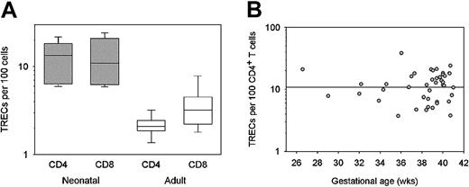
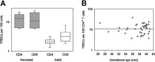
TRECs are episomal DNA byproducts of TCR α-chain rearrangement that are not replicated during cell division.23,24 The half-life of these episomes is unknown, but in neonates it is safe to assume that cellular proliferation is the major mechanism of TREC loss. Nearly all T cells in the newborn have a naive phenotype.25,26 Under these conditions, the frequency of TREC+ T cells should be an excellent marker for thymic capacity and should also inversely correlate with clonal size. To estimate clonal sizes of naive T cells, we purified CD4+ and CD8+ T-cell populations from cord blood of 14 full-term newborns, and we quantified TREC numbers by quantitative PCR. Concentrations of TRECs were high, reaching a median of 13.4 per 100 CD4+ and 10.9 per 100 CD8+ T cells. (Figure 1A). Representation of TREC+ T cells was approximately 4-fold to 6-fold higher in the neonates than in the 20- to 30-year-old adults. These data suggested that thymic production did indeed account for a large proportion of the peripheral T-cell pool. Yet, approximately 90% of the cells did not harbor a TREC and represented the progeny of TREC+ cells.
Cord blood TREC levels during the third trimester of gestation. Cord blood samples were collected from 45 newborns of 26 to 41 weeks gestational age. Peripheral blood lymphocytes were harvested from 12 young adults. Lymphocytes were separated into CD4+ and CD8+ subsets using magnetic beads, and TREC concentrations were determined by quantitative PCR. TREC levels are presented as box plots showing medians, 25th and 75th percentiles are presented as boxes, and 10th and 90th percentiles are presented as whiskers. Percentages of CD4+ and CD8+ T cells expressing TRECs were approximately 6-fold and 4-fold higher, respectively, in full-term newborns (filled boxes) than in the young adults (open boxes) (A). TREC levels in the CD4+ T-cell subset were plotted in relation to the gestational age of the infant. Percentages of TREC+CD4+ T cells remained stable throughout the third trimester (B).
Cord blood TREC levels during the third trimester of gestation. Cord blood samples were collected from 45 newborns of 26 to 41 weeks gestational age. Peripheral blood lymphocytes were harvested from 12 young adults. Lymphocytes were separated into CD4+ and CD8+ subsets using magnetic beads, and TREC concentrations were determined by quantitative PCR. TREC levels are presented as box plots showing medians, 25th and 75th percentiles are presented as boxes, and 10th and 90th percentiles are presented as whiskers. Percentages of CD4+ and CD8+ T cells expressing TRECs were approximately 6-fold and 4-fold higher, respectively, in full-term newborns (filled boxes) than in the young adults (open boxes) (A). TREC levels in the CD4+ T-cell subset were plotted in relation to the gestational age of the infant. Percentages of TREC+CD4+ T cells remained stable throughout the third trimester (B).
To estimate clonal size throughout the period of repertoire generation, we correlated TREC concentrations with gestational age in a cohort of 45 healthy infants born between 26 and 41 weeks of gestation (Figure 1B). TREC concentrations varied but were remarkably stable over the time range we examined, suggesting that the ratio between the production of TREC+ T cells by thymic production and the generation of TREC– T cells by proliferative expansion was constant throughout the third trimester. The stability of TREC concentrations also indicated that the contribution of thymic production to the T-cell repertoire formation did not increase with increasing maturity during fetal development.
High proliferative turnover of peripheral T cells in neonates
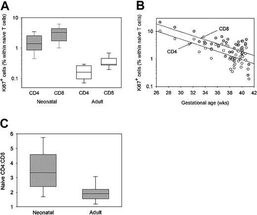
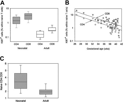
To understand how postthymic proliferation contributes to the formation of the T-cell repertoire, we determined the frequencies of cycling CD4+ and CD8+ T cells in freshly harvested cord blood. Proliferative rates of CD4+ and CD8+ T cells were estimated by measuring the fraction of Ki67-expressing T cells in 39 cord blood samples and PBMCs of 15 young adults. In newborn samples, CD45RO+ T cells account for less than 3% of the T-cell population; almost all T cells have the naive phenotype. To allow for proper comparison between the 2 cohorts, we distinguished CD45RO– naive and CD45RO+ memory T cells. As shown in Figure 2A, infants had a median of 1.4% CD4+ T cells and 3.2% CD8+ T cells in the cell cycle. These rates were 10-fold higher than the Ki67-expressing populations in young adults, who had a median of 0.2% CD4+CD45RO– and 0.3% CD8+CD45RO– T cells undergoing division. Neonatal naive T-cell proliferation rates in the newborns were as high as those measured among antigen-experienced memory T cells of adults.27
Proliferation of postthymic T cells in neonates and young adults. Lymphocytes were isolated from 39 cord blood samples from newborns of 26 to 41 weeks gestation and 15 healthy young adults. T cells in the cell cycle were identified by the expression of the Ki67 nuclear antigen by flow cytometric analysis of CD4+CD45RO– (naive) and CD8+CD45RO– T cells. Results in the adults (open boxes) and the full-term infants (shaded boxes) are shown as box plots. Frequencies of Ki67-expressing naive T cells were 10-fold higher in the infants than in the adults. The CD8+ T-cell subset included a higher proportion of Ki67+ cells in neonates (P < .001) and adults (P = .006) compared with CD4+ T cells (A). Frequencies of Ki67+ cells within the CD4+CD45RO– (open circles) and CD8+CD45RO– cells (filled circles) T-cell populations are given in correlation to the gestational ages of the neonates. Proliferation rates of cord blood CD4+ and CD8+ T cells were highest during the early phase of the third trimester and exponentially declined to maturity (R = 0.81 for CD4+ T cells; R = 0.79 for CD8+ T cells). The number of cycling cells remained elevated in full-term infants compared with adults. Frequencies of Ki67+CD45RO–CD8+ T cells were always higher than in their CD4+ counterparts (B). The ratio of CD4+CD45RO– and CD8+CD45RO– T cells was determined by flow cytometry in lymphocytes isolated from cord blood or peripheral blood of young adults. CD4+ T cells outnumbered CD8+ T cells in newborns and adults, but the ratios were significantly different (P < .001), with a relative reduction of CD4+ T cells over the first 2 decades of life (C).
Proliferation of postthymic T cells in neonates and young adults. Lymphocytes were isolated from 39 cord blood samples from newborns of 26 to 41 weeks gestation and 15 healthy young adults. T cells in the cell cycle were identified by the expression of the Ki67 nuclear antigen by flow cytometric analysis of CD4+CD45RO– (naive) and CD8+CD45RO– T cells. Results in the adults (open boxes) and the full-term infants (shaded boxes) are shown as box plots. Frequencies of Ki67-expressing naive T cells were 10-fold higher in the infants than in the adults. The CD8+ T-cell subset included a higher proportion of Ki67+ cells in neonates (P < .001) and adults (P = .006) compared with CD4+ T cells (A). Frequencies of Ki67+ cells within the CD4+CD45RO– (open circles) and CD8+CD45RO– cells (filled circles) T-cell populations are given in correlation to the gestational ages of the neonates. Proliferation rates of cord blood CD4+ and CD8+ T cells were highest during the early phase of the third trimester and exponentially declined to maturity (R = 0.81 for CD4+ T cells; R = 0.79 for CD8+ T cells). The number of cycling cells remained elevated in full-term infants compared with adults. Frequencies of Ki67+CD45RO–CD8+ T cells were always higher than in their CD4+ counterparts (B). The ratio of CD4+CD45RO– and CD8+CD45RO– T cells was determined by flow cytometry in lymphocytes isolated from cord blood or peripheral blood of young adults. CD4+ T cells outnumbered CD8+ T cells in newborns and adults, but the ratios were significantly different (P < .001), with a relative reduction of CD4+ T cells over the first 2 decades of life (C).
Proliferation of peripheral T cells was high throughout the third trimester (Figure 2B), but it was highest in infants born at younger gestational ages. At the beginning of the third trimester, the fetal peripheral blood compartment contained 10% T cells that were undergoing replication. The frequencies of Ki67+CD4+ and Ki67+CD8+ T cells decreased in an exponential fashion between weeks 26 and 40 (R = 0.81 for CD4+ T cells; R = 0.79 for CD8+ T cells), but they remained more than 10-fold elevated even in full-term infants when compared with young adults. Throughout the entire third trimester, the CD8+ subset contained a higher proportion of cycling cells than the CD4+ subpopulation (P < .001). This difference persisted into adult life (P = .006). The increased likelihood of CD8+ T cells to proliferate may explain why the dominance of CD4+ T cells decreases over a lifetime. CD4+ T cells outnumbered CD8+ T cells by a factor of 3.3 in the cord blood samples (Figure 2C). In 20- to 30-year-old donors, the naive CD4+/CD8+ T-cell ratio was 1.9 and was significantly different from that in infants (P < .001).
Postthymic T cells in newborns do not erode but rather elongate telomeric sequences
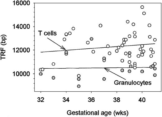
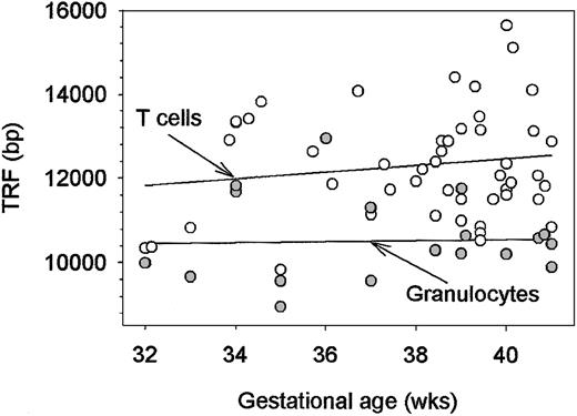
Rates up to 10% of peripheral T cells undergoing replication raised the question of whether the high demand for lymphopoiesis imposed proliferative stress on peripheral T cells compared with the hematopoietic precursor cell. The replicative history of hematopoietic stem cells can be estimated by measuring telomeric lengths in neutrophils. Neutrophils are terminally differentiated; they do not divide, and they have emerged from their precursor cells within the last 6 to 7 days. Telomeric repeats of cord blood neutrophils and lymphocytes were determined and correlated with the gestational ages of the infants (Figure 3). As expected, cord blood neutrophils possessed relatively long telomeres (median, 10 294 bp in a cohort of 19 donors). Neither the neutrophils nor the lymphocytes showed any evidence of telomeric erosion throughout the last trimester. Surprisingly, telomeric sequences in CD4+ lymphocytes were 1000 to 2000 bp longer (median, 12 139 bp in a cohort of 48 donors), suggesting an active mechanism of telomeric elongation. To address whether CD8+ T cells, which have an even higher proliferative rate, have shortened telomeres, paired samples of CD4+ and CD8+ T cells were compared. There was no difference (median, 12 592 vs 12 623; P = .94) between the 2 T-cell subsets.
Lengths of telomeric sequences in neonatal granulocytes and CD4+ T cells. Granulocytes and CD4+ T cells were purified from fresh cord blood samples. DNA was isolated, and the lengths of telomeres were determined by hybridization with a telomere repeat-specific probe. Median TRF lengths from each cell population are shown in correlation to the gestational age of the neonate. TRF lengths in granulocytes (filled circles) were stable throughout the third trimester. Although granulocytes and T cells derive from the same hematopoietic precursor cells, telomeric ends in CD4+ T cells (open circles) were elongated by 1500 to 2000 bp.
Lengths of telomeric sequences in neonatal granulocytes and CD4+ T cells. Granulocytes and CD4+ T cells were purified from fresh cord blood samples. DNA was isolated, and the lengths of telomeres were determined by hybridization with a telomere repeat-specific probe. Median TRF lengths from each cell population are shown in correlation to the gestational age of the neonate. TRF lengths in granulocytes (filled circles) were stable throughout the third trimester. Although granulocytes and T cells derive from the same hematopoietic precursor cells, telomeric ends in CD4+ T cells (open circles) were elongated by 1500 to 2000 bp.
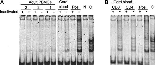
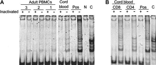
Hematopoietic stem cells have the capability of telomeric repair and self-renewal. Nevertheless, they show progressive telomeric loss over a lifetime.28,29 The proliferative stress of early hematopoiesis is not sufficient to shorten telomeres. To investigate the mechanism leading to telomere elongation in lymphocytes, we analyzed the expression of constitutive telomerase activity in newborn and adult CD4+ and CD8+ T cells. As shown in Figure 4, CD4+ and CD8+ T cells from cord blood had telomerase activity. Adult CD4+ and CD8+ T cells no longer expressed telomerase activity.
Spontaneous expression of telomerase activity in neonatal T cells. Lymphocytes from adult blood and cord blood were assayed for telomerase activity. Representative results from 3 adults and one cord blood sample are shown (A). CD4+ and CD8+ T cells obtained from cord blood were purified and tested for telomerase activity (B). Telomerase activity was assayed with (+) and without (–) heat inactivation. Results are representative of 8 experiments. A positive (HeLa cells, Pos) and a negative (N) control and an assay control (C) were included in all assays.
Spontaneous expression of telomerase activity in neonatal T cells. Lymphocytes from adult blood and cord blood were assayed for telomerase activity. Representative results from 3 adults and one cord blood sample are shown (A). CD4+ and CD8+ T cells obtained from cord blood were purified and tested for telomerase activity (B). Telomerase activity was assayed with (+) and without (–) heat inactivation. Results are representative of 8 experiments. A positive (HeLa cells, Pos) and a negative (N) control and an assay control (C) were included in all assays.
IL-7 drives proliferation of newborn postthymic CD4+ and CD8+ T cells
If the replication of peripherally seeded T cells is a necessary component of fetal T-cell lymphopoiesis, which T-cell growth factors control expansion in the periphery? Constantly higher proliferation rates among CD8+ T cells suggested subset-specific regulation, possibly a reflection of selective growth-factor responsiveness. IL-7 is a potent growth factor for thymocytes.30-32 We examined whether IL-7 contributed to the postthymic expansion of neonatal T cells and whether CD8+ T cells responded preferentially to this growth factor.
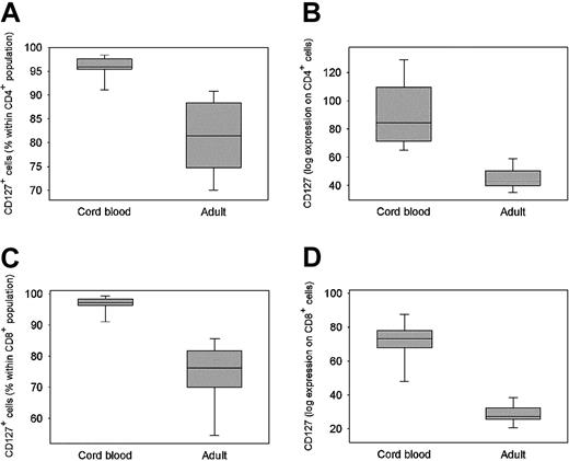
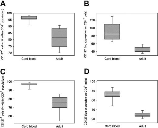
Flow cytometric analysis demonstrated that the IL-7 receptor α-chain (CD127) was highly expressed on cord blood CD4+ and CD8+ T cells; more than 95% of fetal T cells were positive for CD127 (Figure 5A-C). CD127+ T cells were less frequent among adult naive CD4+ and CD8+ T cells, but most of each T-cell population continued to be potentially IL-7 responsive. With advancing age, the cell surface density of CD127 also decreased markedly. As shown, (Figure 5B, D) mean fluorescence intensity of CD127 was 84.4 in the cord blood CD4+ T cells and 44.8 in the adult counterparts (P = .002). Similarly, mean peak expression of CD127 on fetal CD8+ T cells was measured at 73.2, but it was many times lower on adult naive CD8+ T cells (mean, 28.6; P < .001). Unexpectedly, CD127 expression was generally higher on CD4+ T cells than on CD8+ T cells in neonates (P = .06) and in adults (P = .008).
Expression of the IL-7R (CD127) on neonatal and adult lymphocytes. PBMCs were separated from 6 cord blood samples and 8 peripheral blood samples of young adults. Expression of the IL-7 receptor α-chain (CD127) was determined by 2-color flow cytometry of CD4+ (A, B) and CD8+ T cells (C, D). Data are presented as box plots of frequencies (A, C) or mean fluorescence intensities (B, D) with medians, 25th and 75th percentiles are presented as boxes, and 10th and 90th percentile are presented as whiskers.
Expression of the IL-7R (CD127) on neonatal and adult lymphocytes. PBMCs were separated from 6 cord blood samples and 8 peripheral blood samples of young adults. Expression of the IL-7 receptor α-chain (CD127) was determined by 2-color flow cytometry of CD4+ (A, B) and CD8+ T cells (C, D). Data are presented as box plots of frequencies (A, C) or mean fluorescence intensities (B, D) with medians, 25th and 75th percentiles are presented as boxes, and 10th and 90th percentile are presented as whiskers.
To test the functional relevance of CD127 expression, PBMCs were isolated from cord blood samples and from peripheral blood of the adult donors, labeled with CSFE, and cultured in the presence of a panel of growth factors. To assess the spontaneous responsiveness of freshly isolated T cells, no TCR triggering was provided in the cultures. The presence of proliferating CD4+ and CD8+ T cells was determined after 7 days of culture, and the percentage of responding cells was calculated. Optimal concentrations for each of the growth factors were identified in preliminary experiments. Results from experiments with 4 different donor pairs are summarized in Table 1. Insulin-like growth factor-1 (IGF-1) could not elicit a growth response in either newborn or adult T cells. IL-2 functioned as a weak stimulator for fetal CD4+ T cells (median, 34.3% of cells proliferating). A small fraction of cord blood CD8+ cells (median, 7.4% of cells proliferating) responded to IL-2. IL-2 was not sufficient to drive the proliferation of adult T cells.
IL-7 was a powerful stimulator of neonatal T cells and drove most CD4+ and CD8+ T cells into the cell cycle. Adult naive T cells responded to optimal concentrations of IL-7 in the absence of a TCR signal, though less efficiently. An average of 21.2% of adult naive CD4+ T cells and 51.2% of adult naive CD8+ cells proliferated in response to 10 ng/mL IL-7. IL-7–induced proliferation was not associated with the acquisition of CD45RO; the naive phenotype persisted on all fetal CD4+ and CD8+ T cells, even though they progressed through several cell cycles (data not shown).
Postthymic neonatal CD8+ T cells respond to IL-15
Beginning in the fetal period, peripheral CD8+ T cells proliferate more extensively than their CD4+ counterparts. The growth-promoting capability of IL-7 could not explain the preferential proliferation of CD8+ T cells in vivo. Among the growth factors tested, IL-15 was the only one that preferentially stimulated the replication of CD8+ T cells (Table 1). In all experiments, IL-15 was sufficient to initiate cell cycling of most cord blood CD8+ T cells (median, 63.2%). Adult naive CD8+ T cells remained responsive to this growth factor, with 40.5% of the cells proliferating. Only a small fraction of neonatal and none of the adult CD4+ T cells were responsive to IL-15. Monitoring of CD45RO expression in cultures of CSFE-labeled cord blood cells revealed that after 4 divisions, proliferating cells started to express the memory phenotype.
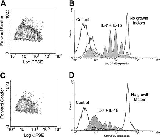
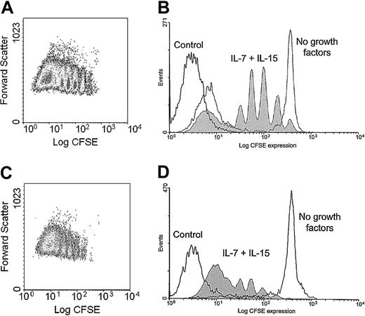
In cultures combining the 2 growth factors, IL-7 and IL-15, at optimal concentrations of 10 ng/mL and 24 ng/mL, respectively, essentially all CD8+ T cells and 75% of the CD4+ T cells entered the cell cycle (Figure 6).
Proliferative response of neonatal CD4+ and CD8+ T cells to IL-7 and IL-15. Lymphocytes were purified from cord blood, labeled with CSFE, and cultured in the absence or presence of IL-7 (10 ng/mL) and IL-15 (24 ng/mL). Cell division patterns of CD4+ and CD8+ T cells were evaluated by flow cytometric analysis on day 7. Representative results from 4 experiments are shown. Results gated on CD4+ (A, B) and CD8+ T cells (C, D) are shown as scatter plots (A, C) and histograms (B, D). Cells were cultured in the absence (gray line) and the presence of IL-7 and IL-15 (shaded area). The dark line represents autofluorescence.
Proliferative response of neonatal CD4+ and CD8+ T cells to IL-7 and IL-15. Lymphocytes were purified from cord blood, labeled with CSFE, and cultured in the absence or presence of IL-7 (10 ng/mL) and IL-15 (24 ng/mL). Cell division patterns of CD4+ and CD8+ T cells were evaluated by flow cytometric analysis on day 7. Representative results from 4 experiments are shown. Results gated on CD4+ (A, B) and CD8+ T cells (C, D) are shown as scatter plots (A, C) and histograms (B, D). Cells were cultured in the absence (gray line) and the presence of IL-7 and IL-15 (shaded area). The dark line represents autofluorescence.
Discussion
Immunocompetence requires a highly diverse repertoire of self-tolerant T cells that respond with clonal burst when the need arises. Thymic function is critical for generating diversity. Thymopoiesis must be complemented by the expansion of selected T cells to achieve an optimal clonal size. Single-copy representation of each T cell would minimize the probability of recognizing antigen in the lymphoid space of a host. Here we have examined how thymopoiesis and clonal expansion are balanced during the critical phase of T-cell repertoire generation in late fetal development. We found that both mechanisms work together in filling the developing T-cell pool. The ratio between thymic output and T-cell generation by the proliferation of mature T cells is controlled as indicated by the stability of the frequencies of TREC+ T cells. The proportion of the T-cell compartment that is occupied by TREC+ T cells is held at approximately 10% throughout the third trimester. During this period, the fetal body triples in size, imposing the need for the T-cell compartment to expand proportionally. We conclude that initial T-cell generation is not simply regulated by the lymphopenia of the growing host but that thymic T-cell release and postthymic expansion are interlinked events, leading to stability of clonal size in the T-cell compartment.
In the current study, neonates had high frequencies of proliferating T cells in the circulation. We did not find evidence that this increased turnover was a consequence of fetal stress. Mothers and babies were screened for infections and pre-existing inflammatory disorders. In addition, there was no correlation between the type of delivery (cesarean section versus vaginal) or the reason for premature birth and the frequency of proliferating cells. Notably, proliferation rates up to 10% of naive T cells are generally not observed under physiologic or pathologic conditions in humans.27 Peripheral T-cell expansion could be considered a necessary element of T-cell homeostasis during a period of life when the demand for T-cell generation is high and the thymus is possibly not fully matured. The T-cell pool is under homeostatic control, and lymphopenia induces the proliferation of naive T cells to fill the space.33-36 If homeostatic expansion occurs without the influx of novel T cells, clonal size increases and T-cell diversity contracts, as is described for patients after chemotherapy or experimental T-cell depletion9,37 and for patients with autoimmune diseases such as rheumatoid arthritis.16,20,38 Decreasing T-cell proliferation rates during the third trimester would fit the model that the need for T-cell reconstitution is the greatest early in the third trimester and levels off as the fetus reaches term.
Peripheral expansion of T cells in response to “empty space” results in the dilution of TREC+ T cells.39 Instead, TREC concentrations remained stable over the entire period of prenatal T-cell repertoire formation without a corresponding increase associated with fetal maturation and declining peripheral expansion.40 It can be assumed that during the short 10-week period of the third trimester, TRECs were predominantly lost by cell division and that TREC frequencies inversely reflected clonal size. TREC concentrations in CD1a+CD3–CD4+CD8+ thymocytes have been measured at approximately 150 per 100 cells, demonstrating a frequent deletion on both alleles.32 TREC concentrations in mature thymic CD4+ and CD8+ T cells are 60 and 40 per 100 cells, respectively.32 TREC concentrations in the blood of newborns of approximately 10 per 100 T cells would therefore indicate that T cells have undergone 4 divisions after TCR rearrangement, at least 2 of them in the periphery. This rate was independent of gestational age, the rate of peripheral proliferation, and the changing size of the lymphoid compartment. The stability of TREC frequencies is compatible with the notion that the system strives for a stable clonal size for each T-cell specificity instead of proliferative expansion functioning as a compensatory mechanism for insufficient thymic T-cell release.
The next question is which regulatory mechanisms coordinate thymic T-cell release and T-cell proliferation such that the proportion of TREC+ T cells is maintained and clonal size is kept stable. One possibility is that recent thymic emigrants have a predetermined probability to enter the cell cycle and that this probability is dependent on the age of the T cell. A stable ratio of novel T cells and their offspring would be generated if all neonatal thymocytes passed through 4 replications. The model implies that only the fraction of youngest T cells in newborns divide. In vitro, essentially all newborn T cells were capable of responding. Possibly, in vitro culture conditions do not sufficiently mimic conditions in vivo because of excessive levels of IL-7 and IL-15, such that most T cells enter the cell cycle. In vivo, under conditions of limited IL-7, only newly released thymocytes may have the ability to replicate. Another possibility is that “new” T cells emigrating out of the thymus home preferentially to anatomically defined microenvironments, where they are likely to encounter IL-7. In monkeys, TREC+ recent thymic emigrants home initially to lymph nodes.41 The gut, the skin, and the liver have also been described as sites of high IL-7 production.42,43 Whether selected regions in lymph nodes provide the IL-7 necessary to accomplish proliferative expansion of new thymic emigrants is unknown.
A stable relationship between thymic export and proliferation of postthymic T cells could also result from a coordinated response of both T-cell production mechanisms to empty space. In mice, the rate of thymic production is fixed and does not respond to peripheral control mechanisms.44,45 On the other hand, Mackall et al46 have suggested that peripheral T-cell expansion as a mechanism of restoring the pool is suppressed in the presence of a functional thymus, favoring de novo production over peripheral replication.
The impact of clonal size on immunocompetence is unknown, but given the challenge that at least one T cell must find its specific antigen, optimization of immune responses may require a certain clonal size of naive T cells. Based on the model that TREC concentrations in the neonate are mostly determined by clonal expansion, we estimated an average clonal size of 10 to 20—close to the findings of Casrouge et al,47 who report that the mouse spleen harbors 2 × 10⁶ clones of approximately 10 cells each. TREC concentrations in young adults are 4-fold to 6-fold lower than in neonates. If TREC loss by any mechanism other than division is neglected, 2 doublings would result in such a dilution factor. Experimental approaches that have directly analyzed the diversity of TCR chains in human adults have arrived at estimates of clonal sizes of 100 to 1000 for each TCR, slightly higher than the estimates based on TREC concentrations.15,16 Different donor ages, definitions of naive T cells, and estimates on the likelihood of TCR α-β chain pairing could explain these discrepancies.
Absolute thymic production rates in newborns and adults are unavailable; however, these can be estimated based on the finding that thymic production and peripheral expansion in the newborn are balanced. Assuming that a neonate has approximately 1 × 10¹⁰ T cells, a frequency of 2% cycling cells would correspond to the production of 2 × 10⁸ T cells from peripheral replication. Because TREC concentrations remain stable at approximately 10% in the last 10 weeks of fetal development, thymic output can be estimated at 2 × 10⁷ cells/d. In mice, the daily export of thymocytes is estimated to be 1% to 2% of all thymocytes.48 This would correspond to a daily production of 2 to 4 × 10⁷ cells for a neonatal thymus of 2 × 10⁹ thymocytes, close to the estimate based on the data presented here in the newborn.
Our study confirms the effect of IL-7 on the expansion of neonatal T cells.30,31,49-51 In the absence of TCR triggering, 70% to 80% of the neonatal T cells are induced to proliferate.52 In contrast to the findings of Dardalhon et al,51 we found that adult T cells can still respond to IL-7. We detected not only incomplete transition, but also multiple divisions through the cell cycle, in 20% of the adult CD4+ T cells and 50% of the adult CD8+ T cells. It is important to note that our adult donors were young, which might explain this difference. IL-7 responsiveness may not be abruptly terminated after the perinatal period, but it may steadily decrease during the first decades of life. In line with this interpretation, the density of CD127 on the cell surface decreased, but most T cells from young adults still expressed this receptor.53
Understanding the mechanisms by which T-cell compartments are filled in fetal development and in the early postnatal period can provide insight into the principles of T-cell homeostasis and T-cell repertoire formation. This study demonstrated that the peripheral T-cell pool in newborns is in high proliferative turnover with a combination of maximal thymopoiesis and high proliferative expansion of mature naive T cells. During fetal repertoire formation, the system uses random and cytokine-driven expansion. Protective mechanisms, particularly telomeric elongation, are in place to prevent premature exhaustion of replicative potential. Understanding the molecular basis of these principles could have potential therapeutic implications. Reconstituting the immune system, particularly improving T-cell immunity, is a pressing therapeutic issue not only in patients who have undergone cancer chemotherapy or bone marrow transplantation but also in the growing population of elderly adults.
Prepublished online as Blood First Edition Paper, April 24, 2003; DOI 10.1182/blood-2002-11-3591.
Supported by National Institutes of Health grants R01 AG15043, R01 AR42527, and R01 AR41974 and by the Mayo Foundation.
S.O.S. and J.K.Z. contributed equally to this project and should be considered co-first authors.
The publication costs of this article were defrayed in part by page charge payment. Therefore, and solely to indicate this fact, this article is hereby marked “advertisement” in accordance with 18 U.S.C. section 1734.
We thank Daniel C. Douek for his generous gift of the standards for TREC quantification, James W. Fulbright for figure preparation and assistance with manuscript preparation, and Linda H. Arneson for secretarial support.

