Abstract
Acute myeloid leukemia (AML) cases with 11q23 abnormalities involving the MLL gene comprise one category of recurring genetic abnormalities in the WHO classification. In an unselected series of 1897 AML cases, 54 patients with an 11q23/MLL rearrangement were identified, resulting in an incidence of 2.8%. The incidence of AML with MLL rearrangement was significantly higher in therapy-related AML (t-AML) than in de novo AML (9.4% vs 2.6%, P < .0001). The frequency of MLL rearrangements was significantly higher in patients younger than 60 years (5.3% vs 0.8%, P < .0001). While the incidence of MLL rearrangements in AML M4, M5a, and M5b was 4.7%, 33.3%, and 15.9%, respectively, it was found in only 0.9% of all other French-American-British (FAB) subtypes (P < .0001). Compared with AML with intermediate karyotype, AML with 11q23/MLL rearrangement had a worse outcome, which was rather comparable with AML with unfavorable karyotype. Compared with t-AML, the median overall survival (OS) of de novo AML with MLL rearrangement was significantly better (2.5 vs 10 months, P = .0143). No significant differences in median OS were observed between cases with t(9;11) compared with all other MLL rearrangements (10.0 vs 8.9 months, P = .36). In conclusion, the category AML with 11q23/MLL abnormalities accounts for 2.8% of unselected AML, is closely associated with monocytic differentiation, and has a dismal prognosis. (Blood. 2003;102:2395-2402)
Introduction
Classification of acute myeloid leukemia (AML) has been based on cytomorphology and cytochemistry since the introduction of the French-American-British (FAB) classification in 1976.1 As other techniques such as immunophenotyping as well as cytogenetics and moleculargenetics contributed to the definition of AML subtypes, the FAB classification was updated accordingly.2-5 In parallel, the MIC classification was published in 1986, which used morphology, immunophenotype, and cytogenetics to define subgroups in AML.6 This system already proposed a category M5a/t(11q), although no detailed studies are quoted to underline this decision. In 2001 the WHO classification for tumors of hematopoietic and lymphoid tissues was proposed.7 In an attempt to define biologic homogenous entities that have clinical relevance, morphologic, immunophenotypic, genetic, and clinical features were incorporated into the classification of AML.8 The WHO classification of AML encompasses 4 major categories: (1) AML with recurring genetic abnormalities, (2) AML with multilineage dysplasia, (3) AML, therapy related, and (4) AML not otherwise categorized. In the first category the following subcategories are defined: (a) AML with t(8;21)(q22;q22); AML1/ETO, (b) AML with abnormal bone marrow eosinophils inv(16)(p13q22) or t(16;16)(p13;q22); CBFB/MYH11, (c) acute promyelocytic leukemia (AML with t(15;17)(q22;q12); PML-RARA and variants), and (d) AML with 11q23/MLL abnormalities. While a, b, and c are homogenous entities on the cytogenetic as well as at the molecular level and show close correlations with morphology, AML with 11q23/MLL abnormalities is heterogeneous due to the different partner chromosomes/partner genes. More than 50 partners of the MLL gene have been described and more than 30 partner genes have already been cloned and analyzed at the molecular level.9,10 Therefore, it was the aim of this study to characterize this subcategory of AML on the basis of a series of 1897 unselected and cytogenetically analyzed AML cases at diagnosis with special emphasis on the overall incidence, the different partner chromosomes, FAB subtype, age distribution, and prognostic impact.
Patients, materials, and methods
Patient material
Between January 1996 and August 2001, cytogenetics were successfully performed in our laboratory on 1897 patients with newly diagnosed AML who were the basis for this study without any further selection. This cohort represents 98.2% of all AML cases sent to our laboratory for cytogenetic analysis. Because no or only an insufficient number of metaphases was obtained (< 15 metaphases if no clonal chromosomal abnormalities were detected), 1.8% were scored not evaluable. Altogether 54 cases with an 11q23 abnormality involving the MLL gene were identified. In each case the rearrangement of the MLL gene was proved either by reverse transcription-polymerase chain reaction (RT-PCR) and/or fluorescence in situ hybridization (FISH) with an MLL probe.
Treatment
Of 92 patients, 54 (58.7%) with t-AML and 1037 (95.1%) of 1091 cases with de novo AML were treated within the German AML Cooperative Group (AMLCG) trials of 1992 and 1999 or the AMLCG acute promyelocytic leukemia trial, while the others were treated with comparable therapies.11,12
In detail, patients within the AMLCG 1992 trial received the following treatment. For remission induction, patients were treated according to the double induction strategy. The first course consisted of thioguanine, AraC, and daunorubicin TAD. The high-dose AraC and mitoxantrone (HAM) course was scheduled to be started on day 21 unless patients had severe life-threatening nonhematologic toxicity in case of which chemotherapy was postponed until resolution of toxicity. The second course of the double induction therapy was applied to patients older than 60 years only if they had residual leukemic blasts of 5% or more in the bone marrow on day 16. Consolidation therapy consisted of one course of TAD, which was applied 2 to 4 weeks after achievement of complete remission. Patients younger than 60 years with HLA-identical sibling donors subsequently underwent allogeneic bone marrow or peripheral blood stem cell transplantation. All other patients received further treatment according to the randomization performed at study entry. Patients were randomized upfront to 3 years of myelosuppressive maintenance therapy or to a second course of intensive consolidation therapy following TAD consolidation. The second course of consolidation therapy consisted of the sequential high-dose AraC and mitoxantrone. The dose per application of high-dose AraC was 1 g/m2 in patients younger than 60 years and 500 mg/m2 in older patients.
Patients treated within the AMLCG 1999 trial received the identical treatment as described for the 1992 trial with regard to induction and postremission therapy with the following exceptions: patients were randomized (1) to receive TAD/HAM or HAM/HAM as double induction therapy and (2) to undergo autologous stem cell transplantation or 3 years of maintenance after consolidation therapy (patients < 60 years only; all patients ≥ 60 years received maintenance treatment). In addition, patients from 25 of the 52 participating study centers were randomized to receive or not priming with granulocyte colony-stimulating factor (G-CSF) 2 days before and during chemotherapy.
Cytomorphology
In 1293 of the 1897 patients, cytomorphologic analysis based on FAB criteria was carried out in parallel in the same laboratory. In total, FAB subtypes were available in 1490 cases.
Cytogenetics
Bone marrow or peripheral blood cells were cultured in RPMI 1640 medium supplemented with 20% fetal calf serum at 37°C for 24 and 48 hours. Unstimulated as well as cytokine-stimulated cultures as described elsewhere were performed in parallel. Standard cytogenetic preparations were made, a modified chromosome banding technique (GAG, Giemsa bands by acetic saline Giemsa) was used, and 15 to 30 metaphases were analyzed and classified according to the International System for Human Cytogenetic Nomenclature.13,14 In 91.4% of cases with normal karyotype, between 20 to 30 metaphases were analyzed, while in the other cases at least 15 metaphases were analyzed. Cases with fewer than 15 analyzable metaphases and no clonal chromosomal aberrations were defined as failures.
FISH
FISH on interphase nuclei and/or metaphases was performed using commercially available probes for flanking the breakpoints within the MLL according to the protocol of the manufacturer (VYSIS, Downers Grove, IL). In detail, the probe labeled in SpectrumGreen covers a 350-kb portion centromeric of the MLL gene breakpoint cluster region and the Spectrum-Orange-labeled probe a 190-kb portion largely telomeric of the breakpoint cluster region. The signals were viewed with a Zeiss Axioskop (Zeiss, Jena, Germany). For documentation the analyzing system ISIS (MetaSystems, Altlussheim, Germany) was used. For each case 100 interphase nuclei were evaluated.
To determine the rate of false-positive interphases (cutoff level) 5 bone marrow smears and 5 cytogenetic preparations obtained from healthy donors or patients with non-Hodgkin lymphomas were analyzed. On each slide, 500 interphase nuclei were scored. The cutoff level was determined as mean plus 2 standard deviations (2.9%).
RT-PCR
Mononucleated bone marrow cells were obtained by Ficoll Hypaque density gradient centrifugation. Total RNA was extracted from 107 cells with RNeasy (Qiagen, Hilden, Germany) in cases diagnosed from 1997 to 2000. Since January 2001 in all cases mRNA was extracted with the MagnaPureLC mRNA Kit I (Roche Diagnostics, Mannheim, Germany). The cDNA synthesis of 1 to 2 μg total RNA or mRNA from an equivalent of 107 cells was performed in a 50-μL reaction using 300 U Superscript II (GibcoBRL/Invitrogen, Karlsruhe, Germany) and random hexamer primers (Pharmacia, Freiburg, Germany). MLL fusion transcripts were amplified as described previously.15-18 For each sample an ABL-specific RT-PCR was performed to control the integrity of DNA or RNA as described.19 Water instead of cDNA was included as a blank sample in each experiment. Amplification products were analyzed on 1.5% agarose gels stained with ethidium bromide.
Strict precautions were taken to prevent contamination. Water instead of cDNA was included as a blank sample in each experiment. Amplification products were analyzed on 1.5% agarose gels stained with ethidium bromide.
Statistics
Overall survival (OS) was defined as time from start of therapy until death and was calculated according to Kaplan-Meier,20 and the differences between groups were analyzed using the log-rank statistics.21 Patients undergoing bone marrow transplantations were not censored at bone marrow transplantation. Multivariate analysis was performed applying the Cox model. All calculations were performed using SPSS 11.01 (Chicago, IL).
Results
Incidence of 11q23/MLL rearrangements in de novo AML versus t-AML versus AML after an antecedent hematologic disorder
AML occurred de novo in 1632 patients (86%), after an antecedent hematologic disorder in 148 (7.8%), and was therapy related in 117 cases (6.2%). In 54 AML cases an 11q23 rearrangement was identified by chromosome banding analysis and an involvement of the MLL gene was confirmed by FISH analysis and/or RT-PCR in all cases. Therefore, the incidence was 54 (2.8%) of 1897. Of the cases, 43 were de novo AML and 11 were t-AML. Preceding tumors in patients with t-AML were Hodgkin disease (n = 3), breast cancer (n = 2), testicular cancer (n = 2), gastric cancer (n = 1), thymoma (n = 1), neuroendrocrine tumor (n = 1), and non-Hodgkin lymphoma (n = 1). The median latency period between therapy for the primary tumor and diagnosis of t-AML was 24 months (range, 9-100 months). The incidence of AML with MLL rearrangement was significantly higher in t-AML than in de novo AML (9.4% vs 2.6%; P < .0001). No case with 11q23/MLL rearrangement occurred in AML after an antecedent hematologic disorder.
Correlation with cytomorphology
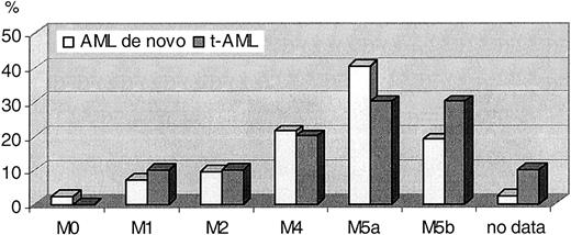
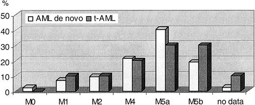
FAB subtype distribution of cases with 11q23/MLL rearrangement was M0: 1, M1: 4, M2: 4, M4: 11, M5a: 20, M5b: 11, no data: 2 and did not differ between de novo AML and t-AML (Figure 1). In total 81% showed an involvement of the monocytic lineage. While the incidence of MLL rearrangement in AML M4, M5a, and M5b was 4.7%, 33.3%, and 15.9%, respectively, it was found in only 0.9% of all other FAB subtypes (P < .0001).
FAB subtype distribution in 54 AML with 11q23/MLLrearrangement: 43 de novo AML and 11 t-AML.
FAB subtype distribution in 54 AML with 11q23/MLLrearrangement: 43 de novo AML and 11 t-AML.
In Table 1 the FAB subtype distribution in the whole AML cohort is compared with AML with an 11q23/MLL rearrangement.
Age distribution
The median age of cases with 11q23/MLL rearrangements was 48.5 years (48 years in de novo AML, 53 years in t-AML; range, 14-74 years), while in the total cohort the median age was 57.9 years (range, 14-84 years). Thus, the incidence of MLL rearrangements was significantly higher in patients younger than 60 years (5.3% vs 0.8%, P < .0001). The age distribution in AML with MLL rearrangement compared with the total cohort of AML is shown in Table 2.
Partner chromosomes
The partner chromosomes of the MLL gene in the 54 cases were 9p22 (n = 19; 35.2%), 10p12 (n = 8; 14.8%), 6q27 (n = 5; 9.3%), 19p13 (n = 5; 9.3%), 17q21 (n = 4; 7.4%), 17q25 (n = 3; 5.6%), 1q21 (n = 2; 3.7%), 15q15 (n = 2; 3.7%), 22q12 (n = 2; 3.7%), and 2q37, 10q22, 12q24, and 14q32 in one case each (1.9%). In Table 3 the different partner chromosomes are shown for de novo AML and t-AML as well as the distribution with respect to age and FAB subtype. No specific association was observed between partner chromosomes and de novo versus t-AML or age or FAB subtype. Table 4 provides information on the respective fusion genes as assessed by RT-PCR. FISH was carried out with whole chromosome painting probes for chromosomes 11 and the respective partner chromosome in those cases without an amplifiable fusion gene to prove the partner chromosome.
Additional chromosomal aberrations
In 22 cases additional chromosome abnormalities were observed. More than 2 additional abnormalities were found in 8 cases (in 3 cases only gains of whole chromosomes, in 5 cases numeric and structural abnormalities). Karyotypes of the 22 cases with abnormalities in addition to the translocation involving the MLL gene are shown in Table 5. Recurring additional abnormalities were +8 (n = 10), +6 (n = 3), +20 (n = 3), -7 (n = 3), and del(3)(q21) (n = 2). Showing additional chromosomal aberrations were 1 (9%) of 11 t-AML and 21 (48.9%) of 43 de novo AML.
Clinical outcome
For 51 of 54 patients with MLL rearrangement and for 1132 of 1843 AML cases without an MLL rearrangement, clinical follow-up data were available. Median follow-up time was 16 months (range, 1 to 75 months). Patients with clinical follow-up data were younger and more frequently had favorable karyotype characteristics compared with cases without available clinical follow-up data (Table 6). There were 2 cases with t(9;11) and 5 patients with other 11q23/MLL rearrangements who underwent an allogeneic stem cell/bone marrow transplantation (6 in first complete remission, 1 case with a t(1;11) in second complete remission).
AML without MLL rearrangement were grouped according to karyotype into 4 categories: (1) AML with t(15;17), (2) AML with t(8;21) or inv(16)/t(16;16), (3) AML with intermediate karyotype (normal cytogenetics, other abnormalities than t(15;17), t(8;21), inv(16)/t(16;16), or abnormalities defined as unfavorable), and (4) AML with unfavorable karyotype (5q-/5, 7q-/-7, inv(3)/t(3; 3), 12p-, 17p abnormalities, and complex aberrant karyotypes [3 or more chromosomal aberrations]).
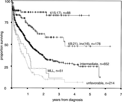
The median overall survival was 8.9 months in the group with MLL rearrangement compared with 40.6 months in cases with t(8;21) or inv(16)/t(16;16), 13.7 months in the intermediate group and 5.7 months in the unfavorable group, respectively, the median was not reached in AML with t(15;17) (P < .001, Figure 2). The data for de novo AML only (n = 1090) were as follows: median overall survival not reached in cases with t(8;21) or inv(16)/t(16; 16), and in cases with t(15;17), 13.8 months in the intermediate group, 5.7 months in the unfavorable group, and 10.0 months in the group with11q23/MLL rearrangement (P < .001).
Univariate Cox regression analyses in the cohort of patients with intermediate karyotypes or with 11q23/MLL rearrangements using age, white blood cell count, 11q23/MLL rearrangements (vs intermediate karyotype), and t-AML versus de novo AML as covariates resulted in significant correlations of all tested parameters with overall survival (Table 7). A multivariate analysis confirmed the prognostic impact of all these parameters (Table 8).
In univariate Cox regression analyses of overall survival in the cohort of patients with unfavorable karyotypes or with 11q23/MLL rearrangements using age, white blood cell count, 11q23/MLL rearrangements (vs unfavorable karyotype), and t-AML (vs de novo AML) as covariates, only age and t-AML were significantly correlated with overall survival (P < .001 and P = .032). This was confirmed in a multivariate analysis (P < .001 and P = .031, respectively).
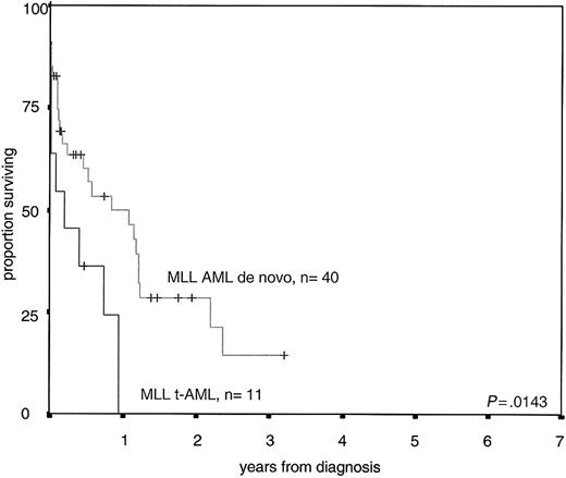
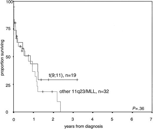
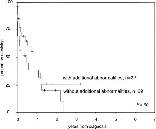
Further evaluations were performed within the cohort of AML with 11q23/MLL rearrangements. In univariate Cox regression analysis for overall survival using age, white blood cell count, t(9;11) (vs other 11q23/MLL rearrangements), additional abnormalities (vs no additional abnormalities), and t-AML (vs de novo AML) as covariates, only t-AML was significantly correlated with overall survival (P = .01). If only de novo AML cases were analyzed all other parameters were also not significant. The median overall survival of AML de novo with MLL rearrangement was 10 months compared with 2.5 months in t-AML with MLL rearrangement (P = .0143, Figure 3). The median overall survival was 10.0 months in cases with t(9;11) compared with 8.9 months in AML with other MLL rearrangements in the whole group (P = .36, Figure 4). Median age was 47 years (range, 18-67 years) in AML with t(9;11) and 46 years (range, 18-65 years) in AML with other MLL rearrangements. The median white blood cell count was 27 450/μL (range, 250-220 000/μL) in t(9;11) cases and 24 550/μL (range, 740-270 000/μL) in cases with other MLL rearrangements. In de novo AML the median overall survival was 10.6 versus 12.8 months (P = .67). The median overall survival was 11.3 months in cases with MLL rearrangement as the sole abnormality compared with 5.3 months in AML MLL rearrangements and additional chromosomal aberrations (P = .90, Figure 5).
Overall survival of 40 de novo AML with 11q23/MLLrearrangement compared with 11 t-AML withMLLrearrangement.
Overall survival of 40 de novo AML with 11q23/MLLrearrangement compared with 11 t-AML withMLLrearrangement.
Overall survival of 19 AML with t(9;11) compared with 32 AML with other 11q23/MLLrearrangements.
Overall survival of 19 AML with t(9;11) compared with 32 AML with other 11q23/MLLrearrangements.
Overall survival of 29 AML with 11q23/MLLrearrangements without additional chromosomal abnormalities compared with 22 AML with 11q23/MLLrearrangements with additional chromosomal abnormalities.
Overall survival of 29 AML with 11q23/MLLrearrangements without additional chromosomal abnormalities compared with 22 AML with 11q23/MLLrearrangements with additional chromosomal abnormalities.
Discussion
The MLL gene is fused to a large variety of more than 50 different partner genes in acute myeloid leukemia.10 In childhood as well as in adult leukemia t(9;11) is the most frequent translocation involving the MLL gene and accounts for about one third of cases with MLL rearrangement.22,23 In t-AML an even higher proportion of 9p22 as translocation partner (48%) has been reported.24 Rearrangements involving AF10 on the short arm of chromosome 10 are observed in 15% to 20% in de novo AML with MLL rearrangement but were reported only in 1% of t-AML in a large series.24 All other partner genes are found with lower frequencies. Partner genes in 19p seem to be more frequently involved in t-AML than in de novo AML.24 In our large series of 54 AML cases with proven MLL rearrangement, 13 different partner genes were observed.
Clonal chromosomal aberrations in addition to the 11q23 rearrangement were observed in 41% of our total cohort and in 48.8% of de novo AML. This frequency is comparable with data reported in other adult and childhood series.22,23,25 The most frequently reported additional aberration is trisomy 8.22,26
With regard to cytomorphology, our data confirm the correlation of 11q23/MLL rearrangements with AML of the monocytic lineage; however, also single cases with AML M0, M1, and M2 were detected. A correlation between the partner chromosomes and FAB subtypes was not observed in our series.
Based on their recent evaluation on the prognostic impact of cytogenetics, the Cancer and Leukemia Group B (CALGB) assigned cases with t(9;11) to the intermediate group, while cases with other balanced 11q23 abnormalities were grouped as unfavorable.27 Evaluation of 2 other large trials from the Medical Research Council in younger and in elderly patients revealed that patients with 11q23 abnormalities showed an intermediate outcome with an overall survival rate of 45% at 5 years (n = 60; median age, 17 years) in the younger cohort and an overall survival rate of 0% at 5 years (n = 11; median age, 64 years) in the cohort of elderly cases.28,29 In contrast, in a combined analysis of Southwest Oncology Group and Eastern Cooperative Oncology Group trials including adult patients (age, 16-55 years) 11q abnormalities were assigned to the unfavorable cytogenetic subgroup.30 Our data show that de novo AML patients with 11q23/MLL rearrangements show a worse outcome compared with the intermediate group. Their prognosis is rather comparable with the unfavorable group.
We found that the median overall survival of patients with t(9;11) was not significantly different from patients with other MLL rearrangements both in de novo and in t-AML. This is in contrast to data from the CALGB indicating a median overall survival of 13.2 months in 23 cases of t(9;11) compared with 7.7 months in 24 cases with other 11q23 rearrangements (P = .009).25 In a recent update of this series of AML from CALGB including 54 cases with balanced 11q23 rearrangements, 27 cases with t(9;11) showed a median overall survival of 13 months compared with 7.2 and 8.4 months for cases with other partner genes of 11q23. Based on these differences, t(9;11) cases were assigned to the intermediate group and the other 11q23 rearrangements to the unfavorable cytogenetic group.27 In our cohort of de novo AML, 14 cases with t(9;11) had a median overall survival of 10.0 months compared with 12.8 months of 26 patients with other MLL rearrangements. Thus, there is no indication for a better outcome of AML with t(9;11) compared with AML with other MLL rearrangements. Supporting the validity of the present analyses with regard to prognostic impact of 11q23 abnormalities in all cases the MLL rearrangement was proved by FISH or RT-PCR. This may be crucial, particularly in cases with MLL-AF10 rearrangements, which often are complex on the cytogenetic level and not distinguishable in certain cases from CALM-AF10 rearrangements on the cytogenetic level.31,32 In addition, the description of balanced translocations involving 11q23 but not the MLL gene33 and the reported differences in clinical outcome between cases with MLL rearrangement and other 11q23 abnormalities34 further underline the need for molecular analyses when evaluating the group of AML with 11q23/MLL rearrangements.
In a collaborative analysis of 108 patients with AML and t(9;11), prognosis was related to age with children 1 to 14 years old showing a more favorable outcome than infants and adults 15 to 49 years of age; the poorest outcome was observed in patients 50 years and older.22 With respect to age the CALGB cohort does not differ from our cohort (median age in cases with t(9;11) 44 years and in cases with other 11q23 translocations 39 years compared with 47 and 46 years, respectively). Rubnitz et al compared the outcome of t(9;11)-positive cases versus other 11q23/MLL rearrangements in childhood AML. They observed a more favorable outcome in cases with t(9;11) than in patients with other 11q23/MLL rearrangements (event-free survival at 5 years 65% [n = 23] vs 24% [n = 33]).23 But also in childhood AML results vary. In a large trial of the Pediatric Oncology Group no differences in outcome were observed between cases with t(9;11) and cases with other 11q23/MLL rearrangement. Both groups showed a quite poor outcome.35 The differences observed so far within the different published trials with respect to the prognostic impact of t(9;11) versus other 11q23 translocations might be due to the still small number of patients or the impact of different therapy protocols. The impact of allogeneic bone marrow or stem cell transplantation on prognosis of cases with 11q23/MLL rearrangement has to be further evaluated as it has been reported that in younger patients those with an HLA-identical donor had a better outcome compared with those without a donor, especially in patients with unfavorable cytogenetics.36 Whether the reported differences in outcome between cases with t(9;11) versus other 11q23/MLL rearrangements reflect differences in the frequency of allogeneic transplantation in the respective trials remains unclear as the number of reported patients who received an allograft is low in most series.
In our series a significant difference in outcome was demonstrated in AML with 11q23/MLL rearrangement between de novo and therapy-related cases. Karyotype abnormalities in addition to the 11q23/MLL rearrangement had no influence on prognosis. This confirms the data of Byrd et al who showed a comparable outcome in cases with t(9;11) with and without additional aberrations.27 In t-AML no differences in overall survival according to partner genes were reported, but patients with additional karyotype abnormalities showed a worse outcome.24
From a morphologic and a cytogenetic point of view, AML cases with 11q23/MLL translocations are more heterogeneous than AML with t(8;21), AML with t(15;17), and AML with inv(16)/t(16; 16). Furthermore, MLL rearrangements also occur in acute lymphoblastic leukemia (ALL). Therefore, it can be speculated that they do not comprise a biologic entity. However, it has been shown in AML as well as in ALL that cases with MLL rearrangements have specific gene expression profiles that allow the distinction from other ALL subtypes as well as from other AML subtypes.37,38 The ALL-MLL group included cases with at least 5 different translocation partners, while in the analysis of AML the 15 cases with 11q23/MLL translocation included rearrangements with 7 different partner genes.39,40 Therefore, it seems appropriate to regard AML with 11q23/MLL rearrangement as an entity as suggested by WHO as long as no clear data are available that give evidence for major differences in biology. This is in line with the distinct clinical outcome that has been demonstrated in the present analysis.
In addition to rearrangements with a large variety of different partner genes MLL rearrangements can be found within MLL as the result from partial tandem duplication (MLL-PTD) usually spanning exons 3 through 9 or exons 3 through 11, which are also mentioned in the WHO classification. Translocations of the MLL gene and MLL-PTD seem to be mutually exclusive as they do not occur together.41 In contrast to AML with translocations involving MLL, AML cases with MLL-PTD usually are not associated with a typical morphology.41-44 Clinical outcome in this AML subgroup is also unfavorable.42,43,45 Similar to translocations of 11q23 involving the MLL gene, the MLL-PTD occurs significantly more frequently in AML secondary to a previous cytotoxic therapy. In a study analyzing AML with normal karyotype with MLL-PTD (n = 10) and AML with MLL translocations (n = 15) using gene expression analysis, differentially expressed genes were identified, which allowed a clear distinction between both AML subtypes.46 Therefore, both groups have to be regarded as 2 different entities.
In conclusion, the present data suggest that AML with 11q23/MLL rearrangement and different partner genes seem to comprise a biologically and clinically homogeneous entity despite a large variety of different partner genes of MLL. In the majority of cases the monocytic lineage is involved. Overall survival is dismal and comparable with AML with unfavorable karyotype. No influence of the presence of t(9;11) or additional chromosomal abnormalities on outcome were observed in AML with 11q23/MLL rearrangement.
Prepublished online as Blood First Edition Paper, June 12, 2003; DOI 10.1182/blood-2003-02-0434.
The publication costs of this article were defrayed in part by page charge payment. Therefore, and solely to indicate this fact, this article is hereby marked “advertisement” in accordance with 18 U.S.C. section 1734.
We thank all participants of the AMLCG study group for sending bone marrow and/or blood for cytogenetics and for clinical data, especially Thomas Büchner (coordinator of the trial) and Maria Cristina Sauerland and Achim Heinecke (statisticians).

