Abstract
Nuclear factor κB (NF-κB) transcription factors are key regulators of immune, inflammatory, and acute-phase responses and are also implicated in the control of cell proliferation and apoptosis. While perturbations in NF-κB activity impact strongly on B- and T-cell development, little is known about the role for NF-κB in natural killer (NK) cell differentiation. Inhibitors of NF-κB (IκBs) act to restrain NF-κB activation. We analyzed the cell-intrinsic effects of deficiencies in 2 IκB members (IκBα and IκBϵ) on NK cell differentiation. Neither IκBα nor IκBϵ deficiency had major effects on NK cell generation, while their combined absence led to NF-κB hyperactivation, resulting in reduced NK cell numbers, incomplete NK cell maturation, and defective interferon γ (IFN-γ) production. Complementary analysis of transgenic mice expressing an NF-κB-responsive reporter gene showed increased NF-κB activity at the stage of NK cell development corresponding to the partial block observed in IκBα × IκBϵ-deficient mice. These results define a critical window in NK cell development in which NF-κB levels may be tightly controlled. (Blood. 2004;103:4573-4580)
Introduction
Natural killer (NK) cells are an essential component of the innate and adaptive immune responses. Their major weapons include secreted cytolytic proteins (perforin, granzymes) and proinflammatory cytokines (tumor necrosis factor α [TNF-α] and interferon γ [IFN-γ]). NK cells provide a first line of defense against infections by viral and bacterial pathogens and play important roles in the immunosurveillance of tumors (reviewed in Trinchieri1 and Colucci et al2 ). NK cells distinguish between healthy and diseased cells via activating and inhibitory receptors that sense the quality and quantity of self-major histocompatibility complex (MHC) class I molecules expressed at the cell surface (reviewed in Ljunggren and Karre3 ). While the intracellular pathways that control NK cell activation are being unraveled,4 the molecular signatures that drive their development are still unclear. Soluble factors including growth factors (Flt3L and stem cell factor [SCF]) and cytokines like interleukin 15 (IL-15) appear important for NK cell generation from hematopoietic precursors.2 On the other hand, the role for transcription factors (TFs) in orchestrating T-cell, B-cell, and NK cell development is less well defined: deficiencies in several TFs affect all lymphoid lineages (Ikaros, purine rich box-1 [PU.1], E2b transformation specific [Ets-1]), whereas others (such as inhibitors of DNA binding-2 [Id2]) affect NK cell generation.2
The nuclear factor κB (NF-κB) family of transcription factors is composed of several DNA binding proteins that play pleiotropic roles in the regulation of immunity, cell proliferation, and death (reviewed in Caamano and Hunter5 and Li and Verma6 ). The NF-κB family is characterized by the presence of a Rel homology domain and comprises 5 evolutionary conserved and structurally related members, including c-Rel, RelA (p65), RelB, NF-κB1 (p50/p105), and NF-κB2 (p52/p100). NF-κB/Rel members associate to form homodimers or heterodimers. NF-κB dimers are sequestered in an inactive form in the cytoplasm through interactions with inhibitors of NF-κB (IκB) proteins, which include IκBα, β, γ, δ, ϵ, and ζ as well as the
NF-κB1 and NF-κB2 precursors.5,6 In response to various stimuli (bacteria, viruses, proinflammatory cytokines, or stress), the activation of IκB kinase (IKK) complex (which includes the kinases IKKα and IKKβ and the regulatory subunit IKKγ/NF-κB essential modulator [NEMO]) results in IκB phosphorylation, leading to its ubiquitinylation and subsequent degradation. As a result, released NF-κB dimers translocate into the nucleus to activate or repress target genes. Since different stimuli can activate distinct or overlapping combinations of NF-κB/Rel dimers, the number of potential NF-κB-dependent target genes is extensive and includes cytokines, adhesion molecules, and regulators of apoptosis (reviewed in Pahl7 ).
NF-κB members are expressed in a cell- and tissue-specific pattern: NF-κB1 and RelA are ubiquitous, whereas NF-κB2, RelB, and c-Rel are expressed abundantly in lymphocytes. Gene targeting experiments have helped to elucidate the roles of NF-κB pathways in T- and B-cell differentiation. Considering the well-characterized antiapoptotic role for NF-κB, it is not surprising that deficiencies in NF-κB members are associated with defects in B- and T-cell development. For example, mice deficient in NF-κB1, NF-κB2, and c-Rel develop normally, but all manifest defects in lymphocyte activation.8-11 RelB-deficient mice die after birth from a multiorgan inflammatory disorder that has been traced to defects in dendritic cell development.12 Mice deficient in RelA die in utero due to massive TNF-mediated liver apoptosis.13 Nevertheless, hematopoietic chimeras made using RelA-deficient (RelA°) stem cells have shown that RelA is also required for normal lymphocyte activation.14 Given the structural similarities between different NF-κB members (NF-κB1 vs NF-κB2 and RelA vs c-Rel), some level of functional redundancy may operate during NF-κB activation. Indeed, the combined deficiency in multiple NF-κB proteins often (NF-κB1°NF-κB2°, NF-κB1°RelA°, NF-κB1°RelB°), but not always (NF-κB1°c-Rel°), results in a more severe phenotype.6,15,16
Inactivation of the regulatory IKKα, IKKβ, or NEMO subunits also results in a block in NF-κB activation; IKKβ- and NEMO-deficient mice die in utero due to TNF-dependent liver apoptosis (similar to RelA° mice).17,18 Mice deficient in IKKα are viable but show severe developmental defects and an absence of mature B cells.19 Lastly, expression of a degradation-resistant form of IκBα (which cannot be phosphorylated and is thereby incapable of dissociating from NF-κB family members) results in global inactivation of the NF-κB pathway. When this IκBα inhibitor is expressed in the T lineage, thymic cellularity is decreased due to enhanced apoptosis of early thymocytes.20 Collectively, the data clearly indicate that inhibition of NF-κB activation has severe and detrimental effects on lymphocyte differentiation.
Analysis of mice deficient in IκB members demonstrates that hyperactivation of NF-κB signaling can likewise have a dramatic impact on lymphopoiesis. Mice deficient in IκBα die about 10 days after birth and appear immunodeficient with extensive inflammatory dermatitis and secondary granulocytosis due to constitutive NF-κB activation.21 In contrast, IκBϵ-deficient mice exhibit a subtle immunologic phenotype but demonstrate an up-regulation of IκBα and IκBβ, suggesting that IκB members may functionally compensate for one another.22 This hypothesis has been recently confirmed through the generation and analysis of mice deficient in both IκBα and IκBϵ (IκBα°ϵ°23 ). In contrast to single IκBα or IκBϵ mutants, IκBα°ϵ° mice die at birth, lack mature B and T cells, and their lymphocyte precursors exhibit increased apoptosis due to NF-κB hyperactivation.23 Taken together, it appears that a relatively narrow range of NF-κB activation must be controlled during B- and T-cell development: either hypoactivation or hyperactivation of NF-κB can perturb both B and T lymphopoiesis.
Little is known about the roles of different NF-κB family members in NK cell development. While the ubiquitous expression of RelA and NF-κB1 has been confirmed in human NK cells,24 data relating to expression patterns of other NF-κB or IκB proteins has not been reported. Hypohidrotic ectodermal dysplasia with immune deficiency patients with point mutations in NEMO have NK cells with reduced cytotoxic potential against certain target cells.25 T and NK cells from mice deficient in IL-1 receptor-associated kinase (IRAK) have a partial impairment in IL-18-induced NF-κB activation with a decrease in IFN-γ production.26 However, NK cell development or function has not been studied in the context of other perturbations in the NF-κB signaling pathways. In this report, we characterize steady-state NF-κB activity in NK cells and analyze the cell-intrinsic effects of IκBα and IκBϵ deletion on NK cell differentiation.
Materials and methods
Mice and generation of hematopoietic chimeras
Alymphoid mice deficient in the recombinase-activating gene-2 (Rag°) and the common cytokine receptor γ chain (γ°c) on the H-2k background have been described.27 Mice deficient in IκBϵ (129Sv × C57Bl/6 × DBA/2, H-2b/d) or IκBα (C57Bl/6 × 129Sv, H-2b) have been described21,22 and were intercrossed to generate embryos deficient in both IκBα and IκBϵ expressing H-2b and H-2d. Fetal livers (FLs; embryonic day 14.5 or 15.5 [E14.5/15.5]) were genotyped by polymerase chain reaction (PCR) and used to generate hematopoietic chimeras in Rag°γ°c mice (H-2k) as previously described.23,28
Analysis of endogenous NF-κB activity
Transgenic mice carrying an NF-κB reporter comprising the immunoglobulin (Ig) promoter (containing NF-κB binding sites) fused to LacZ gene have been previously described.29,30 The β-galactosidase activity (an indicator of NF-κB activity) can be monitored using the substrate fluorescein-di-β galactopyranoside (FDG; Sigma, St Louis, MO), which upon cleavage generates fluorescein. Cell suspensions (10 × 106/mL) were prepared in prewarmed complete medium (RPMI-1640 with 10% fetal calf serum [FCS], 10-5 M β- mercaptoethanol [β-ME], 100 μg/mL streptomycin, 100 U/mL penicillin) and an equal volume was mixed with a solution of FDG (2 mM in distilled water) for 1 minute at 37°C. FDG loading was stopped by adding a 10-fold excess of cold medium containing 300 μM chloroquine to inhibit lysosomal degradation of FDG. After 30 minutes of incubation on ice, cells were stained for cell surface markers and analyzed by flow cytometry.
Cell culture
FL cells (E14.5/15.5) were cultured on irradiated (25 Gy [2500 rad]) OP9 stromal cells in complete medium supplemented with IL-7, SCF, Flt3-ligand, and IL-2 as previously described.28,31
Lymphokine-activated killer cells were generated using unfractionated splenocytes cultured in complete medium containing human IL-2 (huIL-2; 1000 U/mL) or murine IL-15 (muIL-15; 20 ng/mL) as described.28 At various time points, cells were harvested and percentages of viable NK cells (DX5+CD3-CD19-) quantified by fluorescence-activated cell sorter (FACS) analysis.
Flow cytometric analysis
Single-cell suspensions were prepared from various organs (spleen, bone marrow [BM], and liver) as described,27 and red blood cells were lysed using hypotonic ammonium chloride. Cells were resuspended in phosphate-buffered saline (PBS) containing 1% bovine serum albumin (BSA) and 0.01% sodium azide. Monoclonal antibodies (Pharmingen, San Diego, CA) were directly coupled to fluorescein isothiocyanate (FITC), phycoerythrin (PE), peridinin chlorophyll A protein (PerCP), PerCP-Cy5.5, allophycocyanin (APC), or biotin (revealed with streptavidin-PerCP-Cy5.5; Pharmingen). Analysis of murine B-cell lymphoma-2 (Bcl-2) was performed using FITC-labeled antibodies (3F11) and isotype controls (Pharmigen) as previously described.32 Stained cells were analyzed using a FACScalibur flow cytometer running Cell Quest 3.3 software (Becton Dickinson, Mountain View, CA). Dead cells were excluded by means of their forward and side scatter profiles and using propidium iodide staining.
We analyzed the mitochondrial membrane potential as an early marker of entry into the apoptotic process.33 Cells were incubated with 3,3′-dihexyloxacarbocyanine iodide (DIOC6; Molecular Probes, Leiden, Netherlands; 40 nM in RPMI 1640 with 5% FCS) and analyzed using a FACScalibur flow cytometer.
Ex vivo cytokine production
Splenocytes or liver cells (5 × 10⁵ cells/200 μL) were cultured in U-bottom microtiter plates in complete medium containing huIL-2 (1000 U/mL) and stimulated with mIL-12 (5 ng/mL; Peprotech, Rocky Hill, NJ) and/or IL-18 (25 ng/mL; R&D Systems, Minneapolis, MN) overnight. Intracellular staining for IFN-γ, IL-13 (BAF13; R&D Systems), or granulocyte-macrophage colony-stimulating factor (GM-CSF; MP1-22E9; Pharmingen) was performed as described34 except cells were incubated in Brefeldin A (20 ng/mL; Sigma) for the last 4 hours of culture. Stimulation with phorbol myristate acetate (PMA) and ionomycin (4 hours) served as a positive control. Cytokine levels in culture supernatants were quantified by a cytokine-specific sandwich enzyme-linked immunosorbent assay (ELISA) as previously described.35
NK cell lytic activity
A standard 51 Cr-release assay was used to measure NK lytic activity ex vivo as described.27 YAC-1 cells (mouse thymoma; H-2a) and Chinese hamster ovary (CHO) cells were used as targets and maintained in complete medium. Effector cells were either freshly isolated splenic cells or cells treated overnight with IL-2, IL-12, and IL-18. Equivalent NK cell numbers were used after normalization based on FACS staining.
Statistical analysis
Data were analyzed with the Microsoft Excel software (Redmond, WA) applying the 2-tailed Student t test. The null hypothesis was rejected and difference assumed significant when P is less than .05.
Results
Visualizing NF-κB activity in NK cells
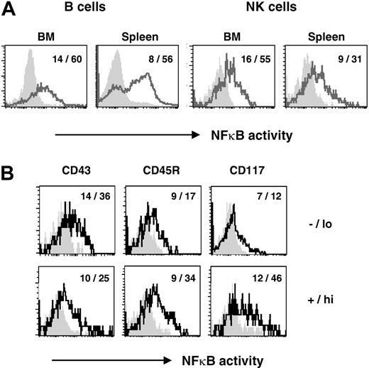
In order to assess NF-κB activity in murine NK cells under steady-state conditions, we analyzed transgenic mice harboring the lacZ reporter gene under the control of the κB sites from the κ light chain gene enhancer (κB-lacZ mice22,29 ). In these mice, NF-κB activity is reflected by β-galactosidase activity and can be visualized in target cell populations by flow cytometry using the vital fluorescent substrate fluorescein-di-β-galactopyranoside. As previously shown,22 a large proportion of BM and splenic CD19+ B cells from κB-lacZ mice exhibit constitutive NF-κB activation (Figure 1A). We further investigated NF-κB expression in NK cells from different organs. Most BM NK cells (defined as CD3-NK1.1+IL-2Rβ+) and mature peripheral splenic NK cells showed evidence of NF-κB activation (Figure 1A). These results suggest that the NF-κB pathway is engaged in freshly isolated NK cells from normal mice.
Steady-state NF-κB activation in BM and splenic NK cells. (A) NF-κB activation was assessed using κB-lacZ transgenic mice having the β-galactosidase gene driven by a κB responsive promoter.29,30 Increasing NF-κB activity is correlated with increasing fluorescence. NF-κB activity is shown on BM and splenic B (CD19+) and NK (CD3-NK1.1+) cells of κB-lacZ mice (bold lines) and nontransgenic littermate controls (shaded histograms). (B) Steady-state NF-κB activity in NK cell subsets. Splenic NK cells (CD3-NK1.1+) were electronically gated and analyzed for expression of CD43, CD45R, or CD117. NF-κB activity is shown on NK cells gated from κB-lacZ mice (bold lines) and nontransgenic littermate controls (shaded histograms) gated for -/lo versus +/hi expression of the indicated markers. Mean fluorescence intensities (MFIs) of cells are indicated (background levels in nontransgenic mice/levels in κB-lacZ mice).
Steady-state NF-κB activation in BM and splenic NK cells. (A) NF-κB activation was assessed using κB-lacZ transgenic mice having the β-galactosidase gene driven by a κB responsive promoter.29,30 Increasing NF-κB activity is correlated with increasing fluorescence. NF-κB activity is shown on BM and splenic B (CD19+) and NK (CD3-NK1.1+) cells of κB-lacZ mice (bold lines) and nontransgenic littermate controls (shaded histograms). (B) Steady-state NF-κB activity in NK cell subsets. Splenic NK cells (CD3-NK1.1+) were electronically gated and analyzed for expression of CD43, CD45R, or CD117. NF-κB activity is shown on NK cells gated from κB-lacZ mice (bold lines) and nontransgenic littermate controls (shaded histograms) gated for -/lo versus +/hi expression of the indicated markers. Mean fluorescence intensities (MFIs) of cells are indicated (background levels in nontransgenic mice/levels in κB-lacZ mice).
We next analyzed NF-κB activation in several splenic NK cell subsets in κB-lacZ mice. Phenotypic changes occur as NK cells develop and mature in the BM prior to their seeding of the peripheral lymphoid tissues.36 For example, expression levels of the Ly49 family of inhibitory and activating receptors, the sialophorin CD43 and the integrin CD11b (macrophage antigen-1 [Mac-1]), increase on NK cells as they mature. In contrast, the receptor for stem cell factor (c-kit or CD117) is expressed by NK precursors but is largely extinguished on mature NK cells (although a minor NK cell subset remains CD117+).28,36,37 Steady-state NF-κB activity was modulated in NK cell subsets depending on their expression of CD43, CD45R, and CD117 (Figure 1B). NK cells (CD3-NK1.1+) with the highest constitutive NF-κB activity were CD43lo, CD45R+, and CD117+ (Figure 1B). Thus, NF-κB activity is regulated during NK cell differentiation and appears highest in NK cells with an immature phenotype.
Combined deficiency in IκBα and IκBϵ perturbs NK cell development
Previous results suggested that normal B and T lymphopoiesis can tolerate only a relatively restricted level of NF-κB: hyperactivation or hypoactivation of NF-κB appears detrimental to B- and T-cell development.23,38 In contrast, whether modulating NF-κB activity will influence NK cell differentiation is not known. Since various NK cell subsets exhibit differential steady-state NF-κB activity (Figure 1A-B), we assessed the effects of constitutive activation of NF-κB pathways on NK cell differentiation by analyzing NK cells in mice deficient in the NF-κB inhibitors IκBα (IκBα°) or IκBϵ (IκBϵ°) or lacking both IκBα and IκBϵ (IκBα°ϵ°). We generated hematopoietic chimeras by reconstituting alymphoid Rag°γ°c mice27 with fetal liver hematopoietic stem cells (FL-HSCs) from IκBα° or IκBϵ° mutant mice or from IκBα°ϵ° double-mutant mice. This strategy (i) eliminated any confounding effects of abnormal NF-κB activation on nonhematopoietic cells and (ii) bypassed the limitations imposed by early death due to the IκBα mutation. Using this approach, we could assess the cell-intrinsic roles of IκBα and IκBϵ in the development of NK cells from HSCs.
As observed in previous work,23 wild-type (WT), IκBα°, and IκBϵ° FL-HSCs could give rise to B cell and T cells after reconstitution of alymphoid mice. IκBα° and IκBϵ° mice were on a mixed genetic background (including 129Sv and DBA/2), and most mice tested did not express the NK1.1 allelic marker (data not shown). We therefore quantified NK cells using a combination of markers including CD3, CD122 (IL-2Rβ), and the pan-NK marker DX5.39 We found equivalent numbers of splenic NK cells (CD3-CD122+DX5+) in WT and IκBϵ° chimeras, whereas they were slightly decreased in IκBα° chimeras (2-fold; Table 1). The cytotoxic potential of NK cells from IκBα° and IκBϵ° chimeras were similar to that of NK cells derived from WT FL-HSCs (data not shown). These results indicate that IκBα and IκBϵ are not individually essential to generate the normal pool of mature lytic NK cells.
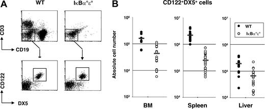
In marked contrast, FL-HSCs from IκBα°ϵ° double mutants demonstrated a differential capacity to generate B, T, and NK cells. Splenic B and T cells were essentially absent (300-fold and 50-fold reduced, respectively, compared with controls; Figure 2A; Table 1). In contrast, NK cells (CD3-CD122+DX5+) were only 3- to 4-fold reduced in the BM and liver and 10-fold reduced in the spleen in the IκBα°ϵ° chimeras compared with WT chimeras (Figure 2A-B). These results suggest that developing NK cells, unlike B and T cells,23 are more tolerant of the combined absence of IκBα and IκBϵ.
Lymphoid reconstitution in HSC chimeras. FL-derived HSCs were transferred into alymphoid mice (Rag°g°c) to generate hematopoietic chimeras as described in “Materials and methods.” (A) Flow cytometric analysis of splenocytes stained for B (CD19+) and T (CD3+) cells. NK cells (CD122+DX5+) were detected in the CD19-CD3- fraction. (B) Absolute numbers of NK cells (CD3-CD122+DX5+) in WT (•) and IκBα°ϵ° ( ) chimeras. Each circle represents values obtained from a single mouse and a gray bar indicates the mean values. There is a significant decrease in BM, liver, and splenic NK cells (P < .001) in the absence of IκBα and IκBϵ compared with controls.
Lymphoid reconstitution in HSC chimeras. FL-derived HSCs were transferred into alymphoid mice (Rag°g°c) to generate hematopoietic chimeras as described in “Materials and methods.” (A) Flow cytometric analysis of splenocytes stained for B (CD19+) and T (CD3+) cells. NK cells (CD122+DX5+) were detected in the CD19-CD3- fraction. (B) Absolute numbers of NK cells (CD3-CD122+DX5+) in WT (•) and IκBα°ϵ° ( ) chimeras. Each circle represents values obtained from a single mouse and a gray bar indicates the mean values. There is a significant decrease in BM, liver, and splenic NK cells (P < .001) in the absence of IκBα and IκBϵ compared with controls.
The reduction in NK cell numbers in IκBα°ϵ° chimeras suggested a cell-intrinsic defect in developing NK cells in the absence of IκBα and IκBϵ. To address this more directly, we used an in vitro culture system whereby NK cells are generated from fetal liver precursors in the presence of cytokines and OP9 stromal cells.28,31 We found that NK cells were present in such cultures derived from either WT or IκBα°ϵ° FL precursors (expressing normal levels of CD2, CD94, and CD122; data not shown), although the absolute numbers of NK cells were reduced by a factor of 5 in the absence of IκBα and IκBϵ (WT, 175 000 ± 95 600 versus IκBα°ϵ°, 35 000 ± 61 500; P = .013). These results demonstrate a cell-intrinsic defect in NK cell generation in IκBα°ϵ° FL precursors.
Survival, proliferation, apoptosis of IκBα°ϵ° NK cells
Transcriptional targets of NF-κB activation include genes controlling cellular homeostasis, ie, survival, proliferation, and apoptosis (reviewed in Pahl7 ). Curiously, NF-κB activation can promote or protect against cell death depending on the cell type and on the pathway of induction. For example, NF-κB inhibits apoptosis by up-regulation of molecules including Bcl-xl, Bfl1/A1, cellular inhibitors of apoptosis protein 1 (c-IAP-1), and -2.40-42 However, in thymocytes, NF-κB activation may up-regulate expression of Fas/FasL molecules and result in increased apoptosis.43,44 The developmental block in B and T lymphopoiesis in IκBα°ϵ° double-mutant mice results from apoptosis of B- and T-cell precursors as they receive antigen receptor signals.23 In order to understand the mechanism for reduced NK cell numbers in IκBα°ϵ° chimeras, we analyzed different parameters of NK cell survival.
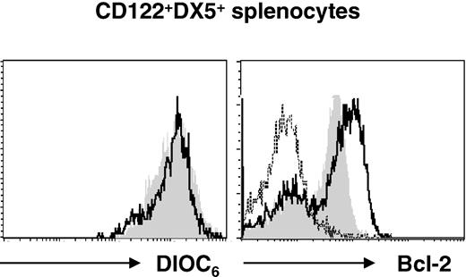
We assessed NK cell death by several cytofluorometric assays using WT and IκBα°ϵ° BM and splenic NK cells. There was no difference in the percentage of annexin V-positive NK cells or in the mitochondrial membrane potential between BM or splenic NK cells from the different genotypes (data not shown; Figure 3), suggesting that the IκBα°ϵ° NK cells were not programmed for apoptosis. Cell cycle analysis demonstrated a similar percentage of splenic NK cells in S-G2/M phase (approximately 4%) in WT and IκBα°ϵ° chimeras, making it unlikely that a block in the cell cycle caused reduced NK cell numbers in IκBα°ϵ° chimeras (data not shown). Interestingly, expression of the antiapoptotic molecule Bcl-2 was clearly up-regulated in IκBα°ϵ° NK cells compared with controls (Figure 3).
NK cells from IκBα°ϵ° chimeras have normal mitochondrial membrane potential but overexpress Bcl-2. (Left) Assessment of mitochondrial membrane potential using DIOC6 staining of splenic NK cells in WT (shaded histogram) and IκBα°ϵ° (bold line) chimeras. (Right) Bcl-2 expression on splenic NK cells from WT (shaded histograms) and IκBα°ϵ° (bold line) chimeras. Dotted line shows staining with isotype control antibody.
NK cells from IκBα°ϵ° chimeras have normal mitochondrial membrane potential but overexpress Bcl-2. (Left) Assessment of mitochondrial membrane potential using DIOC6 staining of splenic NK cells in WT (shaded histogram) and IκBα°ϵ° (bold line) chimeras. (Right) Bcl-2 expression on splenic NK cells from WT (shaded histograms) and IκBα°ϵ° (bold line) chimeras. Dotted line shows staining with isotype control antibody.
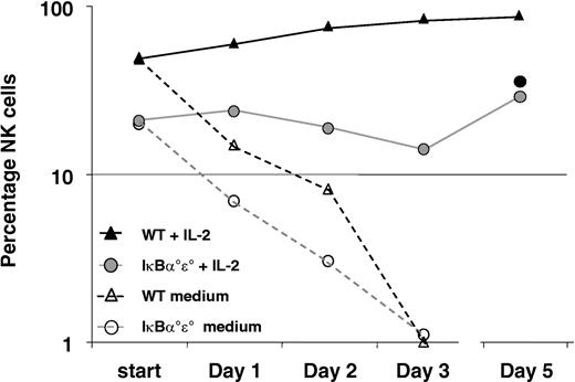
Since the ex vivo analysis of NK cells from IκBα°ϵ° chimeras failed to reveal a major defect in cell survival, we next assessed the proliferative capacity of IκBα°ϵ° NK cells in vitro. Cytokines, including IL-2 or IL-15, can expand murine NK cells in vitro generating lymphokine-activated killer cells with potent cytolytic and cytokine production capacity (reviewed in Colucci et al2 ). We therefore analyzed the fate of WT and IκBα°ϵ° splenic NK cells at various time points in culture with or without IL-2 or IL-15 (Figure 4). Both WT and IκBα°ϵ° NK cells were rapidly lost in culture in the absence of exogenous cytokines, whereas IL-2 could maintain (at day 1) and expand WT NK cells. In contrast, NK cells from IκBα°ϵ° chimeras survived normally in IL-2 but did not expand (Figure 4). Similar results were obtained with IL-15 (Figure 4). By the end of the culture period (day 6), WT cells had expanded 5-fold, while absolute numbers of IκBα°ϵ° NK cells were slightly reduced (1.5-fold). Thus IκBα°ϵ° NK cells manifest a cell-intrinsic defect in their capacity to respond to homeostatic signals that are required to maintain normal steady-state NK cell levels.45
IL-2 or IL-15 maintain but do not expand NK cells from IκBα°ϵ° chimeras. Splenocytes (2 × 10⁵) from WT (Rag° mice, triangles) or IκBα°ϵ° chimeras (circles) were cultured in complete medium with or without IL-2 or IL-15 and percentages of NK evaluated daily. NK cells were rapidly lost in cultures lacking exogenous cytokines (open symbols). IL-2 (or IL-15, •) maintained but did not expand IκBα°ϵ° NK cells (absolute NK cell number was 27 000 on day 5). WT NK cells proliferated vigorously over the same time period (NK cell number was 8 × 10⁵ at day 5).
IL-2 or IL-15 maintain but do not expand NK cells from IκBα°ϵ° chimeras. Splenocytes (2 × 10⁵) from WT (Rag° mice, triangles) or IκBα°ϵ° chimeras (circles) were cultured in complete medium with or without IL-2 or IL-15 and percentages of NK evaluated daily. NK cells were rapidly lost in cultures lacking exogenous cytokines (open symbols). IL-2 (or IL-15, •) maintained but did not expand IκBα°ϵ° NK cells (absolute NK cell number was 27 000 on day 5). WT NK cells proliferated vigorously over the same time period (NK cell number was 8 × 10⁵ at day 5).
Phenotype of NK cells in IκBα°ϵ° chimeras
We next analyzed levels of expression of molecules known to be modulated following NF-κB activation. Classical NF-κB target genes, including MHC class I molecules and the CD69 activation marker, were clearly overexpressed on NK cells from IκBα°ϵ° chimeras compared with WT controls (Figure 5; Table 2), consistent with NF-κB hyperactivation in the absence of IκBα and IκBϵ.
NK cells from IκBα°ϵ° chimeras express increased levels of MHC class I and CD69 molecules. Expression of H-2b,d and CD69 on splenic CD3-CD122+DX5+ NK cells from WT (shaded histograms) and IκBα°ϵ° (bold lines) chimeras.
NK cells from IκBα°ϵ° chimeras express increased levels of MHC class I and CD69 molecules. Expression of H-2b,d and CD69 on splenic CD3-CD122+DX5+ NK cells from WT (shaded histograms) and IκBα°ϵ° (bold lines) chimeras.
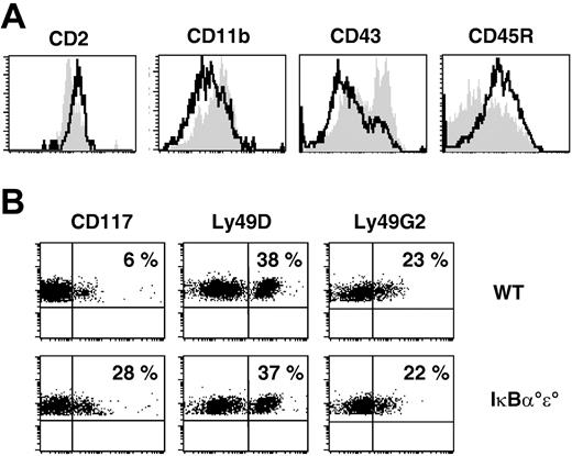
We further characterized the phenotype of the NK cells that developed in the absence of IκBα and IκBϵ. Previous studies have demonstrated that immature BM NK cells differ in the expression of several surface markers (CD11b, CD43, CD45R, CD117, and Ly49 family members) compared with their splenic counterparts.36 For example, BM NK cells are CD11bloCD43lo, while splenic NK cells are mostly CD11bhiCD43hi. In the absence of IκBα and IκBϵ, splenic NK cells expressed low levels of CD11b and CD43 and elevated levels of CD117 (Figure 6A-B). CD45R expression was also higher and more uniform in intensity. In this way, splenic IκBα°ϵ° NK cells more closely resembled immature BM NK cells.36 Nevertheless, expression of inhibitory (Ly49G2) and activating (Ly49D) members of the Ly49 receptor family, and that of CD2, was normal (Figure 6A-B). Collectively, the phenotypic analysis suggests that the maturation of NK cells in the absence of IκBα and IκBϵ may not be complete.
Splenic NK cells from IκBα°ϵ° chimeras have an abnormal phenotype. (A) Expression of CD2, CD11b, CD43, and CD45R on splenic CD3-CD122+DX5+ NK cells from WT (shaded histograms) and IκBα°ϵ° (bold line) chimeras. (B) Expression of CD117, Ly49D, and Ly49G2 on splenic CD3-CD122+DX5+ NK cells from and IκBα°ϵ° chimeras. Percentages of positive cells are indicated.
Splenic NK cells from IκBα°ϵ° chimeras have an abnormal phenotype. (A) Expression of CD2, CD11b, CD43, and CD45R on splenic CD3-CD122+DX5+ NK cells from WT (shaded histograms) and IκBα°ϵ° (bold line) chimeras. (B) Expression of CD117, Ly49D, and Ly49G2 on splenic CD3-CD122+DX5+ NK cells from and IκBα°ϵ° chimeras. Percentages of positive cells are indicated.
IκBα°ϵ° NK cells demonstrate normal cytotoxic activity
We next determined whether the abnormal phenotype in NK cells from IκBα°ϵ° chimeras was associated with defective NK cell effector functions. One of the best-characterized NK cell activities is natural cytotoxicity: the capacity to lyse target cells without prior sensitization (reviewed in Trinchieri1 ). NK cells use a variety of receptors to monitor the level of MHC expression on target cells and to detect stress- or infection-induced molecules.3 We found that the prototypical NK cell target YAC-1 was lysed with the same efficiency by WT and IκBα°ϵ° splenic NK cells (Figure 7A). Moreover, IκBα°ϵ° NK cells were cytolytic against CHO targets (Figure 7B), consistent with expression of a functional Ly49D receptor.46 We also cultured splenocytes from IκBα°ϵ° chimeras overnight in the combination of IL-2, IL-12, and IL-18; this cytokine cocktail has been shown to stimulate NK cell IFN-γ production and cytolytic potential.47 We found that IL-12/IL-18 stimulation of both WT and IκBα°ϵ° NK cells resulted in enhanced lysis of YAC-1 targets compared with their fresh counterparts (Figure 7C), suggesting that IκBα°ϵ° NK cells express functional receptors for IL-2, IL-12, and IL-18. The collective results suggest that despite an apparent immature phenotype, IκBα°ϵ° NK cells had clearly attained cytotoxic potential.
Natural cytotoxicity in the absence of IκBα°ϵ°. (A) Freshly isolated splenocytes from WT (▴) and IκBα°ϵ° (•) chimeras showed similar lytic activity against Cr51 -labeled YAC-1 target cells. NK cell-target cell ratios were normalized using flow cytometric analysis. One representative experiment of 2 is shown. (B) Freshly isolated splenocytes from WT (▴) and IκBα°ϵ° (•) chimeras showed similar lytic activity against CHO target cells. (C) WT (black symbols) and IκBα°ϵ° (gray symbols) splenocytes were stimulated for 12 hours with IL-2 alone (circles) or cytokine combination (IL-2+IL-12+IL-18) (triangles) prior to analysis of lytic activity against YAC-1 target cells. Error bars represent SEM.
Natural cytotoxicity in the absence of IκBα°ϵ°. (A) Freshly isolated splenocytes from WT (▴) and IκBα°ϵ° (•) chimeras showed similar lytic activity against Cr51 -labeled YAC-1 target cells. NK cell-target cell ratios were normalized using flow cytometric analysis. One representative experiment of 2 is shown. (B) Freshly isolated splenocytes from WT (▴) and IκBα°ϵ° (•) chimeras showed similar lytic activity against CHO target cells. (C) WT (black symbols) and IκBα°ϵ° (gray symbols) splenocytes were stimulated for 12 hours with IL-2 alone (circles) or cytokine combination (IL-2+IL-12+IL-18) (triangles) prior to analysis of lytic activity against YAC-1 target cells. Error bars represent SEM.
IκBα°ϵ° NK cells have a selective defect in cytokine production
NK cells are potent and prompt producers of multiple cytokines, most notably IFN-γ.1 Previous reports identified a role for NF-κB in modulating CD4+ T-cell differentiation and cytokine production. NF-κB members (including RelB) stimulate T-helper 1 (Th1) responses (IFN-γ production) by directly increasing promoter activity.48,49 In contrast, NF-κB1 appears necessary for efficient Th2 responses.50 We therefore analyzed whether NF-κB hyperactivation modified NK cell cytokine production capacity.
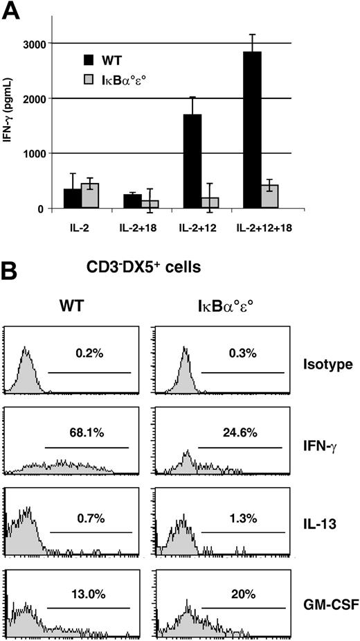
Freshly isolated splenic NK cells were cultured in the presence of IL-2 and stimulated with a combination of IL-12 plus IL-18. This protocol results in a synergistic secretion of IFN-γ from control NK cells (Figure 8A). Surprisingly, splenic NK cells from IκBα°ϵ° chimeras were clearly defective in IFN-γ production after either IL-12 or combined IL-12/IL-18 stimulation; levels of secreted IFN-γ were 7-fold reduced compared with controls (Figure 8A). The defective IFN-γ production from IκBα°ϵ° NK cells was confirmed by intracellular cytokine staining: IκBα°ϵ° splenic NK cells produced quantitatively (3-fold percentage) and qualitatively (2-fold mean fluorescence intensity) less IFN-γ compared with control NK cells (Figure 8B). Since IκBα°ϵ° NK cells could survive in the presence of IL-2 (Figure 4) and respond to IL-12/IL-18 stimulation with increased cytotoxicity (Figure 7A), the abnormal IFN-γ responses in mutant NK cells are not due to defective cytokine signaling through these receptors.
IκBα°ϵ° NK cells have a defect in IFN-γ production. (A) Splenic NK cells (104 cells) from Rag° mice or IκBα°ϵ° chimeras were stimulated for 24 hours with the indicated combinations of IL-12 and/or IL-18 and IFN-γ release measured by ELISA. Error bars represent SEM. (B) Splenocytes from WT or IκBα°ϵ° chimeras were stimulated for 7 hours with a combination of IL-12 and IL-18 (for IFN-γ) or with PMA and calcium ionophore (for IL-13, GM-CSF) and were analyzed by intracellular staining. Analysis was performed on electronically gated CD3-DX5+ cells. Percentages of positive cells are shown in each panel. One representative experiment of 3 to 6 is shown.
IκBα°ϵ° NK cells have a defect in IFN-γ production. (A) Splenic NK cells (104 cells) from Rag° mice or IκBα°ϵ° chimeras were stimulated for 24 hours with the indicated combinations of IL-12 and/or IL-18 and IFN-γ release measured by ELISA. Error bars represent SEM. (B) Splenocytes from WT or IκBα°ϵ° chimeras were stimulated for 7 hours with a combination of IL-12 and IL-18 (for IFN-γ) or with PMA and calcium ionophore (for IL-13, GM-CSF) and were analyzed by intracellular staining. Analysis was performed on electronically gated CD3-DX5+ cells. Percentages of positive cells are shown in each panel. One representative experiment of 3 to 6 is shown.
To assess whether IκBα°ϵ° NK cells had a global inability to produce cytokines, these cells were stimulated with the pharmacologic activators PMA plus calcium ionophore. Previous studies have shown that this type of stimulation (albeit nonphysiologic) can reveal the capacity of human and mouse NK cells to produce IL-5, IL-13, or GM-CSF (reviewed in Colucci et al2 ). We were unable to detect IL-5 or IL-13 production from WT or IκBα°ϵ° splenic NK cells after stimulation with PMA/ionomycin (Figure 8B; data not shown). In contrast, both WT and IκBα°ϵ° NK cells were capable of GM-CSF production (Figure 8B).
Discussion
NF-κB pathways are pleiotropic in nature, impacting on cell survival, apoptosis, and effector functions. Moreover, since NF-κB signals are ubiquitous, identifying roles for NF-κB members in cellular differentiation remains a challenge. In this report, we have begun to assess the roles for NF-κB in NK cell development and function. We observed that a substantial proportion of NK cells show constitutive NF-κB activation and that hyperactivation of NF-κB secondary to the combined absence of IκBα and IκBϵ results in (1) reduced NK cell numbers and (2) defective NK cell maturation, affecting IFN-γ secretion but not lytic capacity. These results offer new insights into the role of NF-κB pathways in controlling lymphocyte differentiation and define a crucial stage in early NK maturation where cells appear sensitive to overall NF-κB activity.
Since all cells use the NF-κB pathway, we used a hematopoietic reconstitution system whereby the effects on NF-κB signaling would be confined to cells of the lymphoid lineage.6 This allowed us to evaluate the cell-intrinsic roles of NF-κB in NK cells. This approach was necessary since previous studies had shown that NF-κB pathways are implicated in relaying the proper cellular and soluble signals derived from the nonhematopoietic (stromal cell) microenvironment to developing lymphocytes. For example, NF-κB pathways are involved in the regulation of interferon regulatory factor 1 (IRF-1)-controlled IL-15 expression and in the signaling through lymphotoxin-β receptors; both are key elements in NK cell development.51-54 By exploiting hematopoietic chimeras generated in alymphoid Rag°γ°c mice, we found that NK cells, in contrast to B and T cells, could develop in the absence of IκBα and IκBϵ. Nevertheless, overall NK cell numbers were reduced in IκBα°ϵ° chimeras; this phenomenon was recapitulated using an in vitro system of NK cell generation from FL precursors. Thus NF-κB hyperactivation has differential effects on B/T versus NK development; the former cells are exquisitely sensitive, while the latter are more tolerant. We observed increased Bcl-2 expression in IκBα°ϵ° NK cells but not in IκBα°ϵ° B or T cells (S.I.S. and J.P.D., unpublished observations, January 2004). Bcl-2 is a target of NF-κB transcriptional activation55,56 ; the increased Bcl-2 expression in the absence of IκBα and IκBϵ may reflect a compensatory mechanism that helps to maintain these IκBα°ϵ° NK cells in face of NF-κB hyperactivation. In contrast, NEMO mutant NK cells develop to normal levels in human blood,25 suggesting that hypoactivation versus hyperactivation of NF-κB differentially affect NK cell development.
NK cell numbers were decreased in all tested organs (BM, spleen, liver) of IκBα°ϵ° chimeras, but the degree of reduction varied according to the site (BM and liver less than spleen). Interestingly, these sites have been shown to harbor varying ratios of immature to mature NK cells (using low versus high expression of CD11b and CD43 as markers of immaturity and maturity, respectively36 ). Taking this into account, it is striking that the reduction in NK cell number in IκBα°ϵ° chimeras is most pronounced in tissues (such as the spleen) having the highest proportion of CD11bhiCD43hi NK cells. In fact, the residual NK cells in the BM, spleen, and liver of mutant mice are similar in absolute terms to the number of immature (CD11bloCD43lo) NK cells found in control chimeras. These observations are consistent with the hypothesis that NK cell maturation (in terms of CD11b and CD43 expression) is partially blocked upon constitutive NF-κB activation.
The inability to establish normal peripheral NK cell numbers in the absence of IκBα and IκBϵ suggests a defect in cellular homeostasis of this lymphoid subset. We did not observe any increased susceptibility of mature NK cells to apoptosis when assessed ex vivo. In contrast, IκBα°ϵ° NK cells did not proliferate in vitro in response to high doses of IL-2, which can robustly expand WT NK cells. Although IκBα°ϵ° NK cells express normal levels of IL-2Rβ and γc (data not shown), we found that the IL-2Rβ/γc complex did not provide adequate proliferative signals to IκBα°ϵ° NK cells, although IL-2 or IL-15 could maintain their survival in culture. This defect in response to homeostatic signals could explain the failure of IκBα°ϵ° NK cells to achieve normal cell numbers in vivo. A partial defect in IL-2/IL-15 signaling would be predicted to have detrimental effects on NK cell development.57
An alternative mechanism to account for the defective IL-2 responses in IκBα°ϵ° NK cells could involve induction of apoptotic mechanisms. It is known that IL-2 can stimulate NF-κB activation.25 Since NF-κB activity is already elevated in IκBα°ϵ° NK cells, additional NF-κB stimulation might not be well tolerated and could induce cell death after long-term culture. The IL-2 response of NK cells in the context of NF-κB hyperactivation (as in IκBα × IκBϵ deficiency) contrasts sharply with that of NF-κB hypoactivation, as in the case of NEMO mutations, in which treatment with IL-2 normally expands NK cells and allows for a partial recovery of their functional defects.25
Several reports have demonstrated that an inability to normally activate NF-κB pathways results in abnormal development of NK cell effector functions. Patients with mutations in NEMO develop normal numbers of NK cells, however, ex vivo natural cytotoxicity is reduced. This defect can be corrected in vitro following expansion in IL-2, which can activate NF-κB pathways.25 Whether NEMO mutant NK cells have other defects in cytokine production is not known. Tato and colleagues48 recently observed that NK cell cytotoxicity and IFN-γ production were defective in transgenic mice expressing a dominant-negative mutant form of IκBα. These effects were also apparent under physiologic conditions in the context of Toxoplasma gondii infection. Finally, defects in IRAK affect IL-18-induced NK cell cytotoxicity and cytokine production in vitro as well as during the course of an infection with Propionibacterium acnes.26 The collective results indicate that adequate NF-κB activation is essential for the acquisition of NK cell effector functions.
In this work, we found that NF-κB hyperactivation results in selective defects in NK cell effector functions. Residual IκBα°ϵ° NK cells develop a normal cytolytic program but have reduced capacity for IFN-γ production. Since NK cell cytotoxicity does not require new gene transcription, this effector function may be less sensitive to NF-κB levels. Although the molecular mechanism for this selective defect remains unclear, we can speculate that an excess of NF-κB might activate a repressor of IFN-γ transcription or diminish transcription factors implicated in IFN-γ synthesis, such as T-box expressed in T cells (T-bet) or H2.0-like homeo box (Hlx) (reviewed in Szabo et al58 ). Alternatively, IκBα°ϵ° NK cells may manifest an imbalance between different NF-κB members. For example, an excess of NF-κB1 relative to RelB would be predicted to cause a defect in IFN-γ production (the former inhibits IFN-γ transcription in the contrast to the latter). In any event, NK cell IFN-γ expression appears tightly regulated by overall NF-κB levels, since both hypoactivation and hyperactivation have the same negative effect. In contrast, NK cell production of GM-CSF was not affected by the combined absence of IκBα and IκBϵ, ruling out a generalized defect in cytokine production by these cells.
Our results support the notion that NF-κB levels may be critically regulated during NK cell maturation. Uncontrolled NF-κB activation during lymphopoiesis (as in the case in IκBα°ϵ° chimeras) results in the development of NK cells with an immature phenotype, expressing low levels of CD11b and CD43 but high levels of CD117 and CD45R. As this phenotype has been previously described for BM NK cells,36 we further analyzed NF-κB activity in various NK cell subsets using κB-lacZ mice. These results identified elevated NF-κB activity in NK cells expressing low levels of CD43 and high levels of CD45R and CD117. These observations are consistent with a critical stage in early NK cell development defined by NK cells bearing the CD43loCD45hiCD117hi phenotype. We would propose that NF-κB levels during this developmental window would need to be maintained within a tolerable range. In this model, developing NK cells would normally receive signals that up-regulate NF-κB levels during their differentiation. In the context of excessive NF-κB activation (IκBα°ϵ°), a threshold may be passed with detrimental consequences including arrest of maturation and/or induction of apoptosis. Immature NK cells would therefore need to fine-tune NF-κB activity in order to traverse this critical checkpoint.
Prepublished online as Blood First Edition Paper, February 5, 2004; DOI 10.1182/blood-2003-08-2975.
Supported by grants from the Institut Pasteur, Institut National de la Santé et la Recherche Medicale (Inserm), the Ligue National Contre le Cancer (LNCC), and the Association pour le Recherche contre le Cancer (ARC). S.I.S. was supported by fellowships from the MENRT, the LNCC, and the ARC. C.A.J.V. was supported by a Poste Vert fellowship from Inserm.
The publication costs of this article were defrayed in part by page charge payment. Therefore, and solely to indicate this fact, this article is hereby marked “advertisement” in accordance with 18 U.S.C. section 1734.
We would like to thank Erwan Corcuff and Thomas Ranson for help in the realization of this work.

