Abstract
Patients with B-cell chronic lymphocytic leukemia (B-CLL) segregate into subgroups with very different survival times. Because clinical observations suggest that leukemic cells accumulate at different rates, we measured telomere length and telomerase activity in B-CLL cells to distinguish differences in cellular replication. Our data indicate that the telomeres of B-CLL cells are shorter than telomeres of B cells from healthy subjects, indicating that the leukemic cells have a prolonged proliferative history. Leukemic cells of the immunoglobulin V gene mutation subgroups differ in telomere length and telomerase activity. B lymphocytes from the subgroup with poor outcome and with limited IgV gene mutations have uniformly shorter telomeres and more telomerase activity than those from the subgroup with better outcome and with considerable mutations. Differences in telomere length appear to largely reflect the proliferative histories of precursors of the leukemic cells, although differences in cell division, masked by the action of telomerase, cannot be excluded. These results may provide insight into the stages of maturation and the activation pathways of the cells that give rise to B-CLL. In addition, they reinforce the concept that B-CLL is not simply an accumulative disease of slowly dividing B lymphocytes but possibly one of B cells with extensive proliferative histories.
Introduction
B-cell chronic lymphocytic leukemia (B-CLL) is a clonal disease of an apparently slowly dividing B lymphocyte that expresses a distinct cell surface phenotype (CD19+, CD5+, CD23+ cells with diminished surface membrane immunoglobulin).1 Subgroups of B-CLL exist, defined by differences in lymphocyte doubling times in vivo,2 variable degrees of somatic mutation of the immunoglobulin V (Ig V)–region genes in the leukemic cells,3,4 and expression of cell surface5 and intracellular6,7 proteins. Each of these subgroups is accompanied by significant differences in clinical course and outcome.2,6-9
Membership in one of these subgroups may reflect differences in the antigenic experiences of the precursor B cells (naive vs memory), the manner in which these precursor cells were triggered (T-cell dependently or T-cell independently), or the level of spontaneous or induced cell cycling.10 Defining the relative importance of these issues may provide clues to the development and evolution of B-CLL.
Telomere lengths can serve as indicators of the replicative history of individual cells because these chromosomal segments shorten with each cell division.11 As a consequence of the inability to adequately maintain telomere length, a slow but inexorable decrease in a cell's replicative potential accompanies cellular division,12,13 and is also part of the normal aging process.14-16 Two processes counteract telomere shortening and restore telomeric regions in normal and transformed cells: the activity of the enzyme telomerase17-20 and an alternative lengthening of telomere (ALT) pathway.21,22 Of special note is the observation that during a classical T-cell–mediated germinal center (GC) reaction, normal B cells produce high levels of telomerase and exhibit telomere lengthening.23-26 This process presumably permits those B lymphocytes that develop newly diversified and advantageous B-cell receptors to maintain viability and to participate further in adaptive immune responses.
As observed in many solid27 and hematologic18-20 cancers, B-CLL cells have short telomere lengths.28 In this study, we analyzed telomere length as a means to infer the number of cell divisions that individual leukemic lymphocytes underwent and the relationship between telomere length and telomerase activity in the same cells. We were especially interested in trying to define differences in telomere length between the 2 Ig V gene mutation-defined B-CLL subgroups and to decipher to what degree this shortening reflects preleukemic events compared with leukemic events. We found that B-CLL cells have shorter telomeres than B cells or isolated CD5+ and CD5– B cells from a group of healthy subjects matched for age. Moreover, we identified differences in telomere length and telomerase activity within the 2 Ig V gene mutation B-CLL subgroups that possibly indicate different replicative and telomere restorative histories for the cells and may help to explain their different clinical courses and outcomes. Preliminary reports of these findings were presented earlier.29,30
Patients, materials, and methods
Patients and healthy donors
The Institutional Review Board of North Shore University Hospital (Manhasset, NY) and Long Island Jewish Medical Center (New Hyde Park, NY) approved these studies. Venous blood was collected from 70 patients with B-CLL after informed consent was obtained. Laboratory data and Ig VH and VL gene DNA sequences were available for all patients, and clinical information was available for most.4,5,8,29 Thirty-five patients were chosen randomly from a group who expressed Ig V genes with less than a 2.0% difference from the most similar germline gene of both VH and VL (unmutated) and 35 from a group who had V genes with a 2.0% or greater difference from the most similar germline gene in either VH or VL (mutated). The only patients included in this study were those expressing surface membrane IgM (sIgM) and exhibiting allelic exclusion of Ig V genes. Leukocyte-enriched fractions from blood donated by 30 healthy volunteers matched for age against the B-CLL patients were purchased from Long Island Blood Services (Melville, NY). Before use, these samples were tested and found negative for HIV and hepatitis B virus (HBV) antigens. In addition, venous blood from 18 younger, healthy volunteers was collected with informed consent.
Isolation of peripheral blood mononuclear cells
Peripheral blood mononuclear cells (PBMCs) were separated from heparinized venous blood of the B-CLL patients and from the leukocyte fractions of healthy donors by density gradient centrifugation using Ficoll-Hypaque (Pharmacia LKB Biotechnology, Piscataway, NJ). PBMCs were used after thawing samples that had been cryopreserved using a programmable cell-freezing machine (Cryomed, Mt Clemens, MI).
Isolation of B cells and polymorphonuclear leukocytes
Approximately 3 mL leukocyte-enriched fraction from each healthy donor or a similar volume of heparinized blood from each B-CLL patient was mixed (1:1 vol/vol) with 3% Dextran T-500 (Amersham Biosciences, Uppsala, Sweden) in physiological saline to remove red blood cells (RBCs). After allowing the cells to sediment at room temperature for 20 minutes, the RBC-depleted supernatant was collected, washed once with phosphate-buffered saline (PBS), and then subjected to Ficoll-Hypaque (Pharmacia LKB Biotechnology) density gradient centrifugation. Polymorphonuclear leukocytes (PMNLs) were collected from the cell pellet obtained after centrifugation and elimination of RBCs with RBC lysis buffer (Roche Diagnostics, Indianapolis, IN). The interphase comprising PBMCs was collected and washed with PBS. B cells were isolated from this suspension or from cryopreserved PBMCs using a B-cell isolation kit (Miltenyi Biotec, Auburn, CA) according to the manufacturer's instructions. Isolated B cells from 8 healthy donors were stained with phycoerythrin-conjugated anti-CD5 and allophycocyanin-conjugated anti-CD19 and sorted into CD19+CD5+ and CD19+CD5– populations using the FACS Vantage sorter (Becton Dickinson Immunocytometry Systems, San Jose, CA).
The purity of B cells and PMNLs was ascertained by staining aliquots of cells with allophycocyanin-conjugated anti-CD19, phycoerythrin-conjugated anti-CD3, phycoerythrin-conjugated anti-CD15, and fluorescein isothiocyanate (FITC)–conjugated anti-CD14 monoclonal antibodies (mAbs) (all purchased from Becton Dickinson-PharMingen, San Jose, CA). Stained cells were fixed with 1% formaldehyde and were analyzed on a FACScalibur flow cytometer (Becton Dickinson Immunocytometry Systems). PMNLs were identified as CD15+CD14– cells that exhibited high amounts of forward and side scatter, whereas B cells were identified as CD19+ cells with lower levels of forward and side scatter.
Quantification of telomere length
A Flow-fluorescence in situ hybridization (Flow-FISH) protocol, as detailed elsewhere,31 was used to quantify telomere lengths in purified populations of B cells or PMNLs. This test uses a peptide nucleic acid (PNA) probe (C3TA2)3 conjugated with FITC. PNAs are nucleic acid analogues that are capable of strongly and specifically interacting with the targeted DNA and in which the lateral backbone is replaced by a 2-aminoethyl-glycine unit.32 Briefly, 1 × 10⁵ cells in 100 μL hybridization buffer containing 70% deionized formamide (Gibco/BRL, Grand Island, NY), 20 mM Tris (Sigma, St Louis, MO), pH 7.0, and 1% bovine serum albumin (BSA; Sigma) were placed in duplicate in microcentrifuge tubes. In addition, one of every duplicate tube received 0.3 μg/mL telomerespecific FITC-conjugated (C3TA2)3 PNA probe (PerSeptive Biosystems, Framingham, MA). Samples were subjected to heat denaturation of DNA for 10 minutes at 80°C in a circulating water bath followed by hybridization for 2 hours at room temperature in the dark. Cells were then washed twice by centrifugation in swing-out rotors with 1 mL wash buffer containing 70% formamide, 10 mM Tris, 0.1% BSA, and 0.1% Tween 20 (all from Sigma), and once in PBS containing 0.1% BSA and 0.1% Tween 20. Cells were suspended in PBS containing 0.1% BSA, 10 μg/mL RNase A (Boehringer Mannheim, Laval, CA), and propidium iodide (Sigma) at a final concentration of 0.06 μg/mL. The cells were analyzed on a FACScalibur (Becton Dickinson Immunocytometry Systems) flow cytometer.
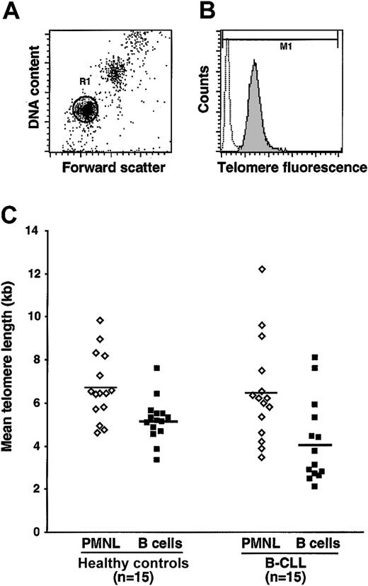
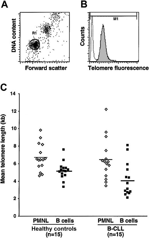
To compare data from different experiments and to correct for daily shifts in the linearity of the flow cytometer and fluctuations in the laser intensity and alignment, FITC-labeled fluorescent beads (Flow Cytometry Standards, Fishers, IN) in PBS were analyzed at the beginning of each experiment. The resultant calibration curve was used for correction of experimental fluorescence values in each experiment. In addition, an aliquot of mononuclear cells from a human umbilical cord sample that had been cryopreserved previously was thawed and used at the time of each experiment. Variations in telomere length between multiple samples of the same cell type in separate experiments were 10% or less. The data acquired on the flow cytometer were plotted as forward scatter versus DNA content (propidium iodide staining; Figure 1A). A region (R1) encompassing the cells under study (B cells or PMNLs) was marked. The amount of fluorescent probe that bound specifically to the telomeric region of the cells contained in R1 was then plotted as a histogram (Figure 1B). The mean equivalence of soluble fluorescence and the mean telomere fluorescence (length) were computed based on the amount of probe bound specifically and, by extrapolation of the calibration values, were generated from the standard beads used in the assay.
Comparisons of telomere lengths in B cells and PMNLs using Flow-FISH. (A) Region R1 identifies the B-cell and PMNL populations under study based on forward scatter. (B) Dotted open histogram represents background fluorescence of cells subjected to Flow-FISH in the absence of the telomere-specific PNA probe; the shaded histogram represents telomere-specific fluorescence of cells. The difference between the mean fluorescence intensity of cells stained with and without probe (as deduced from marker M1) yields mean telomere fluorescence and is used to calculate telomere length in kilobases (see “Patients, materials, and methods”). (C) Mean telomere lengths of paired PMNLs and B-cell populations in 15 B-CLL patients and 15 age-matched healthy donors. The differences in telomere lengths between PMNLs and B-cell populations from B-CLL patients were statistically significant (P < .01), whereas telomere lengths did not differ in B cells and PMNL populations from healthy subjects.
Comparisons of telomere lengths in B cells and PMNLs using Flow-FISH. (A) Region R1 identifies the B-cell and PMNL populations under study based on forward scatter. (B) Dotted open histogram represents background fluorescence of cells subjected to Flow-FISH in the absence of the telomere-specific PNA probe; the shaded histogram represents telomere-specific fluorescence of cells. The difference between the mean fluorescence intensity of cells stained with and without probe (as deduced from marker M1) yields mean telomere fluorescence and is used to calculate telomere length in kilobases (see “Patients, materials, and methods”). (C) Mean telomere lengths of paired PMNLs and B-cell populations in 15 B-CLL patients and 15 age-matched healthy donors. The differences in telomere lengths between PMNLs and B-cell populations from B-CLL patients were statistically significant (P < .01), whereas telomere lengths did not differ in B cells and PMNL populations from healthy subjects.
Preparation of cell extracts and estimation of telomerase activity
The TRAPeze telomerase detection kit (Intergen, Purchase, NY) with the telomere repeat amplification protocol (TRAP)33 was used to quantify telomerase activity in cell extracts from purified cell populations. Briefly, purified cell populations were washed 2 to 3 times with PBS to remove extracellular protein. One million cells were lysed using the cell lysis buffer provided in the kit, kept on ice for 30 minutes, and centrifuged at 14 000 rpm (approximately 12 000g) for 20 minutes at 4°C. The supernatant, comprising the cell-free extract, was flash frozen and stored at –80°C until use in the TRAP assays.
Detection of telomerase activity by TRAP involves the enzymatic addition by telomerase of TTAGGG repeats to an oligonucleotide and the subsequent polymerase chain reaction (PCR) amplification of these extension products using the forward and reverse primers. The ability to detect products generated from cell extracts was initially standardized using dilutions of the cell extract that corresponded to 102, 103, 104, and 105 cell equivalents. Based on these findings, in all subsequent experiments cell extracts from 104 cells were analyzed for telomerase activity. Initially, the TS primer was end labeled with γ32P-dATP (New England Nuclear Biolabs, Boston, MA) using T4 polynucleotide kinase (10 U/mL; Gibco/BRL). For PCR, extract from 104 cells in 2 μL was combined with 48 μL reaction mixture supplied with the kit and 2 U Taq DNA polymerase (Perkin Elmer Life Sciences, Boston, MA). The telomerase extension reaction was carried out at 30°C for 30 minutes followed by PCR at 94°C for 30 seconds and 59°C for 30 seconds for 27 cycles and finally a 7-minute elongation step. Control reactions were included in each assay using an aliquot of heat-treated (80°C for 10 minutes) extract for each sample tested, a telomerase-positive control (cell lysate), a telomerase-negative control (buffer), and 2 quantitation controls (1 and 2 attomoles of a control template). PCR products were electrophoresed on 10% nondenaturing polyacrylamide gels, and the gels were analyzed using a PhosphorImaging system and ImageQuant software from Molecular Dynamics (Sunnyvale, CA). The activity of telomerase in the cell extracts was calculated on the basis of the intensity of the bands corresponding to 50, 56, 62, 68, and 74 bp (extension products) and continuing from there and was based on the intensity of the internal controls (36 bp and the quantification controls). Telomerase activity is expressed as total product generated (TPG) units. Each TPG U corresponds to the number of TS primers extended with at least 4 telomeric repeats by telomerase in the extract in a 30-minute incubation at 30°C.
Statistical analyses
Differences in values of telomere length and telomerase activity between groups were evaluated using the Mann-Whitney U test. To assess the variability of telomere lengths within individual B-CLL cases, the SD of the mean fluorescence intensity of the histogram generated using the PNA probe (and the control) was deduced from the coefficient of variation (CV). Statistical significance of the difference between values of SD for the different groups was assessed using the Mann-Whitney U test. Spearman correlation was determined between telomere length and V gene mutation or telomerase activity. Survival analyses were performed using the Kaplan-Meier product-limit method and the log-rank test.
Results
Comparison of telomere lengths of cells from B-CLL patients and healthy donors
A Flow-FISH method was used to determine telomere lengths of B lymphocytes and PMNLs from patients with B-CLL and from healthy aging subjects. The method is based on the quantitative binding of a fluorescent PNA probe specific to the hexanucleotide repeats that comprise telomeres (TTAGGG). In an initial set of experiments, we compared mean telomere lengths of B cells and autologous PMNLs isolated from 15 B-CLL patients and from 15 healthy subjects matched for age. Mean telomere lengths of PMNLs from 15 B-CLL patients were comparable to those from age-matched controls (Figure 1C).
However, telomere lengths of B-CLL cells were significantly shorter than autologous PMNLs (P < .01). A statistically significantly difference in telomere lengths did not exist between B cells from healthy donors and autologous PMNLs, although a trend in this direction was found (Figure 1C).
Next we analyzed mean telomere lengths of B lymphocytes from 70 B-CLL patients and 33 elderly healthy subjects (the 15 patients and 15 controls already mentioned plus 55 and 18 additional subjects, respectively) and of isolated CD5+ and CD5– B cells from 8 healthy subjects older than 60 years of age. Telomere lengths among the B-CLL cells (mean, 3.41 kb) were significantly shorter (P < .0001) than those of B cells from elderly subjects (mean, 5.69 kb; Figure 2A) and less than those of CD5+ (mean, 6.70 kb) and CD5– (mean, 7.46 kb) subsets (P < .001 and .0001, respectively).
Telomeres are shorter in B-CLL cells, particularly in those with unmutated IgVHgenes. (A) Telomere lengths of negatively selected B cells from 70 B-CLL patients and 33 age-matched healthy donors were determined by Flow-FISH. Telomeres of B cells from healthy donors were significantly longer than those from B-CLL patients (P < .0001). The 70 B-CLL patients were also segregated based on Ig V gene mutation. B cells from the unmutated subgroup expressed significantly shorter telomeres than those from the mutated subgroup (P < .0001). Both unmutated and mutated cells in B-CLL patients displayed significantly reduced (P < .0001) telomere lengths compared independently with telomere lengths of age-matched healthy donors. (B) Mean telomere lengths of fluorescence-activated cell sorter (FACS) B-cell subsets (CD19+CD5+ and CD19+CD5–) from 8 aging healthy subjects compared with B cells from unmutated and mutated cells of B-CLL patients (n = 35 each). B cells from unmutated subsets show significantly shorter telomeres compared with normal B-cell subsets (P < .001) and with mutated cells (P < .0001). Horizontal bars indicate mean values for each set of data points. (C) The positive correlation (r = 0.51; P < .001) between mean telomere length and percentage Ig V gene mutation in the 70 B-CLL patients is illustrated in a scatter plot.
Telomeres are shorter in B-CLL cells, particularly in those with unmutated IgVHgenes. (A) Telomere lengths of negatively selected B cells from 70 B-CLL patients and 33 age-matched healthy donors were determined by Flow-FISH. Telomeres of B cells from healthy donors were significantly longer than those from B-CLL patients (P < .0001). The 70 B-CLL patients were also segregated based on Ig V gene mutation. B cells from the unmutated subgroup expressed significantly shorter telomeres than those from the mutated subgroup (P < .0001). Both unmutated and mutated cells in B-CLL patients displayed significantly reduced (P < .0001) telomere lengths compared independently with telomere lengths of age-matched healthy donors. (B) Mean telomere lengths of fluorescence-activated cell sorter (FACS) B-cell subsets (CD19+CD5+ and CD19+CD5–) from 8 aging healthy subjects compared with B cells from unmutated and mutated cells of B-CLL patients (n = 35 each). B cells from unmutated subsets show significantly shorter telomeres compared with normal B-cell subsets (P < .001) and with mutated cells (P < .0001). Horizontal bars indicate mean values for each set of data points. (C) The positive correlation (r = 0.51; P < .001) between mean telomere length and percentage Ig V gene mutation in the 70 B-CLL patients is illustrated in a scatter plot.
Comparison of telomere lengths of B cells from B-CLL patients segregated by Ig V gene mutation
B-CLL cells with unmutated Ig V genes showed significantly (P < .0001) and uniformly shorter telomeres (mean, 2.45 kb; range, 0.9-3.4 kb) than those with mutated V genes (mean, 4.39 kb; range, 0.9-9.7 kb; Figure 2A). Mean telomere lengths in patients with unmutated and mutated cells were shorter than those from healthy subjects (P < .0001 and P < .001, respectively; Figure 2A), despite the heterogeneity in length in the subgroup with mutations. Furthermore, telomere lengths of both subgroups were shorter than those in normal CD5+ and CD5– B cells (P < .0001 and P < .0001, respectively; Figure 2B). Finally, there was a moderate direct correlation between the number of Ig V gene mutations and the telomere lengths of B cells from B-CLL patients (Figure 2C; r = 0.51; P < .001).
Telomerase activity in lysates of B-CLL cells and normal B cells
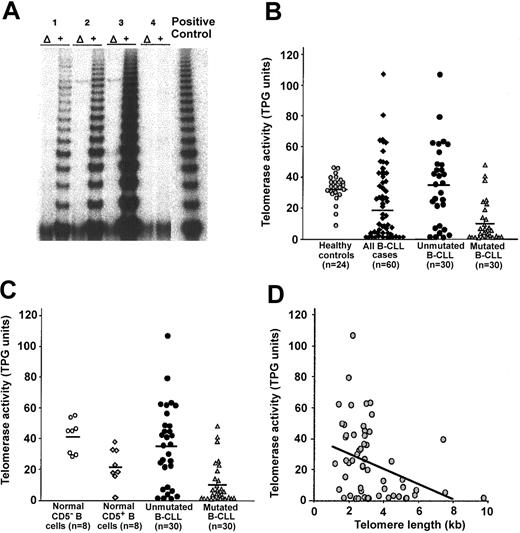
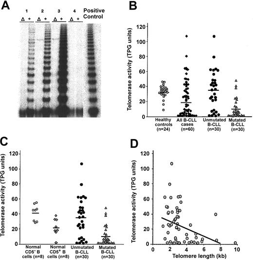
We used TRAP to quantify telomerase activity in cell lysates from purified B-CLL cells and B cells from healthy donors. Figure 3A shows representative autoradiographic profiles of amplified, elongated templates from 4 different B-CLL patients identified with a 32P-labeled probe.
Estimation of telomerase activity by TRAP. (A) Representative profiles of TRAP assay performed using B-cell lysates prepared from 4 B-CLL patients. Each lysate was tested before (+) and after (Δ) heat inactivation. Samples 1, 2, and 3 expressed various levels of telomerase, whereas sample 4 lacked telomerase activity. A positive control assay was performed with each assay. (B) Telomerase activity was quantified in purified B-cell populations from 60 B-CLL patients and 24 aging healthy donors. Telomerase activity of B-CLL cells grouped as a whole was not different than that detected in B cells from healthy subjects. Unmutated cells from B-CLL patients expressed significantly elevated telomerase activity compared with mutated cells from B-CLL patients (P < .001). Telomerase activity of unmutated B cells from B-CLL patients did not differ significantly from that observed in the group of healthy donors. When compared independently, the mutated cells displayed significantly lower telomerase activity than B cells from healthy donors (P < .001). (C) Telomerase activity of flow-sorted B-cell subsets (CD19+CD5+ and CD19+CD5–) from 8 aging healthy subjects compared with that from B cells of unmutated and mutated B-CLL (n = 30 each). Mutated B-CLL exhibited significantly lower telomerase activity than unmutated B-CLL (P < .001) and normal B-cell subsets (P < .01). (D) Scatter plot of mean telomere length compared with telomerase in entire cohort of B-CLL patients. There was a significant indirect correlation between mean telomere length and telomerase in the entire cohort of B-CLL patients (P = .043); however, the correlation of these 2 parameters did not reach statistical significance when the B-CLL patients were analyzed as individual subgroups (P = .067 for unmutated and P = .079 for mutated).
Estimation of telomerase activity by TRAP. (A) Representative profiles of TRAP assay performed using B-cell lysates prepared from 4 B-CLL patients. Each lysate was tested before (+) and after (Δ) heat inactivation. Samples 1, 2, and 3 expressed various levels of telomerase, whereas sample 4 lacked telomerase activity. A positive control assay was performed with each assay. (B) Telomerase activity was quantified in purified B-cell populations from 60 B-CLL patients and 24 aging healthy donors. Telomerase activity of B-CLL cells grouped as a whole was not different than that detected in B cells from healthy subjects. Unmutated cells from B-CLL patients expressed significantly elevated telomerase activity compared with mutated cells from B-CLL patients (P < .001). Telomerase activity of unmutated B cells from B-CLL patients did not differ significantly from that observed in the group of healthy donors. When compared independently, the mutated cells displayed significantly lower telomerase activity than B cells from healthy donors (P < .001). (C) Telomerase activity of flow-sorted B-cell subsets (CD19+CD5+ and CD19+CD5–) from 8 aging healthy subjects compared with that from B cells of unmutated and mutated B-CLL (n = 30 each). Mutated B-CLL exhibited significantly lower telomerase activity than unmutated B-CLL (P < .001) and normal B-cell subsets (P < .01). (D) Scatter plot of mean telomere length compared with telomerase in entire cohort of B-CLL patients. There was a significant indirect correlation between mean telomere length and telomerase in the entire cohort of B-CLL patients (P = .043); however, the correlation of these 2 parameters did not reach statistical significance when the B-CLL patients were analyzed as individual subgroups (P = .067 for unmutated and P = .079 for mutated).
Viewed as a single group or as unmutated and mutated subgroups, the mean telomerase activity (in TPG U) of the B-CLL cells was heterogeneous, and this heterogeneity was more marked in the unmutated subgroup (Figure 3B). Despite this, the activity of unmutated cells and normal B cells was similar, and their activities were significantly greater than cells with mutated Ig V genes (P < .001; Figure 3B). Telomerase activity of unmutated cells was comparable to that of isolated CD5– B cells (P > .05) but higher than that of isolated CD5+ B cells (P < .01; Figure 3C). On the other hand, the mutated cells showed significantly lower activity compared with isolated normal CD5– and CD5+ B cells (P < .01; Figure 3C).
We then analyzed whether telomerase activity correlated with telomere length in B-CLL patients viewed as a group and in the individual Ig V gene-defined subgroups. As illustrated in Figure 3D, there was a direct relationship between telomerase activity and telomere length in the entire B-CLL cohort (P = .046) that did not reach statistical significance when the unmutated or the mutated cells were analyzed independently (P = .067 and P = .079, respectively), presumably because of insufficient sample size.
Changes in telomere length and telomerase activity in samples collected over time
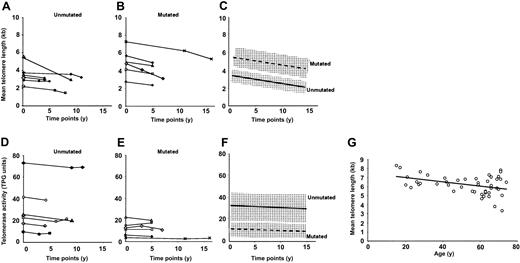
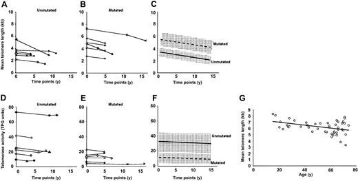
PBMCs from 12 B-CLL patients, stored frozen at 2 or more time points over a period of 4 to 17 years, were available. Therefore, we thawed samples from different dates and determined telomere length and telomerase activity for each. The plots of telomere length compared with time after first analysis are shown in Figure 4A-B.
Changes in telomere length and telomerase activity over time. B cells were purified from PBMCs of 12 B-CLL patients, stored frozen at 2 or more time points during the course of their disease, and subjected to analysis of telomere length by Flow-FISH. (A-B) Changes in mean telomere length in 6 patients from the unmutated subgroup (A) and 6 patients from the mutated subgroup (B). (D-E) Changes in telomerase activity in the same (matched symbols) sets. The rate of change in telomere length and telomerase activity (slope) in each of the 12 patients was estimated by linear regression. (C,F) Average rate of decline of telomere length (C) or change in telomerase activity (F). Shaded areas indicate the range of standard deviation for the values of the 6 individual cases. The average rate of decline in telomere length was 98 bp/y for patients in the unmutated compared with 111 bp/y for patients in the mutated subgroups. Average rate of change in telomerase activity was 0.21 TPG U/y for the unmutated cells compared with 0.19 TPG U/y for the mutated cells. Neither the rate of decline in telomere length nor the rate of change in telomerase activity differed significantly between the 2 subgroups. (G) Telomere lengths of B cells from 48 healthy donors ranging in age from 15 to 74 years were analyzed using Flow-FISH. The rate of decline in telomere length of the normal B cells was computed to be 22 bp/y.
Changes in telomere length and telomerase activity over time. B cells were purified from PBMCs of 12 B-CLL patients, stored frozen at 2 or more time points during the course of their disease, and subjected to analysis of telomere length by Flow-FISH. (A-B) Changes in mean telomere length in 6 patients from the unmutated subgroup (A) and 6 patients from the mutated subgroup (B). (D-E) Changes in telomerase activity in the same (matched symbols) sets. The rate of change in telomere length and telomerase activity (slope) in each of the 12 patients was estimated by linear regression. (C,F) Average rate of decline of telomere length (C) or change in telomerase activity (F). Shaded areas indicate the range of standard deviation for the values of the 6 individual cases. The average rate of decline in telomere length was 98 bp/y for patients in the unmutated compared with 111 bp/y for patients in the mutated subgroups. Average rate of change in telomerase activity was 0.21 TPG U/y for the unmutated cells compared with 0.19 TPG U/y for the mutated cells. Neither the rate of decline in telomere length nor the rate of change in telomerase activity differed significantly between the 2 subgroups. (G) Telomere lengths of B cells from 48 healthy donors ranging in age from 15 to 74 years were analyzed using Flow-FISH. The rate of decline in telomere length of the normal B cells was computed to be 22 bp/y.
Telomere lengths of all samples decreased over time. When the slope of decrease for each patient was calculated, the average rate of decline in telomere length for the unmutated subgroup was 98 bp/y compared with 111 bp/y for the mutated subgroup (Figure 4C). The average rates of telomere erosion did not differ significantly between unmutated and mutated subgroups (averaged slopes represented in Figure 4C).
To compare these data with an approximation of the rate of telomere shortening over time in normal B cells, we quantified telomere lengths of B lymphocytes from 48 controls ranging in age from 15 to 74 years (Figure 4G). These data, of necessity, are single time-point determinations. Based on linear regression analyses, the rate of decline of telomere length of B cells in healthy subjects was estimated to be 22 bp/y, which is in keeping with previous determinations.34
Although there was a consistent decline in telomere lengths in all 12 patients, telomerase activity in the corresponding cell lysates did not change significantly over time (Figure 4D-E). In addition, although unmutated cells had significantly more telomerase activity than mutated cells, the average change of activity over time was similar in both subgroups.
Analyses of survival and lymphocyte doubling time in patients classified by telomere length and telomerase activity
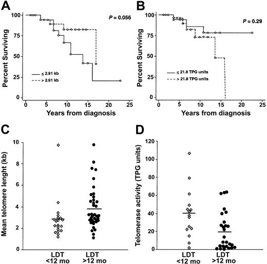
Clinical data were available for 56 of the 70 B-CLL patients studied, allowing us to compare the survival of patients classified as “high” or “low” based on median telomere length (≥ 2.91 kb vs < 2.91 kb, respectively) or median telomerase activity (≥ 21.8 TPGUvs < 21.8 TPG U, respectively). There was no difference in the survival curves of patients based on either of these parameters (P = .056 and P = .29, respectively; Figure 5A-B).
Telomere length and telomerase activity of B-CLL cells in relation to patient survival and lymphocyte doubling time. (A) Kaplan-Meier plots of survival in 56 B-CLL patients stratified by median value of telomere length (2.91 kb). (B) Kaplan-Meier plots of survival in 42 B-CLL patients stratified by median value of telomerase activity (21.8 TPG U). (C) Scatter plot of telomere length in 57 patients stratified by differences in LDT. Note that patients with LDT less than 12 months exhibited significantly shorter telomeres (mean, 2.81 kb) compared with those with LDT of 12 months or more (mean, 3.88 kb; P < .01). (D) Scatter plot of telomerase activity in 43 patients stratified by differences in LDT. Note that patients with LDT less than 12 months exhibited significantly higher telomerase activity (mean, 40.3 TPG U) than those with LDT of 12 months or more (mean, 19.1 TPG U; P < .01).
Telomere length and telomerase activity of B-CLL cells in relation to patient survival and lymphocyte doubling time. (A) Kaplan-Meier plots of survival in 56 B-CLL patients stratified by median value of telomere length (2.91 kb). (B) Kaplan-Meier plots of survival in 42 B-CLL patients stratified by median value of telomerase activity (21.8 TPG U). (C) Scatter plot of telomere length in 57 patients stratified by differences in LDT. Note that patients with LDT less than 12 months exhibited significantly shorter telomeres (mean, 2.81 kb) compared with those with LDT of 12 months or more (mean, 3.88 kb; P < .01). (D) Scatter plot of telomerase activity in 43 patients stratified by differences in LDT. Note that patients with LDT less than 12 months exhibited significantly higher telomerase activity (mean, 40.3 TPG U) than those with LDT of 12 months or more (mean, 19.1 TPG U; P < .01).
When patients were grouped on the basis of lymphocyte doubling time (LDT; less than 12 months or 12 months or more), mean telomere lengths of the leukemic cells of these subgroups showed no significant difference (Figure 5C). However, patients with shorter LDTs (less than 12 months) exhibited significantly higher telomerase activity than patients with longer LDTs (Figure 5D; P < .01). We did not detect a significant relationship between telomere length and either of the LDT groups.
Discussion
In this study, we report on the telomere length and telomerase activity of circulating B cells from patients with B-CLL and their prognostically relevant Ig V gene subgroups. Several approaches were used to derive information about the cycling history of B-CLL cells and their precursors and, by extension, about their maturation status.
We analyzed telomere length and telomerase activity in isolated CD5+, CD19+ (leukemic) B cells from the blood of each patient, thereby avoiding the confusion that other circulating non-B cells, some of which may be oligoclonal,35 could introduce into the analyses. In addition, we compared telomere lengths of leukemic cells with autologous PMNLs, which are terminally differentiated, nondividing cells that turn over rapidly in the blood.36 Telomere lengths of PMNLs are believed to reflect those of bone marrow stem cells; therefore, our comparisons between PMNLs and autologous B-CLL cells helped define the relative “ages” of the leukemic cells. Because the mean telomere lengths of PMNLs from patients with B-CLL were comparable to those from aging controls (Figure 1C), the normal stem cell elements in both populations were similar in their cycling histories. This ensured that our analyses were well controlled for the changes in telomere length that occur as a function of age.34 Moreover, we compared telomere length and telomerase activity in B-CLL cells with purified circulating CD19+ B cells and also with CD5+ and CD5-B cells from healthy subjects matched for age. Furthermore, the use of Flow-FISH31 permits quantification of mean telomere lengths of individual B cells. This method estimates telomere length more accurately than the Southern blotting approach37 used in previous studies of B-CLL cells28,38,39 because it allows statistical analysis of the differences of averages and respective coefficients of variation between individual cells. Finally, we measured telomerase activity in the same purified B-cell populations by determining the ability of telomerase to extend the ends of a standard DNA template.33,40 This assay is useful because it defines the functional capacity of the enzyme, not merely its presence as an RNA species or as a protein. Although telomerase activity is up-regulated in mature B cells by various stimulatory signals in vitro41 and in vivo,23-26 it is unlikely that the levels detected in the normal cells resulted from artifacts of cell handling given that these cells were isolated by negative selection and without in vitro culture and stimulation.
B-CLL cells, considered as a single group, had significantly shorter telomeres than autologous PMNLs (P < .01; Figure 1C) and B cells from healthy subjects matched for age (P < .0001; Figure 2A). Based on the relationship between telomere lengths of PMNLs and bone marrow stem cells, a considerable number of cell divisions must have occurred in the leukemic cells after their generation. These findings are in line with the belief that B-CLL cells are long-lived lymphocytes that originate well before the disease becomes clinically manifest.42,43 Finally, leukemic cells from unmutated cases had significantly shorter telomeres than mutated cases (P < .0001; Figure 2A). This difference correlated with the degree of V gene mutations detected in individual patients (ie, longer telomeres were found in cells with more Ig V gene mutations; Figure 2B). These data agree with our preliminary studies29,30 and those recently published about telomere lengths of leukemic cells from B-CLL patients39 and their family members.38 In addition, they support the contention that even unmutated B-CLL derives from antigen-experienced and not naive B lymphocytes.4,5,44,45
The level of telomerase activity in unmutated cells was significantly higher than that of mutated cells and was similar to that of B cells from healthy donors (P < .0001; Figure 3B). Telomerase activity in B-CLL cells, regardless of subgroup, was similar and relatively constant over time (Figure 4B).
Correlating telomere length with telomerase activity in B-CLL is complex because these are dynamic processes that can only be measured during the leukemic phase of a B-CLL cell's life and often at only one point in time. Nevertheless, some conclusions can be derived about the relationship between these 2 parameters.
First, because most leukemic cells in the blood of a typical B-CLL patient are not cycling,46,47 our data suggest that telomeres in the leukemic B cells shorten at a comparable rate over time in unmutated and mutated cells (Figures 4A-C). Therefore, much of the telomere length difference between these 2 subgroups might have arisen before the final transforming event. This is consistent with our observation that even at the earliest time tested, the telomere lengths of unmutated cells are shorter than those of mutated cells (Figure 4A-C). However, we cannot rule out the possibility that telomerase corrected some telomere length loss in the unmutated cells, thereby confounding this comparison.
Second, because the restorative process mediated by telomerase is usually activated when telomeres reach a length that endangers the life of an individual cell, this enzyme might have been activated in the Ig V gene unmutated B-CLL subgroup to compensate for excessive losses during the preleukemic and leukemic phases of clonal expansion. The uniformity of mean telomere lengths in this subgroup is consistent with this idea or with the possibility that only cells with compensatory telomerase activity survived long enough to be evaluated. It is conceivable that even greater differences in telomere length have been masked by the activity of telomerase. It is difficult to definitively associate the up-regulation of telomerase activity with progressive shortening of telomere lengths because the temporal evolution of the shortening in relation to the time of telomerase activation is not readily determinable.
Third, in contrast to unmutated cells that have uniformly short telomeres, mutated cells display heterogeneous telomere lengths, some comparable to those with the shortest median lengths in the unmutated subgroup of B-CLL patients and others to the longest lengths of healthy aging subjects (Figure 2A). Because telomeres are extended during a typical T-cell–dependent GC reaction, when Ig V gene mutations can arise, the precursors of these leukemic cells may undergo variable numbers of divisions in response to antigen-mediated, T-cell–dependent GC reactions, during which enhanced telomerase activity coordinately elongates their telomeres.23-26 Similarly, the diversity of lengths among patients within this subgroup could reflect the number of antigenic exposures and GC transits that individual preleukemic clones underwent. These possibilities would be compatible with their high degree of V gene mutations and with their similarity in telomere length to CD5– conventional B cells (Figure 2B) that undergo T-cell–dependent GC reactions. In addition, the longer telomeres in the mutated subgroup might reflect differences in the “starting point” at the time of leukemic transformation because of telomere lengthening from up-regulated telomerase activity during GC transits. With similar rates of decline in telomere length in the 2 subgroups after leukemic transformation, these differences would be maintained and could mirror the preleukemic experiences of the B-CLL precursors.
However, given that Ig V gene mutations also can occur in response to T-cell–independent antigens,48,49 at least some B-CLL cells with V gene mutations may have arisen through a T-cell–independent process that might transiently up-regulate a telomere repair mechanism. If true, then both unmutated and mutated subgroups could have experienced similar antigenic drives, but cells from each subgroup would have dealt with these stimuli differently with regard to somatic mutation and possibly telomere maintenance. This model would be consistent with recent gene profiling data suggesting only minor transcriptional differences between the 2 B-CLL subgroups.44,45,50 These 2 possibilities are not mutually exclusive (see Klein et al45 and Chiorazzi and Ferrarini10 for more detailed discussions of these models).
Finally, an alternative mechanism for telomere lengthening, or ALT, involving DNA recombination at chromosomal ends, does exist.22 This process may also be involved in telomere maintenance in B-CLL, particularly because ALT is characterized by the absence of detectable telomerase activity22 and the presence of heterogeneous telomere lengths51 akin to those seen in the mutated subgroup (Figure 2A). Analyses are under way to identify the characteristic nuclear features of cells in which ALT is active.52,53
It is important to recognize that comparisons between clonal leukemic cells and normal B cells must be made cautiously because the latter are polyclonal and comprise at least 3 subsets, each with varying degrees of clonal heterogeneity. Therefore, we also compared B-CLL subgroups with purified CD5+ and CD5– B lymphocytes from the blood of healthy subjects matched for age (Figure 2B). Although direct comparisons of mean values between each of these normal B-cell populations (CD19+CD5+ and CD19+CD5–) and B-CLL subgroups are difficult, proportionate comparisons may be useful. When analyzed in this fashion, the differences between mutated cells that display the longest mean telomere lengths and unmutated cells with shorter telomeres (Figure 2A-B) are similar to the differences between CD5– conventional B2 cells that predominate the circulating CD19+ pool and the CD5+ subpopulation. Because B-CLL cells express genes45 and surface markers5 characteristic of previously activated “memory” B cells, we have begun to analyze CD5+ cells and naive (CD19+CD5–CD27–) and memory (CD19+CD5–CD27+) cells of the conventional B2 cell pool. Using proportional comparisons, preliminary data on B-cell subsets isolated from 4 healthy aging subjects support the possibility that cells that express mutated Ig V genes and long telomeres with considerable length variability resemble conventional memory B cells (preliminary mean telomere length, 9.09 ± 1.04 kb). In contrast, unmutated cells with uniformly short telomeres resemble either CD5+ (preliminary mean telomere length, 7.13 ± 0.66 kb) or CD5– (preliminary mean telomere length, 7.31 ± 0.76 kb) virgin B cells. Unfortunately, because of the extremely low levels of CD5+CD27+ B cells in the blood of healthy subjects, adequate comparisons with this subset have not been possible to date. Comparisons with other noncirculating cells would also be important (though technically difficult), especially because cells of the marginal zone have been mentioned as the potential source of B-CLL cells.10
Our observations may have potential implications for determining prognosis in B-CLL. Using Flow-FISH and TRAP technologies, we did not identify a statistically significant relationship among telomere length, telomerase expression, and clinical outcome in B-CLL (Figure 5A-B). These findings are in agreement with those from a study of circulating leukemic cells in patients with familial B-CLL38 and at variance with those from studies using Southern blotting to analyze telomere lengths in solid-organ–derived and circulating leukemic cells.28,39 However, we did identify a significant relationship between LDT and telomerase activity and a trend in the relationship between LDT and telomere length (Figure 5C-D). The former is consistent with the observation that in chronic B-cell lymphoproliferative disorders, telomerase activity increases with disease progression.54 Thus, telomerase activity and telomere length may represent markers, in addition to Ig V gene mutation status,8,9 CD388 and ZAP-70 expression,6,7 and LDT,2 that identify subgroups of patients whose clinical outcomes differ. Further work with larger cohorts of patients using comparable methodologies to analyze blood-derived and solid-organ–derived B-CLL cells may be needed to definitively determine these potentially important associations.
Prepublished online as Blood First Edition Paper, September 22, 2003; DOI 10.1182/blood-2003-04-1345.
Supported in part by United States Public Health Service grants CA 81554 and CA 87956 from the National Institutes of Health/National Cancer Institute and by the Jean Walton Fund for Lymphoma and Myeloma Research, the Joseph Eletto Leukemia Research Fund, the S.L.E. (Systemic Lupus Erythematosus) Foundation, Inc, and Associazone Italiana Ricerca sul Cancro.
Presented in part at the 42nd Annual Meeting of the American Society of Hematology, San Francisco, CA, December 1-5, 2000.
The publication costs of this article were defrayed in part by page charge payment. Therefore, and solely to indicate this fact, this article is hereby marked “advertisement” in accordance with 18 U.S.C. section 1734.
We thank Cathy Rapelje and Grace Lee of the North Shore-Long Island Jewish Research Institute's flow cytometry core facility for analysis of immunophenotyping and Flow-FISH and Dr Kirk Manogue for detailed discussion of data and for invaluable suggestions during the preparation of the manuscript.

