Abstract
AC133 is a member of a novel family of cell surface proteins with 5 transmembrane domains. The function of AC133 is unknown. Although AC133 mRNA is detected in different tissues, its expression in the hematopoietic system is restricted to CD34+ stem cells. AC133 is also expressed on stem cells of other tissues, including endothelial progenitor cells. However, despite the potential importance of AC133 to the field of stem cell biology, nothing is known about the transcriptional regulation of AC133 expression. In this report we showed that the human AC133 gene has at least 9 distinctive 5′–untranslated region (UTR) exons, resulting in the formation of at least 7 alternatively spliced 5′-UTR isoforms of AC133 mRNA, which are expressed in a tissue-dependent manner. We found that transcription of these AC133 isoforms is controlled by 5 alternative promoters, and we demonstrated their activity on AC133-expressing cell lines using a luciferase reporter system. We also showed that in vitro methylation of 2 of these AC133 promoters completely suppresses their activity, suggesting that methylation plays a role in their regulation. Identification of tissue-specific AC133 promoters may provide a novel method to isolate tissue-specific stem and progenitor cells.
Introduction
AC133 (CD133; human prominin-1; prominin [mouse]-like 1 [PROML1]) is a member of a novel family of 2 known cell surface glycoproteins carrying 5 transmembrane domains.1-3 The function of AC133 is not known, and its ligand has not yet been identified. Initially, AC133 cDNA was isolated as a result of a search for a gene encoding a novel antigen, expression of which in the hematopoietic system was restricted to CD34+ stem cells derived from fetal liver, bone marrow, and peripheral blood.2,3 AC133+ cells were shown to engraft successfully in a fetal sheep transplantation model of primary and secondary recipients, suggesting that these cells have long-term repopulating potential.2 Later, AC133 expression was demonstrated on undifferentiated epithelium,4 retinoblastoma,3 teratocarcinoma,3 leukemias,5,6 and fetal brain neural stem cells.7 Our group previously showed that AC133 is expressed on vascular endothelial growth factor receptor-2 (VEGFR-2)+ endothelial progenitors, and that maturation of these cells is associated with rapid down-regulation of AC133.8 Thus, the overall data suggest that AC133 could be a specific marker for various stem and progenitor cell populations.
A number of recent studies with a monoclonal antibody to the AC133 protein and its mouse homologue prominin significantly increased our knowledge of their cellular localization and antigen distribution. Mouse prominin was originally discovered as a protein localized to microvilli on the apical surface of mouse neuroepithelial stem cells.9 Later, protrusion-restricted localization of AC133 was shown on human cells as well.4
Until now, nothing was known about transcriptional regulation of the AC133 gene. In the present study, we isolated and characterized the 5′–untranslated region (UTR) and promoter region of the human AC133 gene. We identified novel cDNA sequences, which extend the 5′-UTR of previously published AC133 cDNA. Nine additional exons were found within the 5′-UTR. 5′–rapid amplification of complementary DNA ends (RACE) on different tissues and cell lines provided evidence of alternative splicing, showing the formation of 7 AC133 transcripts with alternative 5′-UTRs, driven by 5 alternative promoters (P1, P2, P3, P4, and P5). We tested the activity of these promoters on the AC133-expressing cell lines Caco-2 and NT-2, and detected significant activity for 2 of them (P1 and P2). Analysis of the promoter regions has indicated that all AC133 promoters are TATA-less and 3 of them (P1, P2, and P3) are located within a CpG-island. In vitro methylation of AC133 promoters P1 and P2 completely suppressed their activity, suggesting that methylation may play a role in their regulation. Expression analysis of AC133 5′-UTR isoforms showed that their expression pattern is tissue specific, suggesting that alternative AC133 promoters could be used to identify and isolate organ-specific stem cells.
Materials and methods
Database searches and computer analysis
Analysis of nucleotide sequences was performed using National Center for Biotechnology information (http://www.ncbi.nlm.nih.gov) and Celera Discovery System (http://publication.celera.com/humanpub/index.jsp).
Potential transcription factor binding sites were mapped to putative AC133 promoters using AliBaba software (http://www.alibaba2.com) and TFsearch (http://molsun1.cbrc.aist.go.jp/research/db/TFSEARCH.html).
Cell culture
All cell lines for this study were obtained from American Type Culture Collection (ATCC; Manassas, VA). Human retinoblastoma cell line WERI-Rb-1 and human teratocarcinoma cell line NTERA-2 cl.D1 (NT-2) were grown in Dulbecco modified Eagle medium, supplemented with 10% fetal bovine serum. Colon cancer cell line Caco-2 was grown in Eagle minimum essential medium supplemented with 20% fetal bovine serum. All media were supplemented with 2 mM l-glutamine, 100 U/mL penicillin, 100 mg/mL streptomycin, and 0.25 μg/mL fungizone (BioWhittaker, Walkersville, MD).
Isolation of CD34+ cells from cord blood
Human cord blood samples were obtained in conjunction with ethical and biohazard guidelines set by the institution. CD34+ cells were purified by magnetic activated cell sorting columns (MACS; Miltenyi Biotech, Auburn, CA) using microbead conjugated antibodies. Two purification cycles were performed, using separate columns. The purity of each batch of CD34+-selected cells was determined by flow cytometry using fluorescein isothiocyanate (FITC; green fluorescence)–labeled anti-CD34 monoclonal antibodies (Miltenyi Biotech).
Rapid amplification of cDNA ends (RACE)
In order to perform 5′-RACE for AC133 on Caco-2 cells, NT-2 cells, and CD34+ cells isolated from cord blood, total RNA was isolated using Concert Cytoplasmic RNA Isolation Reagent (Invitrogen, Carlsbad, CA). RACE-ready cDNA was prepared using the GeneRacer Kit (Invitrogen) according to the manufacturer's protocol. 5′-RACE on human brain, kidney, and testis cDNA was performed using RNA-ligase mediated (RLM)–5′-RACE-ready brain, kidney, and testis cDNA (Invitrogen) according to the manufacturer's protocol. All 5′-RACE experiments were carried out using 2 AC133-specific primers matching sequences in exon 2: 5′-GAGGCATCAGAATAATAAACAGCAGC-3′ and nested 5′-CAGCAGCAACAGGGAGCCGAGTA-3′, with Platinum Taq DNA Polymerase High Fidelity (Invitrogen). Resulting polymerase chain reaction (PCR) products were purified, cloned into the pCR4-TOPO vector (Invitrogen), and sequenced.
Northern hybridization analysis
A fragment of AC133 cDNA (361 base pair [bp]) common to all known isoforms was amplified by reverse transcriptase (RT)–PCR, using AC133-specific primers: 5′-CTAGATACTGCTGTTGATGTC-3′ and 5′-CTGCTCTAGGTTGACACACTT-3′, cloned into pBlueScript KS- (Stratagene, La Jolla, CA), and the digested fragment was used as a probe for AC133 detection on a Northern blot. As an internal control for loading, a 408-bp fragment of glyceraldehyde 3–phosphate dehydrogenase (G3PDH) was amplified by RT-PCR, using G3PDH-specific primers: 5′-CCCATCACCATCTTCCAGGAG-3′ and 5′-AGGGATGATGTTCTGGAGAGCC-3′, cloned into pCR4-TOPO with a digested fragment used as a probe for G3PDH detection on a Northern blot. Total RNA (20 μg) from Caco-2, WERI-Rb-1, and NT-2 cell lines was resolved on a 1.2% formaldehyde agarose gel, transferred to Nytran SuPerCharge (Schleicher & Schuell, Keene, NH), and probed with a 32P-labeled probe.
RT-PCR analysis
Human MTC Panel I, Human MTC Panel II, and Human Immune System MTC Panel (Clontech, Palo Alto, CA) were used as a source of cDNA from 18 human tissues for RT-PCR. Amplification was performed using forward primers 5′-GTCCAATCAGAGTGCGTCCA-3′ and nested 5′-GGCCATGCTCTCAGCTCT-3′ for exon 1A, 5′-GCGGCAGCGGTGACTA-3′ and nested 5′-GAGCAGGAGCGGGAGC-3′ for exon 1B, 5′-GAACTGCGGGGAGAGCGT-3′ and nested 5′-CGGAGCGGGAGTCGGA-3′ for exon 1C, 5′-CCAAAAGCACTCCAGATGACA-3′ and nested 5′-AATCCACTACAAAGCTCTTC-3′ for exon 1D1, 5′-TTCTTCTCTGTGGGCTCCT-3′ and nested 5′-CTTTCTCGTGGATCTGGAC-3′ for exon 1E1, and 2 reverse primers located in common exon 2 of AC133: 5′-TCTTGGGTCTCATAATTTGTTGCAGGC-3′ and nested primer 5′-TTCCCGCACAGCCCCAGCAGCAACAG-3′. PCR was performed using Advantage 2 Taq Polymerase (Clontech). PCR conditions were as follows: 94°C for 1 minute followed by 25 cycles (first round) or 32 cycles (second round with nested primers) of 94°C for 30 seconds, 60°C for 30 seconds, and 72°C for 30 seconds. RT-PCR on Caco-2, NT-2, brain, kidney, and CD34+ human cord blood cell RACE-ready cDNA was performed as indicated above, with one exception: a 5′-RACE outer adapter primer was used instead of gene-specific primers for exons 1A, 1B, 1C, 1D1, and 1E1 in the first round of PCR.
Construction of luciferase reporter vectors
The BAC clone RP11-452J21, containing exons 1A, 1B, 1C, exon clusters 1D and 1E, and exons 2 and 3 of AC133 and their flanking regions, was obtained from Research Genetics (Invitrogen). All promoter fragments were generated by PCR using primers to different regions of AC133 P1, P2, P3, P4, and P5 (Table 1). All primers contained 5′ adapters with restriction sites for KpnI or HindIII. Amplified fragments were double digested with KpnI and HindIII and cloned into KpnI/HindIII sites of the pGL3-enhancer firefly luciferase vector (Promega, Madison, WI). Plasmids were purified using the HiSpeed Plasmid Midi Kit (Qiagen, Valencia, CA) and verified by sequencing.
Transient transfections and luciferase assays
All pGL3 and renilla luciferase vectors were obtained from Promega. All transfections were carried out in 6-well plates (Corning, Corning, NY). Cells were plated 24 hours prior to transfection at a density of 2 × 10⁵ per well, and transfections were carried out in antibiotic-free medium. Reporter construct (1.5 μg) in pGL3-enhancer vector and 30 ng renilla luciferase–null vector (as an internal control for transfection efficiency) were transfected, using 3 μL Lipofectamine 2000 Transfection Reagent (Invitrogen) according to the manufacturer's protocol. At 48 hours after transfection, cells were washed with phosphate-buffered saline and lysed with 500 μL Passive Lysis Buffer (Promega). Activities of firefly and renilla luciferases were measured with a TD-20/20 Luminometer (Turner Designs, Sunnyvale, CA) using the Dual Luciferase Reporter Assay System (Promega) according to the manufacturer's protocol. Firefly luciferase activity was normalized by renilla luciferase activity. pGL3-enhancer (promoterless) vector was used in all experiments as a negative control and as a baseline for data comparisons. pGL3-control (contains SV40 promoter and SV40 enhancer) vector was used in all experiments as a positive control.
In vitro DNA methylation
Plasmid DNA (20 μg) was incubated without (mock methylated) or with (methylated) 20 units SssI methylase (New England BioLabs, Beverly, MA) at 37°C for 24 hours, supplemented every 4 hours with 160 μM S-adenosylmethyonine. Complete methylation at CpG sites was confirmed by HpaII digestion of plasmid DNA. Before transfection, methylated DNA was purified using the Wizard DNA Clean-Up System (Promega). Data from methylated and mock-methylated constructs were normalized by the activity of methylated or mock-methylated pGL3-enhancer plasmids, respectively.
Results
Determination of transcription start point
Comprehensive analysis of preexisting sequences of cDNA and expressed sequence tags (ESTs) for AC133 using NCBI databases revealed the presence of an additional exon (assigned as exon 1A) in the 5′-UTR of the AC133 transcripts upstream of the previously reported first exon (assigned as exon 2). Matching the nucleotide sequence of exon 1A to human genomic DNA showed that this exon is located approximately 8 kilobase (kb) upstream of exon 2 (Figure 1). All together, we found 8 (4 ESTs and 4 cDNA) sequences containing exon 1A in the database. Positioning of the first nucleotide of those 8 sequences to genomic DNA did not reveal a precise transcription start point. We found one group of 2 sequences and another group of 3 sequences with the same transcription start point, while 3 other sequences contain different start points. Unexpectedly, one EST derived from skeletal muscle did not show any homology with exon 1A, instead showing homology to a region of genomic DNA approximately 500 bp downstream of exon 1A. This datum indicates the possibility of an alternative first exon (assigned as a putative exon 1C).
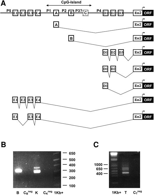
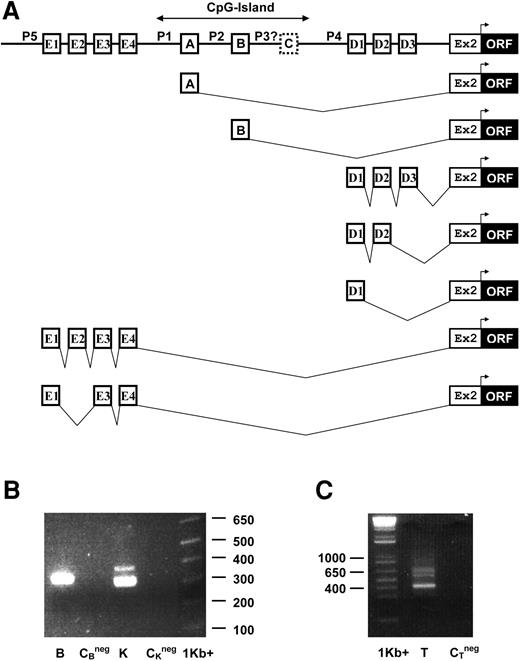
Genomic structure of the promoter region of human AC133 and alternative splicing within its 5′-UTR. (A) Exon 1A, exon 1B, exons D1, D2, and D3 from cluster 1D, and exon E4 from cluster 1E are alternatively spliced to a common exon 2, according to our RACE data. Translation initiation site is located in exon 2. 5′-RACE revealed the presence of exons 1A, 1B, and exon clusters 1D and 1E, while exon 1C was found as a single EST from skeletal muscle. P1, P2, P4, P5, and possibly P3 are alternative promoters for the AC133 gene. Exons 1A, 1B, 1C, and their corresponding promoters are located within a CpG island. (B) 5′-RACE analysis of AC133 mRNA. mRNAs from brain (B) and kidney (K) were subjected to 5′-RACE analysis. The PCR products (second round of amplification; nested primers) were resolved on 1.5% agarose gel, stained with ethidium bromide. Size marker 1 kb plus is shown on the right (1 kb+). CBneg and CKneg are negative controls for brain and kidney, respectively. One band from brain and 2 bands from kidney were isolated, subcloned into pCR4-TOPO, and sequenced. The band from brain was found to comprise exon 1B fragments. The lower band from kidney was found to comprise a majority of exon 1A fragments, with a minority of exon 1B fragments. The upper band from kidney was found to comprise exon 1A fragments from an alternate transcription start point, 55 bp longer than those in the lower band. (C) 5′-RACE analysis of AC133 mRNA in testis (T). Multiple bands were observed for testis 5′-RACE products. Since multiple bands were observed, the entire pool of PCR products was subcloned into pCR4-TOPO directly from the PCR product mix. PCR mix was determined to contain 5 different alternative splice variants containing exons from either cluster 1E or cluster 1D.
Genomic structure of the promoter region of human AC133 and alternative splicing within its 5′-UTR. (A) Exon 1A, exon 1B, exons D1, D2, and D3 from cluster 1D, and exon E4 from cluster 1E are alternatively spliced to a common exon 2, according to our RACE data. Translation initiation site is located in exon 2. 5′-RACE revealed the presence of exons 1A, 1B, and exon clusters 1D and 1E, while exon 1C was found as a single EST from skeletal muscle. P1, P2, P4, P5, and possibly P3 are alternative promoters for the AC133 gene. Exons 1A, 1B, 1C, and their corresponding promoters are located within a CpG island. (B) 5′-RACE analysis of AC133 mRNA. mRNAs from brain (B) and kidney (K) were subjected to 5′-RACE analysis. The PCR products (second round of amplification; nested primers) were resolved on 1.5% agarose gel, stained with ethidium bromide. Size marker 1 kb plus is shown on the right (1 kb+). CBneg and CKneg are negative controls for brain and kidney, respectively. One band from brain and 2 bands from kidney were isolated, subcloned into pCR4-TOPO, and sequenced. The band from brain was found to comprise exon 1B fragments. The lower band from kidney was found to comprise a majority of exon 1A fragments, with a minority of exon 1B fragments. The upper band from kidney was found to comprise exon 1A fragments from an alternate transcription start point, 55 bp longer than those in the lower band. (C) 5′-RACE analysis of AC133 mRNA in testis (T). Multiple bands were observed for testis 5′-RACE products. Since multiple bands were observed, the entire pool of PCR products was subcloned into pCR4-TOPO directly from the PCR product mix. PCR mix was determined to contain 5 different alternative splice variants containing exons from either cluster 1E or cluster 1D.
5′-RACE was performed in order to map the transcription start point for AC133 transcripts. We used commercial RACE-ready cDNA and nested primers located in exon 2 to perform 5′-RACE on human kidney, brain, and testis cDNA. RACE fragments were subcloned into pCR4-TOPO vector and 64 clones were sequenced. Several alternative variants of the 5′ terminal sequences were found. Two bands from the kidney sample were detected, isolated, and sequenced (Figure 1B). Sequence analysis revealed that the lower, more-abundant band contained predominantly sequences corresponding to exon 1A, and trace amounts of DNA corresponding to a novel sequence matching a 60-bp segment of genomic DNA approximately 250 bp downstream of exon 1A and 200 bp upstream of putative exon 1C. We assigned this novel alternative first exon as exon 1B (Figure 1A). Analysis of the upper band revealed a match to exon 1A, with an alternative transcription initiation point 55 bp longer than the sequence derived from the lower band. Only one band was detected in the brain sample, which matched exon 1B (Figure 1B).
Multiple bands were found in the testis 5′-RACE product (Figure 1C). Analysis revealed 2 clusters of 5′-UTR exons. The first cluster, named 1D, was found approximately 3 kb downstream of exon 1C and approximately 4 kb upstream of exon 2. It contained 3 exons (D1, D2, and D3), which form at least three 5′-UTR alternatively spliced isoforms, differing from one another by inclusion of D2 and/or D3 (Figure 1A). The second cluster, named 1E, contained 4 exons (E1, E2, E3, and E4), approximately 36 kb upstream of exon 1A. Exons from cluster 1E generate at least 2 new alternatively spliced forms, differing from each other by the presence or absence of exon E2 (Figure 1A). All newly discovered exons are spliced according to the GT-AG rule (Table 2).
Determination of transcription start point from 5′-RACE data for exon 1A revealed the most common transcription initiation site, although additional initiation points were observed. We were unable to detect any exon 1C–containing transcripts, nor any other new alternative exons, in our RACE experiments (data not shown). The existence of 5 alternative first exons suggested the presence of 5 alternative promoters (Figure 1A), which we named P1 (upstream of exon 1A), P2 (upstream of exon 1B), P3 (upstream of putative exon 1C), P4 (upstream of exon 1D1), and P5 (upstream of exon 1E1).
Expression of AC133 isoforms in human tissues
Sequence-specific forward primers were designed for each first exon (1A, 1B, 1C, 1D1, and 1E1) and a reverse primer for common exon 2 to screen different cell types for the presence of alternatively spliced AC133 5′-UTR isoforms.
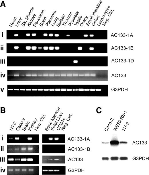
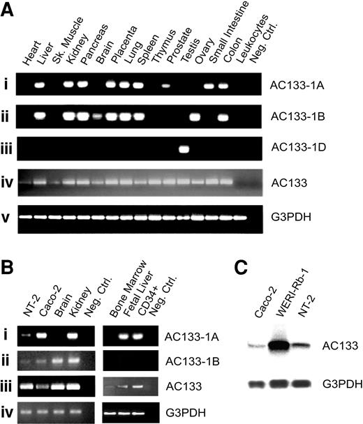
We investigated the expression of AC133 and its isoforms in commercially available cDNA panels for 18 different adult human tissues using RT-PCR (Figure 2). All analyzed tissues, with the exception of peripheral blood leukocytes, were found positive for the AC133 transcript. However, PCR analysis of 5′-UTR isoforms of AC133 revealed a tissue-dependent expression pattern. Liver, kidney, pancreas, placenta, lung, spleen, and colon express both exon 1A– and exon 1B–containing transcripts; brain, ovary, and fetal liver express only the exon 1A–containing isoform; and prostate and small intestine express only the exon 1B–containing transcript. Exon 1D was detected only in testis. Interestingly, 1A, 1B, or 1D isoforms were not detected in heart, skeletal muscle, thymus, or bone marrow. Therefore, it is possible that other isoforms of AC133 5′-UTR may exist. Interestingly, the exon 1C– and exon 1E–containing isoform was not detected in any tested tissues, probably due to low expression level (data not shown).
Tissue distribution of different 5′-UTR isoforms of AC133 mRNA.AC133 isoforms were amplified by PCR and separated on a 2% agarose gel stained with ethidium bromide. Panels Aiv and Biii show expression of AC133 by RT-PCR with primers located in the 3′ distal exons common to all known AC133 isoforms in indicated tissues or cell lines. (A) Expression of exon 1A, exon 1B, and exon 1D containing isoforms of AC133 mRNA in human adult tissues is represented in panels Ai and Aii. (B) Expression of exon 1A–, exon 1B–, and exon 1D–containing isoforms of AC133 mRNA in human brain, kidney, bone marrow, fetal liver, CD34+ cord blood cells, and cell lines Caco-2 and NT-2 are represented in panels Bi and Bii. Results are consistent in at least 3 independent PCR reactions. (C) Northern blot analysis of total RNA isolated from Caco-2, WERI-Rb-1, and NT-2 cell lines. Equal amounts of RNA (20 μg) were loaded in each lane. Blot was hybridized with a 32P-labeled probe for human AC133 mRNA. Size positions of RNA ladder are indicated on the right side. A 32P-labeled probe for human glyceraldehyde 3–phosphate dehydrogenase (G3PDH) was included as an internal control for loading and is shown in the bottom line.
Tissue distribution of different 5′-UTR isoforms of AC133 mRNA.AC133 isoforms were amplified by PCR and separated on a 2% agarose gel stained with ethidium bromide. Panels Aiv and Biii show expression of AC133 by RT-PCR with primers located in the 3′ distal exons common to all known AC133 isoforms in indicated tissues or cell lines. (A) Expression of exon 1A, exon 1B, and exon 1D containing isoforms of AC133 mRNA in human adult tissues is represented in panels Ai and Aii. (B) Expression of exon 1A–, exon 1B–, and exon 1D–containing isoforms of AC133 mRNA in human brain, kidney, bone marrow, fetal liver, CD34+ cord blood cells, and cell lines Caco-2 and NT-2 are represented in panels Bi and Bii. Results are consistent in at least 3 independent PCR reactions. (C) Northern blot analysis of total RNA isolated from Caco-2, WERI-Rb-1, and NT-2 cell lines. Equal amounts of RNA (20 μg) were loaded in each lane. Blot was hybridized with a 32P-labeled probe for human AC133 mRNA. Size positions of RNA ladder are indicated on the right side. A 32P-labeled probe for human glyceraldehyde 3–phosphate dehydrogenase (G3PDH) was included as an internal control for loading and is shown in the bottom line.
Expression of AC133 isoforms on human cell lines
In order to find a suitable AC133-expressing cell line to test promoter activity, we screened previously reported AC133+ cell lines NT-2, Caco-2, and WERI-Rb-13,4 by Northern blot hybridization (Figure 2C). We confirmed that all 3 cell lines express the AC133 transcript, with the highest expression level found in WERI-Rb-1 cells. Caco-2 and NT-2 cells showed similar levels of AC133 expression.
We also performed 5′-RACE on AC133-expressing cell lines Caco-2 and NT-2. Our 5′-RACE data indicate the presence of exon 1A– and exon 1B–containing AC133 transcripts in Caco-2 cells, and exon 1A–containing transcripts in NT-2 cells.
We employed nested PCR amplification, using our specific primers, on preamplified RACE-ready cDNA. Our results indicated that the NT-2 and Caco-2 cell lines utilize 2 alternatively spliced variants of AC133 transcripts: the 1A and 1B isoforms. Applying this method to RACE-ready kidney and brain cDNA confirmed our 5′-RACE results, showing both 1A and 1B transcripts for kidney and only 1B transcripts for brain.
Expression of AC133 isoforms on CD34+ hematopoietic stem cells
Previously published data show that the majority of CD34+ cells also express AC133. In order to investigate the expression of different AC133 isoforms on CD34+ cells, we isolated human CD34+ cells from cord blood and used them to generate RACE-ready cDNA. We analyzed the expression of AC133 5′-UTR isoforms on CD34+ cells by RT-PCR using primers for 1A, 1B, 1C, 1D, and 1E isoforms. We found that CD34+ hematopoietic stem cells express the exon 1A–containing isoform of AC133. In order to check the expression of other possible 5′-UTR isoforms of AC133 on CD34+ cells, we performed 5′-RACE. We were not able to find any new isoforms of AC133 in these cells.
Isolation and analysis of genomic clones
Blast search revealed a genomic bacterial artificial chromosome (BAC) clone which contains exons 1A, 1B, 1C, exon 2, exon 3, and clusters of exons 1D and 1E. In order to clone the putative promoter region of AC133, this BAC clone was obtained and fragments corresponding to putative promoters were amplified, subcloned into the pGL3-enhancer vector, and verified by sequencing. This set of constructs includes an 1810-bp segment of genomic DNA upstream of exon 1A (promoter P1), a 365-bp segment upstream of exon 1B (promoter P2), a 501-bp segment upstream of exon 1C (promoter P3), a 1648-bp segment upstream of exon 1D1 (promoter P4), and a 1575-bp segment upstream of exon 1E1 (promoter P5).
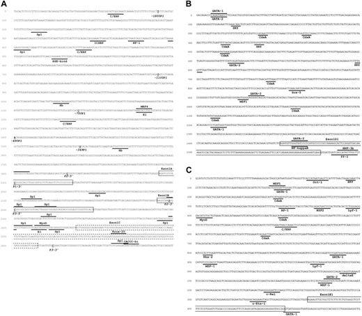
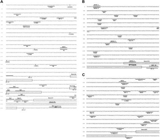
Analysis of the promoter region revealed multiple binding sites for different transcription factors (Figure 3). There were no TATA boxes found in all AC133 promoters. Exons 1A, 1B, and 1C, and also promoters P2, P3, and at least partially P1, are located within a CpG island.
Genomic nucleotide sequence of the 5′-UTR and promoter regions of the AC133 gene. Frames highlight exons. Putative transcription binding sites are underlined or top lined. Bold underlined nucleotide letters indicate either 5′ or 3′ end sites for different AC133 promoter constructs. Mapping based on AliBaba2.1 and TFsearch prediction for 90% to 100% matches only. (A) Genomic nucleotide sequence of promoters P1, P2, and P3. Accession number in GenBank: AY275524. (B) Genomic nucleotide sequence of promoter P4. Accession number in GenBank: AY438641. (C) Genomic nucleotide sequence of promoter P5. Accession number in GenBank: AY438640.
Genomic nucleotide sequence of the 5′-UTR and promoter regions of the AC133 gene. Frames highlight exons. Putative transcription binding sites are underlined or top lined. Bold underlined nucleotide letters indicate either 5′ or 3′ end sites for different AC133 promoter constructs. Mapping based on AliBaba2.1 and TFsearch prediction for 90% to 100% matches only. (A) Genomic nucleotide sequence of promoters P1, P2, and P3. Accession number in GenBank: AY275524. (B) Genomic nucleotide sequence of promoter P4. Accession number in GenBank: AY438641. (C) Genomic nucleotide sequence of promoter P5. Accession number in GenBank: AY438640.
Transcriptional activity of AC133 promoters in the NT-2 and Caco-2 cell lines
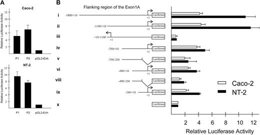
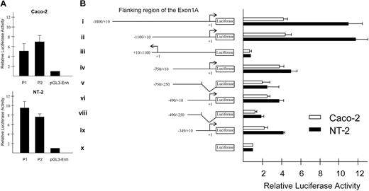
Transient transfection experiments were carried out on the NT-2 and Caco-2 cell lines. Unfortunately, we could not test the WERI-Rb-1 cell line due to difficulties with transfection. Tested AC133 P1 and P2 promoter constructs, normalized by renilla luciferase activity, revealed elevated levels of firefly luciferase activity compared with baseline activity of promoterless pGL3-enhancer vector (Figure 4A). We were not able to detect any significant increase in luciferase activity for the P3, P4, or P5 promoter constructs (data not shown). The promoter segment in the construct with the highest luciferase activity (–1100/+10 P1) was subcloned into the pGL3-enhancer vector in the reverse orientation. We observed no luciferase activity for this construct. Several 5′ and 3′ deletions of P1 were constructed and cloned into the pGL3-enhancer vector, and their activity was assayed using the luciferase system (Figure 4B).
Functional analysis of human AC133 promoters. (A) Functional analysis of promoters P1 and P2 in Caco-2 and NT-2 cell lines. Bars show fold increase in activity of luciferase for the constructs cloned into pGL3-enhancer vector containing promoters P1 or P2, compared with promoterless pGL3-enhancer vector. All data represent the mean of at least 3 independent experiments. (B) Functional analysis of human AC133 promoter 1 (P1) activity using 5′/3′ deletion constructs in Caco-2 and NT-2 cell lines. Diagram on the left side shows different reporter constructs made from the 5′ flanking region of exon 1A, using primers listed in Table 1. Transcription start point is indicated by +1. Luciferase gene is fused to +10 position of P1 in constructs i, ii, iv, vi, and viii; –250 position of P1 in constructs v and vii. In construct iii, the most active fragment of P1 is placed in reverse orientation, luciferase fused to position –1100. Diagram on the right side shows fold increase in activity of the promoter constructs compared with promoterless pGL3-enhancer vector (construct I). All data represent the mean of 3 independent experiments. Error bars represent standard deviation.
Functional analysis of human AC133 promoters. (A) Functional analysis of promoters P1 and P2 in Caco-2 and NT-2 cell lines. Bars show fold increase in activity of luciferase for the constructs cloned into pGL3-enhancer vector containing promoters P1 or P2, compared with promoterless pGL3-enhancer vector. All data represent the mean of at least 3 independent experiments. (B) Functional analysis of human AC133 promoter 1 (P1) activity using 5′/3′ deletion constructs in Caco-2 and NT-2 cell lines. Diagram on the left side shows different reporter constructs made from the 5′ flanking region of exon 1A, using primers listed in Table 1. Transcription start point is indicated by +1. Luciferase gene is fused to +10 position of P1 in constructs i, ii, iv, vi, and viii; –250 position of P1 in constructs v and vii. In construct iii, the most active fragment of P1 is placed in reverse orientation, luciferase fused to position –1100. Diagram on the right side shows fold increase in activity of the promoter constructs compared with promoterless pGL3-enhancer vector (construct I). All data represent the mean of 3 independent experiments. Error bars represent standard deviation.
AC133 promoter activity is methylation sensitive
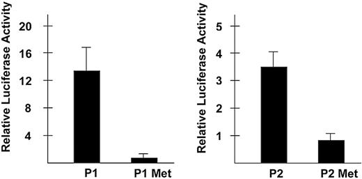
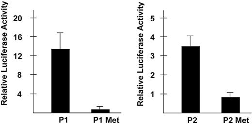
The fact that the 5′ proximal region of the AC133 gene is located within a CpG island suggests a potential role of methylation in its transcriptional regulation. To examine the effect of methylation on AC133 promoter activity, we methylated P1 (–1100/+10) and P2 constructs with SssI methylase in vitro, and measured the luciferase activity of SssI methylated and mock methylated constructs. pGL3-control plasmid, which contains the SV40 promoter, was both mock methylated and methylated and was included in the experiment as a negative control. Methylation completely suppresses the activity of AC133 promoters P1 and P2 in the Caco-2 cell line (Figure 5).
Effect of in vitro methylation on the activity of human AC133 promoters. AC133 promoter 1 (–1100/+10) and promoter 2 luciferase constructs were mock methylated or methylated with SssI methylase. Both methylated and mock-methylated constructs were transiently transfected into the Caco-2 cell line, and luciferase activity was measured and normalized to the activity of the methylated and mock-methylated pGL3-enhancer vector, respectively. All data represent the mean of 3 independent experiments. Error bars represent standard deviation.
Effect of in vitro methylation on the activity of human AC133 promoters. AC133 promoter 1 (–1100/+10) and promoter 2 luciferase constructs were mock methylated or methylated with SssI methylase. Both methylated and mock-methylated constructs were transiently transfected into the Caco-2 cell line, and luciferase activity was measured and normalized to the activity of the methylated and mock-methylated pGL3-enhancer vector, respectively. All data represent the mean of 3 independent experiments. Error bars represent standard deviation.
Discussion
In the present study we have identified and characterized the AC133 (human prominin-1) 5′-UTR and promoter regions. We showed that the structure of the human AC133 gene is more complex than was previously reported. Spanning over 152 kb on chromosome 4, the AC133 gene contains at least 37 exons, with translation initiation sites positioned within exon 2 (previously assigned as exon 1). We also discovered 4 alternative AC133 promoters and demonstrated the activity of 2 of them.
Recently, a novel alternatively spliced variant of human AC133, AC133-2, was reported.10 This novel isoform lacks exon 4 (previously assigned as exon 3), which affects the AC133 open reading frame (ORF) by deletion of 9 amino acids. Furthermore, we showed that additional alternative splicing isoforms exist within the ORF of human AC133 (S.V.S., unpublished data, June 2003).
In addition, we showed that AC133 transcripts can be alternatively spliced, resulting in the formation of mRNAs with different 5′-UTR exons. Nine distinct exons (1A, 1B, D1-3, and E1-4) were found by 5′-RACE, and one additional exon (exon 1C) was discovered by analysis of ESTs for human AC133. We showed the existence of at least 7 alternatively spliced forms (A, B, D1, D1-D2, D1-D2-D3, E1-E3-E4, and E1-E2-E3-E4; Figure 1A). In agreement with these data, we found that the mouse AC133 homologue prominin has 4 alternative 5′-UTR first exons (S.V.S., unpublished data, June 2003). Furthermore, our data suggest that at least 4 promoters are involved in the transcriptional regulation of the AC133 gene. Although we could not detect exon 1C–containing transcripts in the analyzed tissues, and promoter P3 was not active in the NT-2 and Caco-2 cell lines, it is possible that potential promoter P3 could still be involved in the transcriptional regulation of AC133 in other cell types. Exon clusters 1D and 1E were found exclusively in testis. We were unable to detect these alternatively spliced forms in Caco-2 and NT-2 cells. We also could not detect activity of corresponding promoters P4 and P5 in these cell lines. Demonstration of the activity of promoters P4 and P5 in a tissue-specific manner should be a subject of future studies.
It was demonstrated that the AC133-2 transcript expression profile is different from that of the AC133-1 isoform, and that the same organs may express different isoforms in fetuses and adults.10 In addition, we found a tissue-dependent pattern of expression of 1A-, 1B-, and 1D-containing isoforms of AC133 mRNA.
Regulation of certain genes by alternative promoters has previously been reported.11 It is known that alternative noncoding leader exons may play a role in the regulation of the turnover or translation efficiency of mRNA isoforms.11 We believe that alternative AC133 promoters, through inclusion of different 5′-UTRs, might also affect overall alternative splicing and cause the formation of specific isoforms of AC133 mRNA.
Our assessment of AC133 promoter activity using a luciferase reporter system revealed its relatively low activity without additional enhancer elements. We could not detect a significant level of promoter activity using pGL3-basic vector, which contains neither SV40 promoter nor enhancer. Therefore, we used pGL3-enhancer vector (containing SV40 enhancer) as a backbone for the reporter constructs in all our studies. Consequently, we suspect that additional regulatory elements play a crucial role in the proper functioning of the AC133 promoter.
AC133 first exons 1A, 1B, and 1C and their promoters are found within a CpG island, suggesting that methylation plays a role in the regulation of AC133 transcriptional activity. Our data showing the complete suppression of AC133 promoter activity in vitro with a methylated reporter construct support this hypothesis. More extensive research should be performed to understand the importance of methylation in the regulation of AC133 expression in vivo.
Several cell lines, including NT-2 and Caco-2, were previously reported to be AC133 positive by fluorescence-activated cell sorting (FACS) analysis.3,4 To identify a potential negative cell line for our promoter study, we analyzed more than 20 cell lines by RT-PCR and flow cytometry. To our surprise, we found all cell lines to be positive for AC133 by RT-PCR, even those which were negative by flow cytometry (data not shown). In fact, using RT-PCR we detected AC133 expression in most adult human tissues and cell types.
When discovered, the AC133 antigen was thought to be a hematopoietic stem cell–specific marker. Indeed, the AC133 monoclonal antibody recognizes only a subpopulation of CD34+ hematopoietic stem cells, and no immunoreactivity was found in other tissues, despite the detection of AC133 mRNA by RT-PCR and Northern blot.3,12 This finding could be explained by the fact that AC133 is a glycoprotein: it has been reported that the AC133 monoclonal antibody is glycosylation dependent, and glycosylation of the A133 protein may vary in different tissues.2
However, there is another explanation for the discordance between antibody staining data and RT-PCR data. It is possible that the AC133 monoclonal antibody recognizes only one of the possible AC133 isoforms resulting from alternative splicing. The reported finding that the AC133 monoclonal antibody predominantly recognizes AC133-2 supports this hypothesis.10 Previously, we suggested that expression of AC133-2 or other isoforms of AC133 might be regulated by a particular alternative promoter. Thus, it is possible that only one of the alternative AC133 promoters regulates the transcription of a stem cell–specific isoform.
We showed that CD34+ hematopoietic stem cells utilize only exon 1A–containing AC133 mRNA isoforms. Therefore, AC133 promoter P1 may play a key role in the regulation of AC133 expression on CD34+ cells. Considering this, and the fact that expression of AC133 in hematopoietic system is restricted to CD34+ cells, our finding will allow us to follow the fate of CD34+ hematopoietic stem cells in vivo. Identification of a stem cell–specific isoform of AC133 and its concurrent regulatory promoter will ultimately set the stage for the isolation of organ-specific stem cells through the use of AC133 promoter reporter constructs.
Prepublished online as Blood First Edition Paper, November 20, 2003; DOI 10.1182/blood-2003-06-1881.
The publication costs of this article were defrayed in part by page charge payment. Therefore, and solely to indicate this fact, this article is hereby marked “advertisement” in accordance with 18 U.S.C. section 1734.

