Mutations of the GATA1 gene, which is located on chromosome X, have been found in almost all cases of transient myeloproliferative disorder (TMD) and acute megakaryoblastic leukemia (AMKL) accompanying Down syndrome (DS).1-6 No GATA1 mutations have been detected in patients with AMKL who did not have DS, except in AMKL with acquired trisomy 21.1,4,6 However, because only 10 cases with AMKL in non-DS have been analyzed for mutations of the GATA1 gene, it remains unknown whether the GATA1 mutation is exclusively involved in the development of AMKL with DS. Here, we report for the first time a GATA1 mutation in AMKL cells from a patient who did not have DS or acquired trisomy 21.
In October 2002, a 48-year-old woman was admitted to Furukawa City Hospital with complaints of shortness of breath and a bleeding tendency. Peripheral blood analysis showed severe anemia, thrombocytopenia, and mild leukocytosis with the appearance of immature cells: red blood cell count, 87 × 1012/L; hemoglobin, 27 g/L (2.7 g/dL); platelet count, 6 × 10⁹/L; and white blood cell count, 10.8 × 10⁹/L (neutrophils, 29%; basophils, 1%; lymphocytes, 46%; blasts, 16%; metamyelocytes, 2%; myelocytes, 3%; and promyelocytes, 3%). Pathologic examination of her bone marrow showed a mixture of dominantly proliferated small lymphocytic cells, which were positive for leukocyte common antigen (LCA) and CD79α, and scattered blastic cells, which were negative for myeloperoxidase. Chromosomal analysis revealed 47XX, add(17)(p11), +add(17), add(20)(q13). The patient was diagnosed with acute lymphoblastic leukemia, chemotherapy was given, and complete remission was achieved. In February 2003, the disease relapsed and became refractory to chemotherapy. The morphology of leukemic cells at the relapse resembled that of small megakaryocytes, and they were negative for myeloperoxidase and positive for acid phosphatase. Immunohistochemical examination revealed that the leukemic cells were positive for CD33, factor VIII, and CD41a, and dull-positive for CD4 and CD45. In addition, flow cytometric analysis using an anti–GATA-1 antibody revealed that the leukemic cells were positive for GATA-1.7 The same chromosomal anomaly as that detected at onset was detected by chromosomal analysis. Based on these results, she was finally diagnosed as having AMKL.
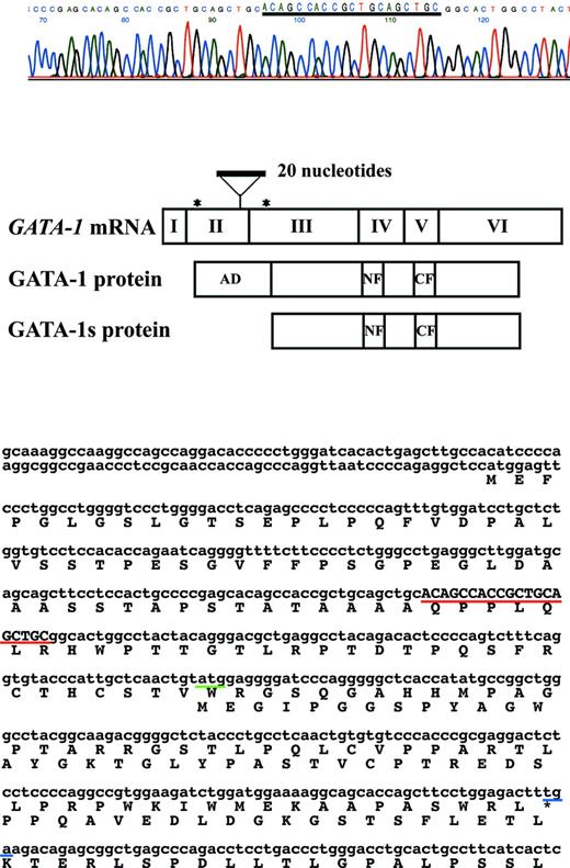
We analyzed the GATA1 mutation in her bone marrow samples. Written informed consent was obtained from the patient. Genomic DNA was extracted and cDNA was constructed, and then polymerase chain reaction was performed and the products were sequenced directly, as described previously.6 An insertion of 20 nucleotides corresponding to a sequence in exon 2 of the GATA1 gene was detected, resulting in the introduction of a premature stop codon in the gene sequence encoding the N-terminal activation domain (Figure 1).
GATA1 mutation in an adult patient with AMKL not accompanying DS. Direct sequence analysis of cDNA and genomic DNA from AMKL blast cells of the adult patient who did not have accompanying DS showed that 20 nucleotides of the duplicated sequence were inserted in exon 2 of the GATA1 gene. The mutation resulted in the introduction of a premature stop codon in the gene sequence encoding the N-terminal activation domain. The predicted protein from a downstream initiation site, GATA-1s, lacks the transactivation domain.
GATA1 mutation in an adult patient with AMKL not accompanying DS. Direct sequence analysis of cDNA and genomic DNA from AMKL blast cells of the adult patient who did not have accompanying DS showed that 20 nucleotides of the duplicated sequence were inserted in exon 2 of the GATA1 gene. The mutation resulted in the introduction of a premature stop codon in the gene sequence encoding the N-terminal activation domain. The predicted protein from a downstream initiation site, GATA-1s, lacks the transactivation domain.
Our results suggest that GATA1 mutations play an important role in the development of AMKL in non-DS individuals. Our discovery warrants further investigation in a larger series of non-DS patients with AMKL.


