Abstract
An understanding of the number and contribution of individual pluripotent hematopoietic stem cells (HSCs) to the formation of blood lineages has important clinical implications for gene therapy and stem cell transplantation. We have been able to efficiently mark rhesus macaque long-term repopulating stem and progenitor cells with retroviral vectors, and track their in vivo contributions to hematopoiesis using the linear amplification mediated–polymerase chain reaction (LAM-PCR) technique of insertion site analysis. We assessed the impact of busulfan on contributions of individual retrovirally marked clones to hematopoiesis. There were 2 macaques that received transplants of retrovirally transduced CD34+ cells 2 years previously that were then treated with 4 mg/kg busulfan. Despite only transient and mild suppression of peripheral blood counts, the numbers of individual stem/progenitor clones contributing to granulocyte production decreased dramatically, by 80% in the first monkey and by 60% in the second monkey. A similar impact was seen on clones contributing to T cells. The clone numbers recovered gradually back toward baseline by 5 months following busulfan in the first monkey and by 3 months in the second monkey, and have remained stable for more than one year in both animals. Tracking of individual clones with insertion-site–specific primers suggested that clones contributing to hematopoiesis prior to busulfan accounted for the majority of this recovery, but that some previously undetected clones began to contribute during this recovery phase. These results indicate that even low-dose busulfan significantly affects stem and progenitor cell dynamics. The clonal diversity of hematopoiesis was significantly decreased after even a single, clinically well-tolerated dose of busulfan, with slow but almost complete recovery over the next several months, suggesting that true long-term repopulating stem cells were not permanently deleted. However, the prolonged period of suppression of many clones suggests that transplanted HSCs may have a marked competitive advantage if they can engraft and proliferate during this time period, and supports the use of this agent in nonmyeloablative regimens
Introduction
Blood cell production is maintained by the differentiation and proliferation of progenitor cells derived from hematopoietic stem cells (HSCs). HSCs are used for many clinical applications including autologous or allogeneic transplantation, gene therapy for acquired or genetic diseases, and regenerative medicine. However, little is known about human or large animal hematopoietic stem cell behavior in steady state or following cytotoxic perturbations. Previously, the analysis of stem and progenitor cell dynamics in vivo, particularly in large-animal models with relevance to humans, has been difficult because the clonal output of individual stem and progenitor cells could be assessed only indirectly, via analysis of X-chromosome inactivation patterns in hematopoietic cells from heterozygous females.1,2 In contrast, marking of hematopoietic stem and progenitor cells with retroviral vectors can identify and track the clonal output of individual hematopoietic precursor cells in vivo, following transplantation of transduced cells.3,4 Retrovirus marking is an ideal tool for studies of hematopoiesis in vivo because these vectors insert semirandomly and permanently into the host cell genome, and thus the vector-genomic DNA junction serves as a unique marker of the initially transduced stem or progenitor cell and all its progeny.5
We have been able to efficiently mark rhesus macaque long-term repopulating stem and progenitor cells with retroviral vectors, and track their in vivo contributions to hematopoiesis using the newly described efficient and sensitive linear amplification mediated–polymerase chain reaction (LAM-PCR) technique to track contributions of individual clones.6 We previously reported that nonhuman primate hematopoiesis with marked cells is highly polyclonal and stable, with individual long-lived retrovirally transduced clones contributing to the myeloid and lymphoid lineages for more than 4 years at most recent follow-up.7
Busulfan is an alkylating agent with probable potent effects on primitive hematopoietic cells, given its known activity and potent myelosuppressive side effects resulting from killing of malignant and normal hematopoietic cells. High-dose busulfan has been extensively used in preparatory regimen for allogeneic hematopoietic stem cell transplantation. Recently, lower doses of busulfan have been incorporated into preparatory regimens for hematopoietic stem cell transplantation and autologous HSC gene therapy protocols, based on its lack of toxicity to other organs at these doses and known stem cell toxicity in murine models.8 In mice, busulfan induces marrow failure with repeated moderate doses, and this effect continues long term.9 After short-term treatment with busulfan, mice recover hematologically, only to die of bone marrow hypoplasia some time later. The overt bone marrow aplasia was preceded by a phase of latent aplasia, characterized by normal mature hematopoietic compartments but progressively shrinking stem cell pools.10,11
Little is known about the effect of busulfan on human or large animal hematopoietic stem and progenitor cell activity because of the difficulties in analyzing these effects in vivo in large animals. We have now used our retroviral tracking approach to assess the impact of low-dose busulfan on contributions of individual retrovirally marked stem and progenitor clones to hematopoiesis. Results from these studies provide insights that should both help in the design of conditioning regimens for transplantation and gene therapy, as well as provide clues as to the mechanism of secondary leukemias and myelodysplasia following alkylating agents such as busulfan.
Materials and methods
Rhesus macaque transplantation model
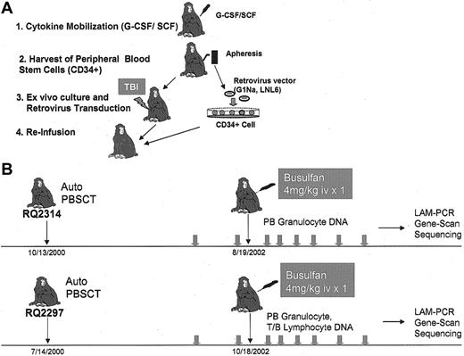
Young rhesus macaques (Macaca mulatta) were housed and handled in accordance with the guidelines set by the Committee on Care and Use of Laboratory Animals of the Institute of Laboratory Animal Resources, National Research Council,12 and the protocol was approved by the Animal Care and Use Committee of the National Heart, Lung, and Blood Institute. The collection of stem cell factor (SCF)/granulocyte colony-stimulating factor (G-CSF)–mobilized peripheral blood (PB) cells, purification, and retroviral transduction of CD34-enriched mobilized peripheral blood cells has been described in detail previously for the 2 animals included in this study.13,14 Both animals received cells transduced with the retroviral vectors LNL6 and G1Na, containing the neomycin resistance gene, for 4 days in the presence of stem cell factor (SCF), fms-like tyrosine kinase-3 ligand (Flt-3L), and megakaryocyte growth and development factor (MGDF) on fibronectin CH-296 fragment, and reinfused after 1000 cGy of total body irradiation (TBI) (Figure 1A).
Experimental design. (A) Autologous transplantation of retrovirally transduced CD34+ cells. Animals were given G-CSF + SCF for 5 days to mobilize CD34+ cells into the peripheral blood, and the cells were collected by leukapheresis. Purified CD34+ cells were transduced with either G1Na or LNL6 retroviral marking vectors.15 (B) Transduced CD34+ cells were reinfused into the monkeys after 500 cGy = 2 TBI. Animals were then followed after engraftment for at least one year before entry into the current studies. Each animal was treated with 4 mg/kg busulfan intravenously. Granulocytes and lymphocytes were collected at multiple time points before and after busulfan, and used for LAM-PCR analysis. iv indicates intravenously.
Experimental design. (A) Autologous transplantation of retrovirally transduced CD34+ cells. Animals were given G-CSF + SCF for 5 days to mobilize CD34+ cells into the peripheral blood, and the cells were collected by leukapheresis. Purified CD34+ cells were transduced with either G1Na or LNL6 retroviral marking vectors.15 (B) Transduced CD34+ cells were reinfused into the monkeys after 500 cGy = 2 TBI. Animals were then followed after engraftment for at least one year before entry into the current studies. Each animal was treated with 4 mg/kg busulfan intravenously. Granulocytes and lymphocytes were collected at multiple time points before and after busulfan, and used for LAM-PCR analysis. iv indicates intravenously.
Busulfan treatment
Animals RQ2314 and RQ2297 were treated intravenously with one dose of 4 mg/kg busulfan 1 to 2 years following transplantation of retrovirally transduced cells. No additional supportive care was required following busulfan treatment at this dose.
Sample collection
PB samples were collected at 2 baseline time points (including the day of busulfan treatment), then every 2 weeks for one month, and then monthly. Mononuclear cells were isolated by density gradient centrifugation over lymphocyte separation medium, and granulocytes were obtained as previously described.16 Peripheral blood mononuclear cells of RQ2297 monkey were stained with fluorescein-conjugated anti-CD2 or phycoerythrin-conjugated anti-CD20 (Immunotech, Marseille, France) or with isotype controls, and positive cells were sorted using a Coulter Epics Elite instrument (Coulter, Hialeah, FL). Sorted populations had purities of more than 99%. Limiting numbers of dilutions of collected cell fractions were made and frozen as cell pellets for subsequent DNA extraction and LAM-PCR analysis.
Linear amplification mediated (LAM)–PCR analysis
Density gradient purification, antibody staining, fluorescence-activated cell sorting, and DNA extraction were performed as previously described.16 Genomic DNA was extracted using the QIAmp blood kit (Qiagen, Chatsworth, CA). The LAM-PCR procedure has been previously described, and only minor modifications were made in the procedure since the original publication17 (Figure 2). The genomic-proviral junction sequence was preamplified by repeated primer extension using 5 pmol of vector-specific, 5′-biotinylated primer LTRa (5′-TGCTTACCACAGATATCCTG-3′; IDT, Coralville, IA) with Taq polymerase (2.5 U; Qiagen) on each 100-ng DNA sample. Linear amplification (100 cycles) was performed with addition of fresh Taq polymerase (2.5 U) after 50 cycles. Selection of biotinylated extension products was performed using 200 μg magnetic beads according to the manufacturer's instructions (Dynal, Oslo, Norway). The samples were then incubated with Klenow polymerase (2U; Roche, Indianapolis, IN), deoxynucleoside triphosphates (dNTPs, 300 FM; Pharmacia, Uppsala, Sweden), and a random hexanucleotide mixture (Roche) in a volume of 20 μl for 1 hour at 37° C. The samples were washed on a magnetic particle concentrator (Dynal) and incubated with TasI endonuclease (4 U in 20 μl; Fermentas, Hanover, MD) for 1 hour at 65° C. After an additional wash step, 10 pmol of a double-stranded asymmetric linker cassette and T4 DNA Ligase (6U; New England Biolabs, Beverly, MA) were incubated with beads in a volume of 10 Fl at 16° C overnight. Denaturing was performed with 5 Fl of 0.1 N NaOH for 10 minutes at room temperature. Each ligation product was amplified with Taq polymerase (5U; Qiagen), 5 pmol of vector-specific primer LTR2 (5′-GGCCTTGATCTGAATTC-3; IDT), and linker cassette primer LC1 (5′-GACCCGGGAGATCTGAATTC-3; IDT) using the amplification conditions described above. Of each PCR product, 0.2% served as a template for a second, nested PCR with internal primers LTR3 (5′-TTCATGCCTTGCAAAATGGC-3′; IDT) and LC2 (5′-GATCTGAATTCAGTGGCACAG-3′; IDT) at identical conditions. Of this final product, 80% was separated on a Spreadex high-resolution gel (Elchrom Scientific, Cham, Switzerland) (Figure 1C). Specific DNA bands were excised and reamplified with primers LC3 (5′-AGTGGCACAGCAGTTAGG-3′; IDT) and LTR4 (5′-CCTTGCAAAATGGCGTTACT-3′; IDT) for a total of 45 cycles. PCR products were then cycle sequenced directly or after cloning into the TOPO TA cloning vector (Invitrogen, Carlsbad, CA)
Clonal tracking analysis via LAM-PCR and Gene Scan. (A) Outline of LAM-PCR methodology. (1) Linear PCR by repeated primer extension from a biotinylated oligonucleotide and capture of the DNA products with avidin-coated magnetic beads. (2) Double-stranded (ds) DNA synthesis by random hexanucleotide priming. (3) DNA restriction digestion with TasI. (4) Ligation of an oligonucleotide ligation cassette (LC) to the overhanging sequence at the TasI site. (5) Nested PCR amplifications using primer pairs LCI-LTRII, LCII-LTRIII. (B) Final PCR amplification using a fluorescent primer allows separation and precise sizing of LAM-PCR products via comparison to size standards (M) on an automated sequencer and analysis using GeneScan software. (C) Repetitive samples of granulocyte DNA (100 ng) from one animal. The absolute number of independent clones detected by performance of each replicate LAM-PCR procedure is shown. By the time 6 replicates are run, the absolute clone number detected begins to plateau, and by 15 replicates, essentially no additional clones are detected.
Clonal tracking analysis via LAM-PCR and Gene Scan. (A) Outline of LAM-PCR methodology. (1) Linear PCR by repeated primer extension from a biotinylated oligonucleotide and capture of the DNA products with avidin-coated magnetic beads. (2) Double-stranded (ds) DNA synthesis by random hexanucleotide priming. (3) DNA restriction digestion with TasI. (4) Ligation of an oligonucleotide ligation cassette (LC) to the overhanging sequence at the TasI site. (5) Nested PCR amplifications using primer pairs LCI-LTRII, LCII-LTRIII. (B) Final PCR amplification using a fluorescent primer allows separation and precise sizing of LAM-PCR products via comparison to size standards (M) on an automated sequencer and analysis using GeneScan software. (C) Repetitive samples of granulocyte DNA (100 ng) from one animal. The absolute number of independent clones detected by performance of each replicate LAM-PCR procedure is shown. By the time 6 replicates are run, the absolute clone number detected begins to plateau, and by 15 replicates, essentially no additional clones are detected.
For Gene Scan analysis of the LAM-PCR products, the LTRIII primers were replaced by fluorescence primers of the same sequence labeled at their 5′-end with 5-carboxyfluorescein (FAM or HEX). Aliquots of PCR products (2 μL) were mixed with loading buffer (12 μL formamide) and 0.5 μL of the internal size standard (GeneScan-1000; Applied Biosystems, Foster City, CA) to allow precise determination of the length of the amplified bands. After denaturation for 2 minutes at 90° C, the products were separated by size and analyzed by automatic fluorescence qualification and area under the curve intensity determination, using the computer program GeneScan 672 (ABI 373A; Applied Biosystems) (Figure 1C).
PCR tracking of individual clones
There were 7 primer sets designed to specifically amplify individual vector/flanking genomic DNA insertions from animal RQ2314. These primers were designed following amplification of granulocyte DNA via LAM-PCR, isolation and sequencing of individual bands, and confirmation of the validity of the band as an insertion site. Each insertion-specific rhesus genomic primer was used together with LTR primers IV and V to analyze genomic DNA (100 ng) isolated from granulocytes of RQ2314, and from another monkey (RQ2265) receiving cells transduced with the same vectors, as a negative control to prove specificity. A 35-cycle PCR reaction with annealing temperature of 56° C was performed. Of this product, 4% was used as a template for the second nested PCR. Except for a 60° C annealing temperature, PCR conditions were identical to the first PCR. The primers used were flanking sequence-specific primers no. 1212-II (5′-ATCCCAGCTACTTGAGAGGC-3′), no. 1247-I (5′-GACATCTTGGTACACGGCCA-3′), no. 1318-I (5′-TGCTTATTTTGCAGATGAAG-3′), no. 1304-I (5′-TTGTATTGCAGCAGTCTCCT-3′), no. 1337-I (5′-CAGGCTGAGAGCCATTCTGT-3′), no. 1315-I (5′-GGCAGGAGGATCACCTGAGCC-3′), or no. 1340-I (5′-GTCAACACACATTTATTGTGAC-3′), each paired with the LTR-specific primer LTR IV (5′-CCTTGCAAAATGGCGTTACT-3′) for the first PCR reaction and flanking sequence-specific internal primers no. 1212-III (5′-TGAGGCAGGAGAATCGCTTG-3′), no. 1247-II (5′-GACTGTGACAAGCCAACAGA-3′), no. 1318-II (5′-AAACTGAGGCTGCAGCTGAC-3′), no. 1304-II (5′-GATACATGAGGCTGGCAGTG-3′), no. 1337-II (5′-CATTATAGCCCCCAGAAAAC-3′), no. 1315-II (5′-GGGAAGTAAAGGCTGCAATGAGC-3′), or no. 1340-II (5′-GGATTCAGTCCATAGATTATG-3′), paired with the LTR-specific internal primer LTR V (5′-CAAACCTACAGGTGGGGTCT-3′) for the second nested PCR. The final PCR products were separated on 2% agarose gels.
Semiquantitative PCR analysis
Genomic DNA was extracted using QIAamp Blood Kit (Qiagen). The primers and conditions used for neo PCR and β-actin PCR have been previously described.18 All neo and β-actin PCR reactions were run under conditions optimized to yield linear results in the range of intensity of the in vivo samples. For the outer reaction, 100 to 200 ng DNA was used, and 18 to 20 cycles were performed for the inner reaction, based on the level of in vivo marking. For every PCR analysis, negative controls included DNA from normal rhesus PB samples extracted with the same methodology and a reagent control. Serial dilutions of G1Na DNA (containing 2 copies of integrated vector per cell) into normal rhesus PB DNA were used as positive controls for generating the control regression curve. Band intensity was quantitated using a PhosphorImager (Molecular Dynamics, Sunnyvale, CA). The neo band intensity was normalized for amplifiable DNA content based on the β-actin signal, and the overall contribution of each vector to in vivo marking was calculated by plotting the signal intensity of each band on a standard curve derived from known copy number controls amplified concurrently.
Statistical methods
The distribution of clones at each visit was described using means, medians, and histograms. To evaluate whether the clonal production was affected by intervention, Wilcoxon signed rank tests were used to compare clonal production between adjacent periods. To examine stability of clonal production, we calculated the rank correlation between detection counts on several pairs of adjacent visits before and after intervention. We tested the equality of these pairs of correlations using a bootstrap procedure.
Results
Study design
Animals RQ2314 and RQ2297 received transplants of SCF- and G-CSF–mobilized transduced CD34+ cells 2 years prior to busulfan treatment (Figure 1). Each animal had very stable marking levels in peripheral blood granulocytes and mononuclear cells, approximately 20% to 30% for animal RQ2314 and 15% to 25% for animal RQ2297. We initially used granulocyte DNA for analysis because granulocytes have a brief half-life in the circulation of 6 to 8 hours and a lifespan in tissues of 3 to 4 days; granulocytes have continual regeneration from primitive precursor cells in the bone marrow, unlike lymphocytes, which may persist for months or even years in the blood or secondary lymphoid organs. Analysis of clonal contributions to circulating granulocytes thus reflects ongoing production from progenitor and stem cells. Following collection of monthly steady-state baseline samples for 2 months in each animal, busulfan at a dose of 4 mg/kg was administered intravenously.
Impact of busulfan on peripheral blood cell counts
There was only transient and mild suppression of peripheral blood counts in both monkeys (Figure 3). The mean white blood cell (WBC) counts before busulfan treatment were 4.95 and 4.92 = 109/L in animals RQ2314 and RQ2297, respectively, falling to nadirs of 2.69 and 2.97 = 109/L 2 weeks following busulfan administration with recovery to baseline by 4 weeks after treatment. Platelets also transiently dropped, but did not fall to less than 200 = 109/L in either animal. Red cell counts were not significantly decreased with busulfan treatment. Neither animal required transfusions or antibiotics, and both remained completely well following this dose of 4 mg/kg busulfan. The animals have now been followed for more than 14 months following busulfan, and no late cytopenias have developed. The impact of 4 mg/kg busulfan on specific lymphoid populations was assessed in 2 independent animals. Peripheral blood CD4 levels fell 69% to 71% and CD8 cells fell 85% to 86%, with recovery by 6 weeks. Absolute B-cell numbers fell by 46% to 60% and recovered by 6 weeks.
Impact of busulfan on peripheral blood counts. (A) Animal RQ2314. (B) Animal RQ2297. RBC indicates red blood cell; PLT, platelet.
Impact of busulfan on peripheral blood counts. (A) Animal RQ2314. (B) Animal RQ2297. RBC indicates red blood cell; PLT, platelet.
Analysis of the number of clones contributing to granulocytopoiesis
In order to accurately quantitate the number of stem and progenitor cell clones contributing to granulocyte production via LAM-PCR amplification of vector insertion sites and Gene Scan identification of specific insertions by band size, we set criteria for identifying peaks on Gene Scan analysis based on reproducible visualization of the same LAM-PCR product bands on Spreadex gel electrophoresis. Peaks with an area under the curve of more than 10 000 and sizes between 100 and 400 base pairs (bp) under these conditions represented true vector insertion sites as confirmed by sequencing, and areas of the gels corresponding to peaks with areas under this value did not reliably represent actual bands on gels, or vector insertions following sequencing of the region excised from the gel (data not shown). Clones were defined as unique band lengths in each animal.
Prior analysis has shown that as few as 5 copies of a single vector insertion can be detected by LAM-PCR within a 100-ng sample containing nontransduced cells or cells with different vector insertions. However, 100 ng DNA corresponds to only about 15 000 cells, and with an overall marking level of on average 10% containing as many as 50 to 100 transduced clones in our prior analysis of steady-state macaques, 100 ng DNA on average may not contain detectable contributions from each contributing clone.19 Increasing the amount of DNA per reaction results in a loss of efficiency and an increase in artifacts, thus we performed multiple replicates of 100-ng samples and found that by increasing the number of replicates, a more complete representation of all clones contributing to a sample could be obtained, with a maximum number of clones detected after approximately 10 to 15 replicates in different animals. Thus we used 6 replicates of 100 ng DNA from each sample to perform LAM-PCR, and each procedure was repeated independently 3 times for a total of 18 replicates to characterize the clones contributing at each time point (Figure 1). Only amplification products between 100 and 400 bp were scored; larger and smaller insertions are less efficiently and consistently amplified, and bands outside this size range are more frequently artifacts.
We analyzed samples before and following busulfan treatments in animals RQ2314 and RQ2297 (Figure 1). At baseline, a total of 83 and 79 clones was detected contributing to granulocytes in RQ2314 and RQ2297, respectively, on the day of busulfan treatment. The baseline values appeared stable, with no change in clone numbers and similar rates of clonal detection from individual clones, as assessed by comparing the mean number of detections per clone at each visit prior to busulfan (P values of .42 and .16, for RQ2314 and RQ2297, respectively). By one week following busulfan, despite the very minimal impact on peripheral blood counts, the number of contributing clones detected decreased dramatically, by 80% in RQ2314 and by 60% in RQ2297 (Figure 4), with P < .001 comparing pre- and postbusulfan clone numbers, with 3 replicates performed at each time point.
Summary of clone numbers identified by LAM-PCR and GeneScan analysis before and after busulfan. (A) Animal RQ2314. (B) Animal RQ2297. Criteria for scoring clones as present are given in “Materials and methods.” A total of 18 replicate LAM-PCR reactions using 100 ng template was performed for each sample, and the total number of individual clones meeting criteria was enumerated and graphed.
Summary of clone numbers identified by LAM-PCR and GeneScan analysis before and after busulfan. (A) Animal RQ2314. (B) Animal RQ2297. Criteria for scoring clones as present are given in “Materials and methods.” A total of 18 replicate LAM-PCR reactions using 100 ng template was performed for each sample, and the total number of individual clones meeting criteria was enumerated and graphed.
The overall level of marking was impacted by the treatment, suggesting no preferential effects of busulfan on transduced versus nontransduced progenitor or stem cells (Figure 5). The number of clones contributing to granulocytes gradually returned toward baseline by 5 months in RQ2314 and by 3 months in RQ2297. A rank correlation analysis for both animals indicated that clones with a high production rate (high frequency of detection out of 18 replicates at a time point) maintained the high production rate following recovery from busulfan, using a bootstrap correlation to compare pairs of pretreatment time points and pairs of posttreatment time points (all P > .05). We also analyzed clone numbers contributing to sorted B and T lymphocytes in animal RQ2297. There was a more pronounced impact on contributing clones in T cells than in B cells (Figure 4), but in both lineages the impact was less than on granulocytes. This was most likely due to survival of long-lived T and B cells following this dose of busulfan, with a gradual return of the diversity of clones contributing to T cells toward baseline, similar to the recovery of clones contributing to granulocytes.
PCR analysis of in vivo marking. Representative PCR of neo and β-actin sequences in PB granulocytes at different time points before and after busulfan treatment in each animal. Concurrent β-actin PCR was performed for each sample to quantitate the amount of amplifiable template DNA. Serial dilutions of G1Na DNA (2 copies of integrated vector per cell) into normal rhesus PB DNA were used as positive controls.
PCR analysis of in vivo marking. Representative PCR of neo and β-actin sequences in PB granulocytes at different time points before and after busulfan treatment in each animal. Concurrent β-actin PCR was performed for each sample to quantitate the amount of amplifiable template DNA. Serial dilutions of G1Na DNA (2 copies of integrated vector per cell) into normal rhesus PB DNA were used as positive controls.
Impact of busulfan on individual clones
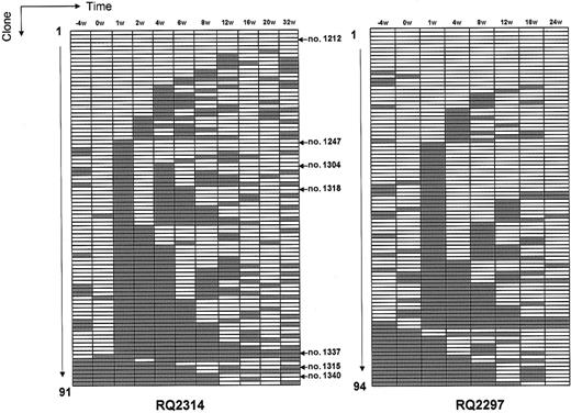
Next, we studied the behavior of individual clones in response to busulfan. Most important, we asked whether the clones appearing during recovery represented output from those clones that contributed to hematopoiesis before treatment, or if the original clones were permanently deleted and instead replaced by hematopoietic contributions from a set of previously inactive clones. Figure 6 summarizes the tracking of individual clones defined by the length of the insertion site isolated following LAM-PCR and sized via GeneScan analysis. As noted above, many individual clones became undetectable beginning 1 to 2 weeks after busulfan treatment. The majorities of these clones were again detected as contributing to granulopoiesis over months 2 to 5 following treatment, suggesting that they were not permanently deleted, but instead that primitive cells from each clone were quiescent at the time of busulfan treatment, and eventually activated to begin contributing randomly and gradually following recovery. A small number of clones detected prior to busulfan treatment was never again detected (2 clones in RQ2314, 3 clones in RQ2297) and could represent complete deletion of transduced clones. A minority of clones was detected for the first time following recovery from busulfan (7 clones in RQ2314, 15 clones in RQ2297).
Summary of individual clones contributing to granulocytes before and following busulfan. Each individual clone detected is shown on the y-axis. White boxes represent the presence of the clone at a time point, and gray boxes represent the lack of detection of a clone at a time point. The presence or absence of individual clones was confirmed using tracking primers as shown in Figure 7. These clones are identified by arrows to the right of the charts.
Summary of individual clones contributing to granulocytes before and following busulfan. Each individual clone detected is shown on the y-axis. White boxes represent the presence of the clone at a time point, and gray boxes represent the lack of detection of a clone at a time point. The presence or absence of individual clones was confirmed using tracking primers as shown in Figure 7. These clones are identified by arrows to the right of the charts.
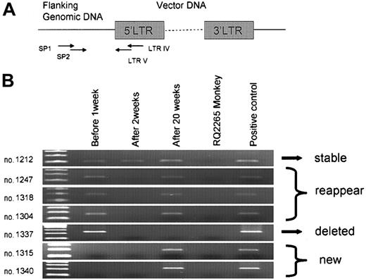
It was critical to confirm that the presence or absence of individual clones defined by sizing of the amplified insertion sites on Gene Scan analysis could be validated by using straightforward clone-specific amplification with a genomic primer designed specifically to amplify only an individual insertion. We designed clone-specific primers for a total of 7 insertion sites following sequencing of LAM-PCR bands in animal RQ2314, specifically choosing clones for this additional analysis that had either stable, deleted, or recruited patterns of contribution following busulfan. For each of these clones, identified in Figure 6, we confirmed the LAM-PCR results demonstrating either presence or absence of a clone contributing to granulocyte production at specific time points (Figure 7).
In vivo clone tracking by conventional PCR. (A) For each individual clone, clone-specific primers (SP1 and SP2) were designed to anneal to genomic sequences 5′ to the LTR insertion, after band isolation and sequencing of LAM-PCR products. Each set of primers was used for nested PCR, in combination with LTR IV and V primers. (B) DNA isolated from granulocytes before (0 week), and 1 and 20 weeks after busulfan treatment of RQ2314 analyzed using clone-specific primers for 7 individual insertions (nos. 1212, 1247, 1318, 1304, 1337, 1315, and 1340). RQ2265 granulocyte DNA was used as negative control; this animal received transduced cells but should not have any insertions identical to animal RQ2314. Cloned plasmid DNA from each insertion was used as a positive control.
In vivo clone tracking by conventional PCR. (A) For each individual clone, clone-specific primers (SP1 and SP2) were designed to anneal to genomic sequences 5′ to the LTR insertion, after band isolation and sequencing of LAM-PCR products. Each set of primers was used for nested PCR, in combination with LTR IV and V primers. (B) DNA isolated from granulocytes before (0 week), and 1 and 20 weeks after busulfan treatment of RQ2314 analyzed using clone-specific primers for 7 individual insertions (nos. 1212, 1247, 1318, 1304, 1337, 1315, and 1340). RQ2265 granulocyte DNA was used as negative control; this animal received transduced cells but should not have any insertions identical to animal RQ2314. Cloned plasmid DNA from each insertion was used as a positive control.
Discussion
Busulfan is an alkylating agent with myelosuppressive effects, and in contrast to other alkylating agents such as cyclophosphamide or nitrogen mustard, busulfan exhibits pronounced activity against noncycling cells.19 The in vitro toxicity of busulfan toward subsets of hematopoietic progenitors defined by their lineage commitment and differentiation stage has been reported. These studies confirmed that busulfan, unlike most other cytotoxic chemotherapy agents, can kill quiescent hematopoietic progenitors.20,21 High doses of busulfan are myeloablative, and in combination with immunosuppressive agents, busulfan conditioning results in complete replacement of host hematopoiesis following allogeneic transplantation.22 In rodent models, even if stem cells survive high-dose busulfan, their proliferative potential is permanently impaired, as evidenced by poor serial transplantation activity and prolonged diminution of colony-forming unit–spleen (CFU-S) frequency.23,24 However, these studies were not able to track the activity of individual stem cell clones, thus actual deletion versus damage of proliferative output of HSCs was not documented.
Much lower chronic dosing of busulfan began to be commonly used to treat chronic myeloid leukemia (CML) during the 1960s and 1970s. This regimen was effective for controlling chronic-phase blood counts, but after prolonged treatment patients regularly developed marked pancytopenia, accompanied by marrow hypocellularity, suggesting toxicity of this agent against stem cells even in the absence of profound prior myelosuppression.25 Repeated doses of busulfan, given weekly to mice and resulting in only moderate peripheral blood cytopenias, produced significant loss of stem cell activity in later repopulation assays, despite complete recovery of peripheral blood counts by the time marrow was harvested for secondary transplantations.26 Mice that survived multiple moderate doses of busulfan with complete blood count recovery developed marrow failure months later, primarily with hypocellular marrows,9 also suggesting an effect on very primitive cells independent from killing of proliferating committed progenitors.
Abkowitz et al reported the effect of busulfan on stem cell kinetics in a feline model by analyzing glucose-6-phosphate dehydrogenase (G6PD) expression in hematopoietic cells in female cats heterozygous for this X chromosome allele.27-29 Following busulfan administration at doses of 2 to 4 mg/kg, repeated 3 times every 2 to 5 weeks, the cats had transient pancytopenia, and upon recovery had marked skewing toward one G6PD allele in both marrow progenitors and mature blood cells. The authors concluded that busulfan was a potent stem cell toxin, with permanent deletion or damage to significant numbers of clones, since a long-term follow-up study demonstrated continued skewing and highly variable patterns over time in individual animals, suggesting very few contributing clones. However, all animals skewed toward the same G6PD allele, and cats followed without busulfan treatment also skewed toward the same allele, suggesting the presence of an X-chromosome gene that confers a selective advantage to stem cells and/or their progeny over time, accelerated by the stress of recovery from busulfan. Conclusions regarding stem cell clone numbers following busulfan based on this approach may not be completely valid, and could account for some differences with our data in macaques, where we have not yet seen frequent permanent clonal deletion with similar doses.
Preparative regimens for HSCs were originally designed with 3 goals: to produce sufficient immune suppression to prevent rejection of allogeneic marrow cells, to kill residual tumor cells in the setting of malignancy, and to provide marrow “space” allowing engraftment and proliferation of donor stem cells. Subsequent studies using little or no ablation and very high transplanted marrow doses indicate that actual marrow space is less important than competition between endogenous and transplanted stem cells. If enough stem cells are given, autologous or syngeneic engraftment can be achieved without any conditioning, and even small doses of irradiation, with little impact on blood counts, can favor engraftment of donor HSCs.30 Recently, single doses or a short course of moderate-dose busulfan has been incorporated into preparatory regimens prior to “nonablative” allogeneic hematopoietic stem cell (HSC) transplantations.31 However, interpretation of the impact of busulfan on endogenous human stem cells at these dosages is clouded by the allogeneic graft-versus-marrow effect that clearly has a major role in establishing full hematopoietic chimerism in this setting.
Aiuti et al gave a single dose of 4 mg/kg busulfan to children with severe combined immunodeficiency (SCID) due to adenosine deaminase deficiency as conditioning prior to reinfusion of genetically corrected autologous bone marrow CD34+ cells.32 Despite only moderate and transient suppression of peripheral blood counts, these patients engrafted genetically corrected cells, with eventual 100% replacement of T cells due to in vivo selective survival and expansion of this deficient lineage. However, even myeloid and B lymphoid engraftment of corrected cells could be detected at significant levels of approximately 1%, suggesting that this dose of busulfan impacted significantly on the ability of endogenous HSCs to compete with infused transduced cells. This study provided the most direct evidence that busulfan could be a very potent conditioning agent for transplantation with significant impact on primitive human hematopoietic cells, at doses associated with little toxicity.
In our current study, we use a novel genetic tracking approach to explore the actual clonal dynamics of in vivo hematopoiesis in response to nonmyeloablative doses of busulfan, not possible in the human transplantation studies. The nonhuman primate model we used has direct relevance and predictive value for human transplantation applications.33 Identification of proviral-genomic junctions allowed tracking of output from individual stem and progenitor cell clones, previously possible in large animals and humans only at a population level by measuring the expression of X-linked polymorphic genes such as G6PD in hematopoietic cells.34,35
In rhesus macaques treated with a single 4-mg/kg dose of busulfan, there was a striking impact on the pattern of clonal contributions to circulating granulocytes. Almost immediately following busulfan administration, the output from 60% to 80% clones was decreased dramatically, to below the level of detection, despite only transient and relatively mild suppression of blood counts. Since both animals had previously undergone a myeloablative conditioning with high-dose total body irradiation, followed by rescue with autologous genetically marked CD34+ cells, their hematopoietic reserve may not have been completely normal, even more than 2 years following transplantation. Therefore, higher doses of busulfan might be required for a similar impact in animals or patients not previously subjected to a hematopoietic stress. Interestingly, even after peripheral blood counts recovered over the next 2 weeks, the number of clones contributing to granulocytes was still markedly decreased. This means that a reduced fraction of primitive stem/progenitor cell clones was contributing to hematopoiesis after busulfan treatment and that only a subset of clones maintained activity in the recovery period. However, we were surprised to then discover that, at most, only a minor subset of clones appeared to be completely deleted, with eventual recovery of contributions from almost all the original clones again, as detected by LAM-PCR and confirmed by PCR using primers specific to individual clones. These findings suggest that a very quiescent primitive cell or cells from each clone survived busulfan treatment, but required a prolonged period of months to again begin to contribute to hematopoiesis. More than one year following busulfan, both animals maintain relatively stable clonal diversity. Longer follow-up is necessary to determine if busulfan-related damage will eventually result in permanent clonal loss. Very few completely new clones were detected at recovery or thereafter, indicating that there does not appear to be a “reserve” of noncontributing clones present in a steady state that are recruited only following stress; this also supports other evidence that all stem cells are active (even if slowly cycling) even in steady state and conflicts with models of continuous hematopoietic “clonal succession.”36
It is conceivable that clones remaining active during the immediate recovery period could be susceptible to secondary mutational events, with eventual progression to secondary myelodysplasia or acute leukemia. These hematologic disorders are known to occur frequently following exposure to alkylating agents, particularly when administered repeatedly.37 In patients who have recieved an alkylating agent as part of combination chemotherapy for Hodgkin or non-Hodgkin lymphomas, the incidence of secondary acute myeloid leukemia (AML) may be as high as 5% to 10%.38 Secondary leukemias have also followed use of alkylating agent therapy for multiple myeloma and ovarian carcinoma.39 Patients treated with alkylating agents for benign diseases also have an increased risk of secondary leukemia, such as patients with nephritis, lupus, psoriasis, rheumatoid arthritis, and Wegener granulomatosis.40,41 However, it is not clear how busulfan or other alkylating agents result in leukemogenesis in vivo. Does progression to clonal hematopoiesis and leukemia depend on prior clonal depletion and increased proliferative stress and associated secondary mutations in remaining clones, or is the primary mechanism directly related only to mutational events in individual cells resulting in abnormal expansion of the clone and risk of additional mutational events? As a first step to answer these questions, in our study we asked whether busulfan actually deletes primitive hematopoietic clones completely or alternatively whether busulfan instead can result in nonlethal mutational events followed by eventual dominance of an aberrant clone, without prior oligoclonality or clonal depletion. Much longer follow-up of these animals is required to answer these questions, along with chronic administration of busulfan to another cohort in an attempt to produce more profound and permanent stem cell damage.
We have for the first time been able to follow the behavior of individual stem and progenitor cell clones in response to a clinically relevant perturbation in vivo. Our results indicate that even low-dose busulfan significantly affects hematopoietic stem and progenitor cell clonal dynamics, although in the current study, the clonal diversity recovered several months following busulfan administration. The potency of moderate-dose busulfan to facilitate engraftment of transplanted HSCs may relate to the drug resulting in an environment for engraftment characterized by inactivity of primitive endogenous cells, allowing initial expansion of infused cells due to availability of niches or microenvironmental signals. Stem cells infused at the time of decreased contributions from most endogenous clones could expand and have a marked competitive advantage. Analysis of B-cell and T-cell compartment in this study revealed that not only the short-lived granulocyte compartment, but also lymphocyte compartments, particularly T cells, had reduced numbers of clones contributing after busulfan treatment. This depression of lymphocyte clone number might reflect effects on either the pluripotent stem cell compartment or a long-lived committed lymphoid progenitor. These and similar investigations in large animals should continue to provide insights into the potential clinical use of busulfan for gene therapy and stem cell transplantation applications, and also provide insights into the pathogenesis of secondary malignant disorders such as AML or myelodysplastic syndrome.
Prepublished online as Blood First Edition Paper, May 4, 2004; DOI 10.1182/blood-2003-08-2935.
The publication costs of this article were defrayed in part by page charge payment. Therefore, and solely to indicate this fact, this article is hereby marked “advertisement” in accordance with 18 U.S.C. section 1734.
We thank Keyvan Keyvanfar and Karin Lore for assistance with flow cytometry and phenotypic analysis.


