Abstract
Factor XI deficiency (MIM 264900) is an autosomal bleeding disorder of variable severity. Inheritance is not completely recessive as heterozygotes may display a distinct, if mild, bleeding tendency. Recent studies have shown the causative mutations of factor XI deficiency, outside the Ashkenazi Jewish population, to be highly heterogeneous. We studied 39 consecutively referred patients with factor XI deficiency to identify the molecular defect. Conventional mutation screening failed to identify a causative mutation in 4 of the 39 patients. Epstein-Barr virus (EBV)–transformed cells from these 4 patients were converted from a diploid to haploid chromosome complement. Subsequent analysis showed that 2 of the patients had a large deletion, which was masked in the heterozygous state by the presence of a normal allele. We report here the first confirmed whole gene deletion as the causative mutation of factor XI deficiency, the result of unequal homologous recombination between flanking Alu repeat sequences.
Introduction
Factor XI is a serine protease zymogen involved in the consolidation phase of blood coagulation by way of a thrombin-generated feedback loop.1-3 The factor XI gene, located on the long arm of chromosome 4 (4q35),4 comprises 15 exons spread over approximately 23 kb.5 Mutations in the factor XI gene are responsible for a variable bleeding phenotype and, although 2 common point mutations exist in the Ashkenazi Jewish population,6 at least 34 different causative mutations, located throughout the gene, have been reported.7 Large deletions have so far not been reported in factor XI deficiency, although they have been in other hemostatic disorders, including hemophilia A and B, and protein C deficiency.7
Alu repeats are the most abundant class of interspersed repeat sequences, and it is estimated that there may be more than 1 million copies in the human genome,8 comprising 5% to 10% of the total genome. Alu elements are particularly common in the noncoding regions of genes, such as the introns and 5′ and 3′ untranslated regions (UTRs), and are acknowledged to contribute significantly to human disease by way of either insertional events (Alu retrotransposition) or unequal homologous recombination resulting in deletions.9,10 Alu repeats are believed to be involved in at least 0.3% to 0.4% of all human genetic disease.9
We studied 2 patients with factor XI deficiency in whom a causative mutation was not identified despite the sequencing of the entire coding region of the factor XI gene. Subsequent quantitative and semiquantitative polymerase chain reaction (PCR) and reverse-transcriptase (RT)–PCR analysis indicated a high probability that 2 of these patients were heterozygous for a large deletion. Epstein-Barr virus (EBV)–transformed lymphocytes from these 2 patients were converted from the diploid to the haploid11,12 state to allow us to study the large deletion(s) further, in the absence of a complicating normal allele.
Study design
Patients
Patients were diagnosed as factor XI deficient as a result of 2 or more consecutive low factor XI:C levels (mean factor XI [FXI] activity levels of 32.1 IU/dL and 37.6 IU/dL, respectively), following exhaustive investigation to exclude any other coagulopathy.
Molecular analysis
Molecular analysis of the factor XI gene was performed as previously described.13-15
EBV-transformed cell lines
Patient blood samples were sent to the European Collection of Animal Cell Cultures (ECACC; Porton Down, United Kingdom) for Epstein-Barr virus (EBV) transformation and establishment of immortalized lymphoblastoid cell lines.
Diploid to haploid conversion
EBV–transformed lymphocytes were sent to GMP Genetics Inc (Fort Lauderdale, FL) where they were electrofused with a recipient rodent cell line (E2). The resulting hybrids were screened, using cytogenetic and molecular genetic techniques, to identify those containing a single copy of human chromosome 4.
Haplotype analysis
Haplotype analysis was performed by using chromosome 4–specific markers from the Linkage Mapping Set v2.5 (Applied Biosystems, Foster City, CA).
PCR amplification across the deletion
Reactions (50 μL) contained 50 mM KCl, 10 mM Tris (tris(hydroxymethyl) aminomethane)–HCl (pH 9 at 25° C), 0.1% Triton X-100, 1.5 mM MgCl2, 0.2 mM dNTPs (deoxynucleoside triphosphates), 500 nM primers, 1.5 units of Taq DNA Polymerase (Promega, Madison WI), and 100 ng DNA. The primer sequences were 5′-TCCGGTTGCTTCTCACTAGGA-3′ forward and 5′-CGAGTTCTGCCAACACCGA-3′ reverse. The amplification reaction included 1 cycle at 92° C for 2 minutes, 35 cycles of 94° C for 35 seconds, 55° C for 40 seconds, and 72° C for 120 seconds, and a final extension at 72° C for 6 minutes.
Results and discussion
Earlier quantitative and semiquantitative analysis had indicated the probable deletion of the entire factor XI gene from one allele in the 2 patients under investigation. Analysis of the haploid hybrids confirmed this, as the “deleted hybrid” failed to amplify any part of the factor XI gene. To define the extent of the deletion a series of PCR primer sets, at various distances 5′ and 3′ of the factor XI gene, were used to amplify DNA from the haploid hybrids. Primer sets were designed by using the sequence from a chromosome 4 bacterial artificial chromosome (BAC) clone RP11-173M11 (GenBank AC110771). Primer sets approximately 15 kb, approximately 10 kb, and approximately 5 kb toward the centromere (5′ to the factor XI gene) all amplified from both allele 1 and allele 2 hybrids in both patients, whereas a primer set at approximately 2.5 kb would amplify only one hybrid for each of the 2 patients. This finding indicated that the deletion breakpoint must lie somewhere between approximately 2.5 kb and approximately 5 kb 5′ of the factor XI gene. Similar experiments 3′ or telomeric of the factor XI gene narrowed the deletion breakpoint to between approximately 4.3 kb and approximately 4.9 kb.
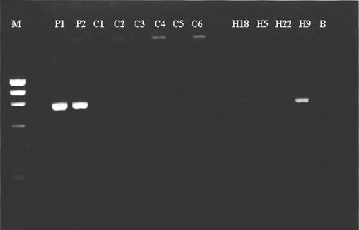
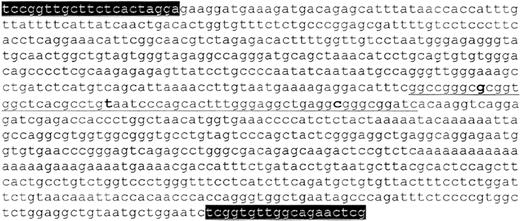
We were able to amplify across the deletion, achieving an 836-bp fragment (Figure 1). This fragment was amplifiable from the 2 deleted hybrids and genomic DNA from the 2 patients but not the normal hybrids or control DNAs. The 836-bp fragment was sequenced, and the resultant sequence contained 353 bp unique to the 5′ region and 421 bp unique to the 3′ region linked by a 62-bp consensus sequence (Figure 2). Both the 5′ and 3′ breakpoints were identified as Alu repeat sequences, one approximately 5.3 kb 5′ of the start of the factor XI signal peptide (codon –18, exon 2)5 and the other approximately 4.7 kb 3′ of the factor XI termination codon (codon 608, exon 15).5 The total size of the deletion on chromosome 4 was approximately 31.5 kb, including the entire factor XI gene. The precise breakpoints for the deletion have been narrowed to a 16-bp sequence, which shows 100% homology in the 2 Alu-repeat regions involved. We have also shown that this is a “true deletion” rather than a recombination, as both quantitative analysis and analysis of the haploid hybrids demonstrate that the “missing” factor XI gene is not located elsewhere within the genome. This finding strongly supports that this is an intrachromosomal recombination rather than one between 2 separate chromosomes.
Amplification of 836-bp fragment across the deletion. P1 and P2 are genomic DNA from the 2 patients, C1-C6 are control DNAs, H18 and H9 are haploid hybrids containing the deletion, H5 and H22 are the hybrids containing the undeleted allele, M is a PhiX174/HaeIII size marker, and B is the negative control.
Amplification of 836-bp fragment across the deletion. P1 and P2 are genomic DNA from the 2 patients, C1-C6 are control DNAs, H18 and H9 are haploid hybrids containing the deletion, H5 and H22 are the hybrids containing the undeleted allele, M is a PhiX174/HaeIII size marker, and B is the negative control.
Sequence across the approximate 31.5-kb deletion. The 62-bp consensus sequence is underlined, with the 3 variant bases in bold. The precise breakpoint lies somewhere in the 16 bp between the first and second variant bases. The consensus region is preceded by 353 bp of 5′ sequence and followed by 421 bp of 3′ sequence. PCR primer sequences are highlighted.
Sequence across the approximate 31.5-kb deletion. The 62-bp consensus sequence is underlined, with the 3 variant bases in bold. The precise breakpoint lies somewhere in the 16 bp between the first and second variant bases. The consensus region is preceded by 353 bp of 5′ sequence and followed by 421 bp of 3′ sequence. PCR primer sequences are highlighted.
Having identified the same large deletion in 2 unrelated patients, we undertook haplotype analysis to investigate whether the patients were ancestrally related. The deleted hybrids did not share haplotypes for the closest markers: D4S2924 (heterozygosity 0.72) located approximately 0.7 cM 5′ to the factor XI gene or D4S3051 (heterozygosity 0.51) located approximately 0.8 cM 3′ to the factor XI gene. This finding suggested that either the 2 patients are not related or that the deletion arose a sufficient number of generations ago to allow linkage with the flanking markers to be lost. Alu repeat regions are known to be hot spots for recombination events, and it is possible that, given the difficulty in detecting large deletions in the heterozygous state and the often mild or even subclinical presentation of heterozygous factor XI deficiency, large deletions are being underreported. The recent rapid increase in the number of non-Ashkenazi Jewish patients with factor XI deficiency undergoing molecular analysis should start to indicate whether a significant number may have a large or whole gene deletion. Suspected whole gene deletions are already being reported.16
Loss of the entire factor XI gene appears no more clinically significant than the presence of a single missense mutation. Both patients had typical heterozygote levels and a history of relatively mild bleeding complications. Paradoxically, it is feasible that certain missense mutations may result in a greater reduction in factor XI levels as a consequence of factor XI dimerization.17 The formation of heterodimers may reduce the amount of wild-type factor XI to nearer 25%, rather than the expected 50%, of normal levels. This again demonstrates the futility of attempting to correlate genetic lesion (alone) and phenotype in factor XI deficiency.
Prepublished online as Blood First Edition Paper, June 29, 2004; DOI 10.1182/blood-2004-04-1318.
The publication costs of this article were defrayed in part by page charge payment. Therefore, and solely to indicate this fact, this article is hereby marked “advertisement” in accordance with 18 U.S.C. section 1734.


