Abstract
In earlier work, we showed that adeno-associated virus–mediated delivery of a Factor IX gene to skeletal muscle by direct intramuscular injection resulted in therapeutic levels of circulating Factor IX in mice. However, achievement of target doses in humans proved impractical because of the large number of injections required. We used a novel intravascular delivery technique to achieve successful transduction of extensive areas of skeletal muscle in a large animal with hemophilia. We provide here the first report of long-term (> 3 years, with observation ongoing), robust Factor IX expression (circulating levels of 4%-14%) by muscle-directed gene transfer in a large animal, resulting in essentially complete correction of the bleeding disorder in hemophilic dogs. The results of this translational study establish an experimental basis for clinical studies of this delivery method in humans with hemophilia B. These findings also have immediate relevance for gene transfer in patients with muscular dystrophy.
Introduction
In previous work, we showed that intramuscular injection of a recombinant adeno-associated virus 2 (AAV-2) vector expressing blood coagulation Factor IX (F.IX) resulted in long-term expression of F.IX, as judged by circulating F.IX levels and by immunohistochemistry of biopsied injected muscle, in mice and hemophilic dogs.1,2 We also showed that safety and efficacy considerations imposed an upper limit on the amount of vector that could be injected into a single site. Skeletal muscle has only a limited capacity to execute essential posttranslational modifications,3 the likelihood of forming neutralizing antibodies to F.IX, an undesirable consequence, rises as the AAV dose per site is increased,4 and vector uptake into target cells is receptor-mediated and saturable.5 This work formed the basis for a clinical trial in which an AAV-F.IX vector was administered by direct intramuscular injection into skeletal muscle of adults with severe hemophilia B.6,7 The study had a dose escalation design; based on preclinical studies, the dose per site in the clinical trial was held to less than 2 × 10¹² vector genomes (vg) per injection site. The first subjects enrolled received only about 10 injections, but the limitation on dose per site meant that subjects in the third and highest dose cohort required close to 100 intramuscular injections in order to receive a dose of 2 × 10¹² vg/kg. Based on preclinical studies in mice and dogs, the dose required for efficacy was approximately 1013 vg/kg, but the number of injections required for this (∼500 sites) seemed prohibitive from a feasibility standpoint, and the target dose was not reached. Instead, the study was stopped after finding subtherapeutic levels of F.IX (an outcome consistent with dose-finding studies in the dog model) in 2 human subjects injected at a dose of 2 × 10¹² vg/kg. Three important conclusions from the human study were that (1) intramuscular injection of AAV-F.IX at doses up to 2 × 10¹² vg/kg in humans was safe, with no evidence of toxicity; (2) the characteristics of skeletal muscle transduction by AAV-2 were similar in mice, dogs, and humans; and (3) transgene expression appeared stable over time, as judged by immunofluorescent staining of muscle biopsies examined up to 10 months after vector injection. We also concluded that administration of the target dose of vector would require a technique that allowed transduction of large numbers of muscle fibers without doing hundreds of intramuscular injections. In this report, we describe and validate such a technique in a large animal model of hemophilia B.
Materials and methods
AAV vector construction and production
Recombinant AAV-2 vectors were produced by triple transfection as previously described, using plasmids expressing canine F.IX (cF.IX) or LacZ under the control of the cytomegalovirus (CMV) promoter/enhancer, a plasmid supplying adenovirus helper functions, and a third plasmid containing the AAV-2 rep and cap genes.1
Animal experiments
All procedures were approved by the Institutional Animal Care and Use Committee at the University of Pennsylvania or at the University of North Carolina at Chapel Hill. Sedation was achieved with sodium pentothal (11 mg/kg to 29 mg/kg) and anesthesia was maintained with 1% to 4% isoflurane.
Intravascular delivery of recombinant AAV to the skeletal muscle of hemophilia B dogs
Four hemophilia B dogs underwent femoral artery and vein isolation. Two overlapping tourniquets were placed transmuscularly at the level of the proximal thigh, followed by systemic heparinization at a dose of 70 U/kg injected intravenously. Heparin effect was confirmed by prolongation of activated clotting time. After tourniquets were tightened, microvascular clamps were placed to occlude the femoral vessels. The hindlimb circulation was then perfused by arterial infusion of phosphate-buffered saline (PBS). An intra-arterial bolus injection of 1 mg/kg body weight of papaverine was followed by 2.5 mL/kg of 10 mM histamine/PBS at pH 7.4. AAV-cF.IX vector was infused 5 minutes later at doses ranging from 1.7 × 10¹² vg/kg to 3.9 × 10¹² vg/kg in a volume of 2.5 mL/kg of a solution of 10 mM histamine/PBS, at pH 7.4, followed by a “chase” with 10 mL/kg PBS. After 15 to 20 minutes of the perfusion/infusion procedure, the limb circulation was flushed with 15 mL/kg PBS. Before release of the tourniquets, animals received an intravenous infusion of 10 mg/kg cimetidine and 2.5 mg/kg benedryl. The vessels were sutured as the cannulae were withdrawn, the clamps on the femoral vessels and tourniquets were removed, and the incision was closed with resorbable suture. Reconnection of limb vessels to systemic vasculature was routinely associated with a brief period of hypotension (1-10 minutes) that responded readily to volume expansion with normal saline. Three dogs received cyclophosphamide at doses of 200 mg/m2 to 250 mg/m2 of body surface 1 day prior to the day of the surgery and weekly thereafter up to 6 doses. One control animal (E60) did not receive cyclophosphamide. Pooled normal canine plasma was infused prior to the surgical procedure at doses calculated to achieve at least 30% normal cF.IX plasma levels. Muscle biopsies of the tibialis anterior of the perfused limb were taken in 2 dogs 2 to 3 years after vector administration.
Peripheral vein delivery of rAAV
To determine whether intravenous vector infusion by peripheral vein resulted in transgene expression, we injected a fifth hemophilia B dog at a dose of 2.9 × 10¹² vg/kg rAAV-cF.IX and cyclophosphamide as described in the previous section.
Regional intravascular delivery of rAAV to the skeletal muscle of normal dogs
Two hemostatically normal dogs underwent an identical surgical procedure prior to experiments in hemophilia B dogs. One dog (6.5 kg) received infusion of rAAV-LacZ at a dose of 3 × 10¹² vg/kg; 4 weeks later muscle was biopsed. A second animal (11 kg) received rAAV-F.IX at a similar dose (3 × 10¹² vg/kg) by limb perfusion. In the contralateral limb, vector from the same lot was introduced by percutaneous injection at one site of the tibialis anterior at a dose of 2 × 10¹² vg. At 8 weeks after injection, the animals were killed and biopsied at the intramuscular injected site. In the perfused limb 18 random muscle biopsies were taken.
Clotting assays and cF.IX antigen
cF.IX concentration was determined by enzyme-linked immunosorbent assay (ELISA) using sheep anti-cF.IX (Affinity Biologicals, Hamilton, ON, Canada) as capture antibody at a dilution 1:800. A rabbit anti-cF.IX antibody (Affinity Biologicals) dilution of 1:1000 was used as secondary antibody, and for detection a 1:2000 swine anti–rabbit immunoglobulin G (IgG) peroxidase-labeled antibody (Dako, Carpinteria, CA) was used. F.IX clotting activity was determined by one-stage activated partial thromboplastin time (aPTT). Plasma test samples were mixed with cF.IX-deficient plasma and the aPTT values were compared with a reference standard consisting of serial dilutions of normal canine plasma mixed with F.IX-deficient plasma. Bethesda assays were carried out for detection of inhibitory antibodies to cF.IX as previously reported.8
Histology and immunohistochemistry
Muscle sections were stained with hematoxylin and eosin for histology. Muscle serial cryosections (5 μm-10 μm) were stained for cF.IX expression by immunofluorescence using a 1:100 dilution of rabbit anti-cF.IX antibody (Affinity Biologicals), and as secondary antibody a fluorescein-conjugated swine anti–rabbit IgG diluted at 1:50 (Dako). For double staining, rat anti–heparan sulfate proteoglycan (HSPG; 1:100; Chemicon, Temecula, CA) was applied simultaneously with rabbit anti-cF.IX at 1:100. The detection of HSPG staining was by a fluorescein-conjugated murine anti–rat IgG (Sigma, St Louis, MO) and F.IX staining was detected by incubating with rhodamine-conjugated goat anti–rabbit IgG (Sigma). Stained sections were viewed with an Eclipse E800 microscope (Nikon, Tokyo, Japan) using a Plan APO × 20/0.75 objective and epifluorescent light (FITC HYQ filter). Images were captured with a CoolSnap Pro camera and analyzed with Image Pro Plus software (Media Cybernetics, Silver Spring, MD).
Biodistribution and toxicity
Serial blood cell counts and biochemical analysis of serum samples for liver and kidney function tests, and muscle enzymes were performed as described before.1
We used a polymerase chain reaction (PCR)9 to detect vector sequences in serum of all dogs and tissues from normal dogs (skeletal muscle of hindlimbs, liver, kidney, lung, heart, and gonads). The sensitivity of detection was 30 vg/μg DNA.
Results
Regional intravascular delivery of rAAV to the skeletal muscle results in widespread transgene expression in a large animal
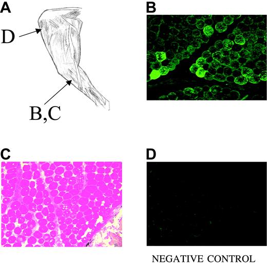
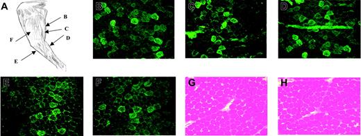
We sought to determine whether a viral vector delivery technique that had been described in hamsters and rats10 could show efficacy in a large animal model. The technique involves vascular delivery of vector to the muscle tissue in a single limb. Briefly, the common femoral vessels are exposed, the animal is heparinized, the vessels are cannulated, and a tourniquet is securely tightened at the level of the hip joint. The now-isolated limb vasculature is perfused with a solution containing papaverine to effect vasodilatation. The limb circulation is infused 5 minutes later with vector mixed with histamine, to induce vascular leakage. After perfusion/infusion, the limb circulation is thoroughly flushed and the tourniquets and clamps removed. Greelish et al10 showed that this technique allowed extensive transduction of skeletal muscle with either adenovirus or AAV vectors in rats and hamsters. In the current study, initial experiments were carried out in normal dogs to determine whether the procedure has relevance for gene transfer in larger animals. The first animal (6.5 kg) was infused with AAV-lacZ at a dose of 3 × 10¹² vg/kg, with heparin, papaverine, and histamine doses scaled up linearly from the rodent studies on a per-kilogram basis. A muscle biopsy taken 4 weeks later was uninformative in terms of gene transfer, showing only a cellular infiltrate and destruction of muscle architecture (data not shown). Based on earlier studies with AAV-lacZ in dogs (V.R.A. and K.A.H., unpublished observations, April 2002), we suspected that this was due to an immune response to β-galactosidase in dogs, and we repeated the experiment using cF.IX, a protein to which normal dogs are fully tolerant. This second dog was injected by isolated limb perfusion (ILP) in one hindlimb with AAV-CMV-cF.IX at a dose of 3 × 10¹² vg/kg, and also received a dose of 2 × 10¹² vg of the same vector at a single site, by direct intramuscular injection in the opposite limb. 8 weeks later, the animal was killed and muscle tissue harvested for immunofluorescence staining with an antibody to canine F.IX. In the hindlimb that received direct intramuscular injection, cF.IX expression was confined to the sites of injection (Figure 1A-B), with a radius of diffusion of approximately 0.5 mm, whereas the hindlimb that received vector by the intravascular infusion process (Figure 2) showed transduction throughout the muscle groups supplied by the injected vessel, with more extensive transduction around the stifle joint (corresponds to knee joint in humans) than at distal sites. Systemic levels of transgene expression could not be quantified in normal dogs, since the endogenous protein and the donated gene product are identical. A biodistribution study was performed on this animal at the time of sacrifice (8 weeks after vector infusion), using a PCR assay that can detect as few as 30 copies in 1 μg DNA. Of all tissues sampled, only perfused skeletal muscle was positive for vector sequences. Liver, lung, kidney, spleen, ovary, and contralateral muscle were all negative. The same assay showed that serum was positive for vector sequences for 5 days after vector infusion but was uniformly negative thereafter (data not shown).
Histology of normal dog muscle 8 weeks after direct intramuscular injection of AAV-CMV-canine F.IX. (A) Anatomy of dog hindlimb, showing sites of biopsy. (B) Immunofluorescence staining for canine F.IX at injection site. (C) Hematoxylin and eosin staining of same site, showing normal histology. Vacuoles within muscle fibers are due to freeze artifact. (D) Immunofluorescence staining of uninjected muscle in same limb. For all panels, original magnification ×100.
Histology of normal dog muscle 8 weeks after direct intramuscular injection of AAV-CMV-canine F.IX. (A) Anatomy of dog hindlimb, showing sites of biopsy. (B) Immunofluorescence staining for canine F.IX at injection site. (C) Hematoxylin and eosin staining of same site, showing normal histology. Vacuoles within muscle fibers are due to freeze artifact. (D) Immunofluorescence staining of uninjected muscle in same limb. For all panels, original magnification ×100.
Histology of normal dog muscle 8 weeks after intravascular vector delivery by isolated limb perfusion. (A) Anatomy of dog hindlimb showing sites of biopsy. (B-F) Immunofluorescence staining for canine F.IX at multiple sites as indicated, showing extensive positive staining. (G-H) Hematoxylin and eosin staining of same samples. For all panels, original magnification ×100.
Histology of normal dog muscle 8 weeks after intravascular vector delivery by isolated limb perfusion. (A) Anatomy of dog hindlimb showing sites of biopsy. (B-F) Immunofluorescence staining for canine F.IX at multiple sites as indicated, showing extensive positive staining. (G-H) Hematoxylin and eosin staining of same samples. For all panels, original magnification ×100.
Long-term correction of hemophilia B phenotype by regional intravascular delivery of rAAV to the skeletal muscle of hemophilia B dogs
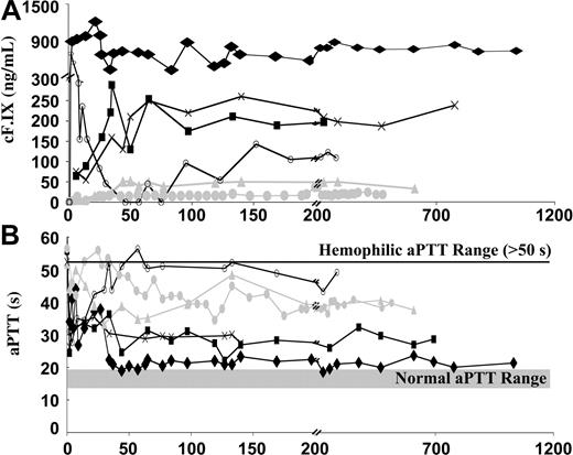
To achieve a more quantitative analysis, and to determine safety and efficacy in an animal model of the human disease hemophilia B, we carried out the same vector delivery procedure in hemophilia B dogs. These animals have disease due to a missense mutation in the portion of the gene encoding the catalytic domain of the protein.11 The animals have no detectable F.IX antigen or activity, so transgene expression can be assessed by both clotting assays, and by ELISA for expressed protein. Performance of the procedure in hemophilic animals required 2 modifications. First, animals were infused prior to and after the procedure with normal canine plasma, to insure adequate hemostasis for the surgical maneuver and for postoperative healing, and second, animals were immunosuppressed transiently with cyclophosphamide, for a period of 6 weeks, because we had shown in earlier studies that this maneuver reduced the risk of formation of inhibitory antibodies to F.IX when vector expressing cF.IX was administered to skeletal muscle in hemophilic animals.4,12,13 Since cyclophosphamide can lead to hemorrhagic cystitis, a complication that can be difficult to control in hemophilic animals, the dogs were also treated with MESNA (sodium 2-mercaptoethane sulfonate), a bladder cytoprotective agent. The first animal (D99) was infused at a dose of 3.7 × 10¹² vg/kg (Figure 3A). This resulted in circulating levels of cF.IX in the range of 600 ng/mL to 800 ng/mL by ELISA, and 15% of normal human plasma levels by activity assay (Table 1) determined in several time points (ranging from 2 to 26 months) following ILP. This level of expression was sufficient to correct the aPTT to a near normal value (Table 1, Figure 3B). These levels have been sustained for approximately 3 years, with observation ongoing. There was no drop in F.IX level and no appearance of neutralizing or nonneutralizing antibodies to F.IX after cyclophosphamide infusions were stopped at 6 weeks after vector infusion. Since prophylaxis against spontaneous bleeding episodes does not require levels of 15%, we infused AAV-CMV-cF.IX at a lower dose (1.7 × 10¹² vg/kg), again accompanied by short-term administration of cyclophosphamide and MESNA, and observed long-term expression of cF.IX in the range of 260 ng/mL by ELISA and 5.2% by activity. This was also accompanied by a shortening of the aPTT (Table 1, Figure 3B). A third experiment resulted in circulating levels of 210 ng/mL (4.2% by activity, with shortening of the aPTT) after a dose of 3.0 × 10¹² vg/kg. Whether the lower F.IX level seen in the third dog results from biologic differences in the canine subjects, or minor variations in surgical technique, is unknown. However, it should be noted that all 3 experimental animals achieved long-term expression of canine F.IX at therapeutic levels. The excellent correlation between antigen and activity levels suggests that, at this level of expression, F.IX synthesized in skeletal muscle is fully biologically active. As further proof of this, it should be noted that, in the 74 months of cumulative observation of the 3 treated dogs, there have been only 2 bleeding episodes requiring treatment. The expected number based on observation of the colony over many years is approximately 5.5 episodes per 12 months, or 34 over this period of observation.14 As a comparison for efficacy, we have shown a historical control in which a dog from the same colony was injected with the same vector (AAV-CMV-cF.IX) at a comparable dose (3.4 × 10¹² vg/kg) by direct intramuscular injection, with resulting circulating F.IX levels of less than 1% (Figure 3). Thus vector delivery by isolated limb perfusion results in higher circulating F.IX levels at comparable doses of vector.
Canine F.IX expression in hemophilia B dogs following ILP delivery of AAV-2 vector. (A) Canine F.IX antigen levels and (B) activated partial thromboplastin times (aPTTs) in plasma samples of hemophilia B dogs as a function of time after delivery of AAV-CMV-cF.IX. Dog D99 ( ) was injected with 3.7 × 10¹² vg/kg, dog F57 (×) with 1.7 × 10¹² vg/kg, and dog H08 (▪) with 3.0 × 10¹² vg/kg, by isolated limb perfusion, accompanied by transient immunosuppression. Dog E59 (▴) was injected by peripheral vein with 2.9 × 10¹² vg/kg. Dog E60 (○) was injected at 3.9 × 10¹² vg/kg by ILP, without immunosuppression. Dog B48 (
 ) was injected by direct intramuscular injection with 3.4 × 10¹² vg/kg. Arrow denotes infusion of canine plasma, resulting in transient spike in cF.IX and drop in aPTT.
Canine F.IX expression in hemophilia B dogs following ILP delivery of AAV-2 vector. (A) Canine F.IX antigen levels and (B) activated partial thromboplastin times (aPTTs) in plasma samples of hemophilia B dogs as a function of time after delivery of AAV-CMV-cF.IX. Dog D99 ( ) was injected with 3.7 × 10¹² vg/kg, dog F57 (×) with 1.7 × 10¹² vg/kg, and dog H08 (▪) with 3.0 × 10¹² vg/kg, by isolated limb perfusion, accompanied by transient immunosuppression. Dog E59 (▴) was injected by peripheral vein with 2.9 × 10¹² vg/kg. Dog E60 (○) was injected at 3.9 × 10¹² vg/kg by ILP, without immunosuppression. Dog B48 (
 ) was injected by direct intramuscular injection with 3.4 × 10¹² vg/kg. Arrow denotes infusion of canine plasma, resulting in transient spike in cF.IX and drop in aPTT.
Inhibitor formation to cF.IX occurs in the absence of transient immunosuppression following regional intravascular delivery to skeletal muscle
As a second control, isolated limb perfusion was repeated at a dose of 3.9 × 10¹² vg/kg, but this time omitting cyclophosphamide and MESNA. In this instance, the animal formed a neutralizing antibody first detected approximately 21 days after vector administration, and persisting approximately 300 days (Figure 4). It should be noted that, even though the ELISA detects a low level of circulating cF.IX (Figure 4A), there is no correction of clotting times, with the aPTT remaining approximately 50 seconds (Figure 4B) and F.IX clotting activity less than 1% (Table 1). This discrepancy between antigen and activity levels is typically seen in the presence of a neutralizing antibody (clinically termed an inhibitor) (Figure 4C). The development of the inhibitor under these circumstances suggests that the transient immunosuppression cannot be omitted.
Coagulation assays in E60, a dog treated by ILP without immunosuppression. (A) Canine F.IX antigen levels, (B) activated partial thromboplastin times (aPTTs), and (C) Bethesda assay (titer of neutralizing antibody) as a function of time after vector injection. Dog E60 was injected at 3.9 × 10¹² vg/kg and did not receive immunosuppresion. Gray band in panel B denotes range of normal aPTT; line at 50 seconds denotes hemophilic values.
Coagulation assays in E60, a dog treated by ILP without immunosuppression. (A) Canine F.IX antigen levels, (B) activated partial thromboplastin times (aPTTs), and (C) Bethesda assay (titer of neutralizing antibody) as a function of time after vector injection. Dog E60 was injected at 3.9 × 10¹² vg/kg and did not receive immunosuppresion. Gray band in panel B denotes range of normal aPTT; line at 50 seconds denotes hemophilic values.
Peripheral intravenous delivery of rAAV to a hemophilia B dog results in subtherapeutic F.IX levels
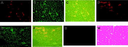
In a control experiment, a hemophilic animal was infused by peripheral vein with AAV-CMV-cF.IX at a dose of 2.9 × 10¹² vg/kg, accompanied by short-term administration of cyclophosphamide and MESNA. The resulting level of F.IX was approximately 50 ng/mL, or approximately 1% of normal circulating levels (Figure 3A), with a corresponding absence of positive fibers on muscle biopsy (Figure 5G). Thus it seems unlikely that the procedure will be successful as a simple intravenous infusion, at least with this AAV serotype.
Immunofluorescence staining of muscle sections of the tibialis anterior of hemophilia B dogs injected 2 (dog F57) or 3 (dog D99) years earlier with AAV-CMV-canine F.IX by isolated limb perfusion. (A, D). Fluorescence of rhodamine (red) showing presence of canine F.IX in muscle fibers of ILP injected dogs F57 (A) or D99 (D). (B, E) Fluorescence of fluorescein isothiocyanate (FITC; green) showing presence of heparan sulfate proteoglycan (HSPG) in fibers of tibialis anterior of dog F57 (B) or D99 (E). (C, F) Simultaneous excitation of both fluorescence tags. (G) Fluorescence of rhodamine showing absence of canine F.IX expression in muscle biopsy from dog E59 (infusion of vector by peripheral vein). (H) Hematoxylin and eosin staining of same samples of dog (F57). For all panels, original magnification ×100.
Immunofluorescence staining of muscle sections of the tibialis anterior of hemophilia B dogs injected 2 (dog F57) or 3 (dog D99) years earlier with AAV-CMV-canine F.IX by isolated limb perfusion. (A, D). Fluorescence of rhodamine (red) showing presence of canine F.IX in muscle fibers of ILP injected dogs F57 (A) or D99 (D). (B, E) Fluorescence of fluorescein isothiocyanate (FITC; green) showing presence of heparan sulfate proteoglycan (HSPG) in fibers of tibialis anterior of dog F57 (B) or D99 (E). (C, F) Simultaneous excitation of both fluorescence tags. (G) Fluorescence of rhodamine showing absence of canine F.IX expression in muscle biopsy from dog E59 (infusion of vector by peripheral vein). (H) Hematoxylin and eosin staining of same samples of dog (F57). For all panels, original magnification ×100.
Route of vector delivery to skeletal muscle does not alter patterns of rAVV-2 transduction
Muscle biopsies of the perfused limb (taken from the tibialis anterior) documented that transduction had occurred in a mosaic pattern (Figure 5A,D) similar to that seen with direct intramuscular injection in mice, dogs, and humans.1,2,7 Immunofluorescent staining of sections for heparan sulfate proteoglycan (HSPG), which acts as a receptor for AAV-2,5 indicates that fibers rich in HSPG are preferentially transduced by AAV-2 (Figure 5B,E), an observation confirmed on the colocalization experiment (Figure 5C,F). As shown previously in mice by Huard and colleagues (Fields et al),15 and in humans by our group,7 HSPG is abundant in slow fibers but not in fast fibers, accounting for the checkerboard pattern of positivity in transduced muscle. These results indicate that this property of AAV-2 transduction is not altered by the regional intravascular delivery method. Hematoxylin and eosin staining of muscle biopsied 2 years after vector administration showed no evidence of muscle injury or inflammation, and continued expressin of cF.IX (Figure 5H).
Biodistribution and toxicity
The procedure was generally well tolerated in both normal and hemophilic dogs, although a transient drop in blood pressure typically occurred on reconnection of the limb vasculature to the systemic circulation, presumably due to diffusion of residual histamine into the systemic vasculature (see “Materials and methods”). Clinical laboratory studies including electrolytes, blood urea nitrogen concentration, creatinine, liver function tests, and complete blood count were all normal in the days and weeks after the procedure. Creatine phosphokinase typically spiked to 3- to 7-fold upper limits of normal immediately after the procedure, with a return to normal within the first week. Transient elevation of creatine phosphokinase is routinely observed in humans undergoing isolated limb perfusion.16 Results of biodistribution studies were similar to those obtained in normal dogs, with serum transiently positive for vector sequences up to day 5 after vector infusion (data not shown).
Discussion
The development of efficient methods for bringing about targeted disruption of genes in murine embryonic stem cells opened a new line of investigation in biomedical research through the generation and characterization of murine models of human disease. Efforts quickly progressed from studies of pathophysiology to generation and testing of novel therapeutic strategies. In the realm of therapeutics, as in pathophysiology, murine models have proven deceptive as well as informative. Their small size guarantees that even difficult-to-produce reagents can be made in sufficient quantities, and it also facilitates delivery of test reagents in high doses. Other aspects of physiology critical for drug delivery,17 therapeutic feasibility,18-21 or toxicity22-26 may be only poorly modeled in mice, and, in the case of gene therapy, inbred strains of mice can be uninformative in terms of immune response to the vector or the transgene product.21,22,25 Thus one can enumerate at least 10 novel therapeutic strategies that have cured hemophilia in mice,18-20,22,27-32 but none have yet done so in humans. The myriad differences in small and large animal physiology underscore the fundamental importance of translational studies of novel therapies in large animal models as a necessary screen before contemplating human studies.
In our initial attempts to develop a clinical protocol for gene transfer in hemophilia B, we used skeletal muscle as the target tissue for an AAV vector expressing blood coagulation Factor IX.1,6,7 Compared to liver, the natural site of synthesis of F.IX, muscle is a desirable target tissue because it can be accessed in a relatively noninvasive fashion (intramuscular injection) that does not give rise to widespread biodistribution of vector.9,33,34 Moreover, the high prevalence of hepatitis B/C in the adult hemophilia population35,36 (due to infection from older plasma-derived products) makes use of liver as the target tissue problematic. Therefore, we chose skeletal muscle as the target tissue in the first clinical studies of parenterally administered AAV vector. These studies established that intramuscular administration of AAV-CMV-F.IX was safe at doses up to 2 × 10¹² vg/kg in men with severe hemophilia B, and that there was long-term persistence of F.IX expression in injected muscle as judged by immunofluorescent staining of the tissue.7 This latter is a particularly important point, since long-term expression may be more difficult to achieve in AAV-2–mediated vector delivery to hepatocytes in humans.37 However, the clinical study and concurrent large animal studies documented limitations to the muscle delivery method that had not been anticipated from the initial studies in hemophilic dogs. First, it became clear in animal models of hemophilia that the risk of forming inhibitory antibodies to F.IX was higher after intramuscular delivery of AAV vector than after delivery of vector to the liver.1,4,12,13,38 Critical determinants included the dose of vector delivered per site, and the underlying mutation in the F.IX gene, which affects the animal's immunologic tolerance to the F.IX protein. To minimize the risk of inhibitor formation in the clinical study, the dose per site was kept below a limit that had been defined in the canine hemophilia model,4 and enrollment in the trial was limited to those with disease due to a missense mutation, a subgroup similar to the canine model, and at lower overall risk of inhibitor formation based on studies with protein infusion therapy.39,40 Other limiting factors included the low efficiency of skeletal muscle at executing certain critical posttranslational modifications of the F.IX protein,3 and the fact that vector uptake is receptor mediated, which sets an upper limit to the amount of vector that can gain access to cells at a single injection site. Using the experimentally determined constraints on vector dose per site (2 × 10¹² vg/site), the procedure required an excessive number of injections merely to achieve a dose of 2 × 10¹² vg/kg, still substantially short of the target dose of 1 × 10¹³ vg/kg. Without a more efficient delivery method, further dose escalation seemed impractical.
A more subtle point about vector delivery was also at play in the scale-up from mouse to dog to human. A dose of 1 × 10¹³ vg/kg yielded circulating F.IX levels of 5% to 7% in the mice, and only 1% to 2% in hemophilic dogs.1,2 The reasons for this decline in efficacy with increasing size were not entirely clear; at the time, we speculated that interspecies differences in promoter activity (ie, a cause not related to size), or diffusion distance of the product to the circulation (more clearly related to size of organism) might account for the difference. The diffusion distance of the vector in solid tissue is fairly constant. As the radius of the muscle bundle increases with increasing size of the animal, a lower fraction of the muscle is covered per injection. Thus, direct intramuscular injection transduces large areas of muscle in a mouse, but relatively much less in a larger animal. Put simply, 2 injections of AAV-lacZ turn all muscles blue in a mouse but certainly not in a dog. To achieve scale-independent dosing, one needs to take advantage of a delivery technique based on fractal rather than Euclidean geometric principles. Regional intravascular delivery, using the vascular tree of the isolated limb as the delivery network, affords such a solution to the problem of scaleable delivery.
The importance of this finding for gene delivery in hemophilia is considerable; this delivery method essentially closes the 10- to 50-fold gap in efficacy between liver-directed and muscle-directed AAV-mediated gene transfer in large animals.1,38 For a disease category in which a substantial percentage (> 80%) of severely affected adults have infection with hepatitis C,35,36 it is crucial to have a treatment strategy that does not require the liver as the target organ for transduction.
Several investigators have shown that, in mice at least, AAV serotypes 1, 6, and 7 result in much more efficient transduction of skeletal muscle.41-43 Confirmation of these results in large animal models has recently been published.8,44,45 We were able to show in the hemophilia B dog model that direct intramuscular injection of AAV-1-cF.IX resulted initially in a high level of F.IX expression, but was rapidly followed by inhibitor formation that, in contrast to isolated limb perfusion with AAV-2, could not be blocked by transient immunosuppression.8 Others have also observed the formation of antibodies to the transgene product after intramuscular injection of more potent AAV serotypes.44,45 It would be of interest in their case to determine whether the transient immunosuppressive regimen that we used could block antibody formation in their model system. All of our findings to date are consistent with the notion that high-level expression of F.IX in the skeletal muscle of an animal not tolerant to the transgene product will, regardless of route of administration, trigger formation of inhibitory antibodies unless transient immunosuppression is administered.4 Also, translational studies in large animal disease models may uncover problems not appreciated in inbred strains of laboratory mice.
The data presented here, combined with data already in hand from earlier clinical studies, argue that this procedure can be safely scaled up to humans. We have documented the safety of administering AAV-2-F.IX vector doses up to 2 × 10¹² vg/kg to skeletal muscle by direct intramuscular injection in humans with hemophilia, and have shown, by immunohistochemistry on injected muscle tissue, that expression persists unabated at time points up to 10 months after vector injection.7 In other clinical studies, we have infused vector doses up to 5 × 10¹² vg/kg into the hepatic artery without systemic symptoms or serious adverse events.37 The surgical procedure outlined here, isolated limb perfusion, has been extensively used clinically for the delivery of high-dose chemotherapy to a single limb.16 A recently described alternative to ILP is isolated limb infusion, a simplified less invasive technique for regional delivery of drugs through percutanenous catheterization of femoral vessels.46,47 Thus the safety of many individual elements of the treatment strategy is already established in humans. An important safety consideration for the procedure we described is the biodistribution of vector following intravascular delivery to skeletal muscle. We documented limited hematogenous dissemination of vector following ILP, and in tissues harvested 8 weeks after procedure vector detection was limited to the perfused muscle. The use of a tight tourniquet and the removal of residual vector from the isolated vessels prior to reconnection to the systemic circulation are likely to restrict vector dissemination.
The current work demonstrates the efficacy of this delivery procedure in a large animal model of hemophilia B. Comparing the data generated here to results obtained in the same animal model with the same vector delivered by direct intramuscular injection,1 it can be seen that the change in method of administration improves expression by as much as one log (compare circulating levels of ∼1% after 3.4 × 10¹² vg/kg by intramuscular injection, to 4%-15% after ∼2-4 × 10¹² vg/kg by ILP). These levels are especially encouraging since the recent studies in liver suggest that doses required for efficacy in the hemophilic dogs were predictive of doses required for efficacy in humans.37 Moreover, this robust level of expression in the dog model allows margin for some loss of efficacy on translation to humans. The use of alternate serotypes which transduce skeletal muscle more efficiently in mice8,41,42 may extend this margin if validity of the observation can be extended to large animals and humans. Although it will be necessary to address details of the immune response to the transgene product, and to identify an approved pharmacologic agent that can induce a vascular leak (ie, a drug other than histamine), the studies here establish the efficacy of the strategy and thus permit us to build on the promising findings from the earlier AAV-F.IX study,7 in which vector was administered by direct intramuscular administration.
Prepublished online as Blood First Edition Paper, October 12, 2004; DOI 10.1182/blood-2004-07-2908.
Supported by National Institutes of Health grants NIDDK-KO1-060 580 (V.R.A.), RR-02512 (M.E.H.), and P01-HL64190 (K.A.H.), and research grants from Hemophilia of Georgia and the Howard Hughes Medical Institute, and grants to H.H.S. from the NIH (NIAMS and NINDS).
V.R.A. and H.H.S. contributed equally to the manuscript.
An Inside Blood analysis of this article appears in the front of this issue.
The publication costs of this article were defrayed in part by page charge payment. Therefore, and solely to indicate this fact, this article is hereby marked “advertisement” in accordance with 18 U.S.C. section 1734.
The authors thank Jian-Hua Liu for excellent technical assistance.


