Abstract
Iron is critical for cell growth and proliferation. Iron chelators are being explored for a number of clinical applications, including the treatment of neurodegenerative disorders, heart disease, and cancer. To uncover mechanisms of action of tachpyridine, a chelator currently undergoing preclinical evaluation as an anticancer agent, cell-cycle analysis was performed. Tachpyridine arrested cells at G2, a radiosensitive phase of the cell cycle, and enhanced the sensitivity of cancer cells but not nontransformed cells to ionizing radiation. G2 arrest was p53 independent and was accompanied by activation of the checkpoint kinases CHK1 and CHK2. G2 arrest was blocked by UCN-01, a CHK1 inhibitor, but proceeded in CHK2 knock-out cells, indicating a critical role for CHK1 in G2 arrest. Tachpyridine-induced cell-cycle arrest was abrogated in cells treated with caffeine, an inhibitor of the ataxia-telangiectasia mutated/ataxia-telangiectasia-mutated and Rad3-related (ATM/ATR) kinases. Further, G2 arrest proceeded in ATM-deficient cells but was blocked in ATR-deficient cells, implicating ATR as the proximal kinase in tachpyridine-mediated G2 arrest. Collectively, our results suggest that iron chelators may function as antitumor and radioenhancing agents and uncover a previously unexplored activity of iron chelators in activation of ATR and checkpoint kinases.
Introduction
Iron is a metal critically important for cell proliferation and survival. It is an essential constituent of hemoglobin and myoglobin, mitochondrial electron transport proteins, hydroxylating enzymes, lipoxygenases, and cyclooxygenases, as well as ribonucleotide reductase, the enzyme that catalyzes the rate-limiting step in DNA synthesis. Tumor cells frequently exhibit increased uptake and utilization of iron, as evidenced by an increase in transferrin receptors at the cell surface.1,2 This has led to the suggestion that agents that deplete intracellular iron may be useful in cancer therapy.3-5
A variety of approaches, including the use of antisense technology6,7 and antibodies8 to target the transferrin receptor, have borne out this prediction. Indeed, gallium, a metal used in combination chemotherapy for the treatment of metastatic urothelial carcinoma9 and non-Hodgkin lymphoma,10 exerts its effects in part by interfering with iron metabolism.
High-affinity, low-molecular-weight metal chelators offer a direct method of achieving intracellular iron deprivation, and the antitumor potential of iron chelators has also been the subject of active investigation. The preponderance of these studies has focused on desferrioxamine (deferoxamine, Desferal, DFO), a hexadentate iron chelator and the drug of choice for the treatment of chronic iron overload. Although desferrioxamine has been shown to retard tumor growth substantially in animal studies11 and some clinical trials,12 results have been mixed13,14 and have led to the identification of chelators with potentially improved antitumor activity. For example, Triapine, a ribonucleotide reductase inhibitor and iron chelator, is currently in phase 1 clinical trials.15 Pyridoxal isonicotinoyl hydrazone,16 O-Trensox,17 desferrithiocin,18,19 desferriexochelin,20 di-2-pyridyl thiosemicarbazones,21 and tachpyridine22-27 are chelators under investigation as antitumor agents.
Tachpyridine is a hexadentate metal chelator.23 It is a member of a chelator family currently in preclinical development at the National Cancer Institute (NCI). Tachpyridine weakly binds divalent cations such as calcium, magnesium, and manganese, but it strongly binds iron, copper, and zinc. The cytotoxic activity of tachpyridine is likely due to its ability to bind one or more of the metals iron, zinc, or copper, because complexes of tachpyridine with these metals (but not others) are nontoxic.23 Tachpyridine exhibits a lower IC50 (inhibitory concentration 50%) than DFO and is preferentially cytotoxic to tumor cells.23 Tachpyridine also retards the growth of implanted tumors in mice (S.V.T., R.P.P., M.W.B., F.M.T., manuscript in preparation).
Mechanisms underlying the anticancer activity of iron chelators are incompletely understood. Ribonucleotide reductase, which contains iron at its catalytic center, is frequently cited as a target of iron chelators, although inhibition of this enzyme by iron chelators can be modest.28-30 Inhibition of proteins required for cell-cycle progression, such as cyclins A, B, and D, and the cyclin-dependent kinases cdk1 and cdk2,31 has also been reported. Chelators have also been reported to exert cytotoxic effects via redox cycling of bound metals and attendant production of free radicals.30,32 Mechanistic studies of tachpyridine have shown that it induces an apoptotic response in several cancer cell lines by activating the mitochondrial pathway.25 In addition, the tumor suppressor protein p53 accumulates in cells treated with tachpyridine, although p53 is not required for tachpyridine-mediated apoptosis.24
To gain further insight into the mechanism of action of iron chelators, and to more completely define the spectrum of tachpyridine's anticancer activity, we assessed the effect of tachpyridine on cell-cycle regulation. We report here that tachpyridine arrests HeLa cervical carcinoma and HCT 116 colorectal cancer cells at the G2 phase of the cell and activates both checkpoint kinase 1 (CHK1) and CHK2. Activation of the CHK kinases in tachpyridine-treated cells is preceded by phosphorylation of p53 at serine 15, implicating ataxia-telangiectasia mutated/ataxia-telangiectasia-mutated and Rad3-related (ATM/ATR) kinases in CHK kinase activation and cell-cycle arrest. ATR-mediated activation of CHK1 is critical to this response, because G2 arrest is blocked by either UCN-01, a CHK1 inhibitor, or genetic knockdown of ATR. Consistent with its ability to induce G2 arrest, tachpyridine selectively sensitized cancer cells to ionizing radiation. Collectively, these results suggest that chelators may function in anticancer therapy as radioenhancing agents as well as cytotoxic agents. These results also indicate a previously unexplored activity of metal chelators in activation of ATM/ATR and the checkpoint kinases.
Materials and methods
Chemicals
Tachpyridine and N-methyl tachpyridine were synthesized from cis-1,3,5-triaminocyclohexane according to methods published previously22,33 and were prepared as a 1-mM stock in phosphate-buffered saline, pH 7.4, 0.22-μm filter sterilized, and stored at 4°C. Purity was greater than 99% as determined by 1H and 13C nuclear magnetic resonance.22,33 UCN-01 was obtained from the National Cancer Institute.
Cell lines and cell culture
HCT 116 human colon carcinoma TP53 wild-type (TP53+/+) and TP53 knock-out (TP53-/-) cell lines and the HCT 116 CHK2-/- cell line were generous gifts from Dr B. Vogelstein (Johns Hopkins University, Baltimore, MD). HCT 116 cells containing a single functional ATR allele into which lox sites flanking exon 2 of the ATR gene were introduced by homologous recombination (termed ATRflox-/+) were kind gifts from the laboratory of Dr S. Elledge (Harvard Medical School and Brigham and Women's Hospital, Boston, MA).34 The human colon cancer cell line RKO was obtained from the American Type Culture Collection (ATCC, Rockville, MD). These cell lines were maintained in McCoys 5A media supplemented with 10% fetal bovine serum (FBS). The simian virus 40-immortalized A-T fibroblast line AT22IJE stably expressing full-length recombinant human ATM (designated YZ-5) or stably transfected with empty vector (EBS-7 cells) were cultured as described.35 The human cervical carcinoma HeLa cell line (Tissue Culture Core Laboratory, Comprehensive Cancer Center of Wake Forest University Health Sciences [WFUSH]) and normal human lung fibroblast MRC5 cells (ATCC) were maintained in Dulbecco Modified Eagle Medium containing 10% FBS plus pen-strep.
Western blot analysis
Cells were lysed in ice-cold NP40 buffer (50 mM Tris [tris(hydroxymethyl) aminomethane], pH 8.0, 5 mM EDTA [ethylenediaminetetraacetic acid], 150 mM NaCl, 0.5% NP40 [nonylphenylpolyethylene glycol]) containing a cocktail of protease inhibitors (Complete; Boehringer Mannheim, Indianapolis, IN) and phenylmethylsulphonyl fluoride (0.1 mg/mL). Primary antibodies used in Western blotting were mouse anti-human p53 monoclonal antibody (Ab-6; Calbiochem, San Diego, CA), rabbit anti-human phosphorylated serine 15 p53, rabbit anti-human phospho serine 345 CHK1, rabbit anti-human phospho threonine 68 CHK2, and monoclonal anti-ATM antibody (Cell Signaling, Beverly MA). ATR immunoblotting was conducted using a rabbit polyclonal antibody that was raised using a bacterially synthesized, purified glutathione-S-transferase-ATR fusion protein as the immunogen.83 Peroxidase-conjugated secondary antibodies were detected using enhanced chemiluminescence (Amersham, Little Chalfont, United Kingdom). Membranes were stained with Ponceau S (Sigma, Saint Louis, MO) and/or probed with mouse anti-human β-actin monoclonal antibody (Sigma) or anti-β tubulin antibody (DM1A; obtained from Dr D. W. Cleveland, University of California, San Diego [UCSD]) to confirm equivalent loading and protein transfer.
Cell-cycle analysis
Cells were fixed and stained in a solution of 25 μg/mL propidium iodide before analysis by flow cytometry (FACStar Plus, Mod-FitLT V2.0; Becton Dickinson, Franklin Lakes, NJ).
Phosphorylated histone H3 immunostaining for mitotic cells
Cells were fixed and permeabilized using Permaflow Intracellular Antigen/DNA/RNA detection kit (55001; Invirion, Frankfort, MI) and immunostained using mouse monoclonal antiphosphorylated serine 10 histone H3 antibody (Cell Signaling) followed by fluorescein isothiocyanate (FITC)-conjugated goat antimouse antibody. Colcemid (Sigma, Saint Louis, MO) trapping experiments involved treatment of HeLa cells with 5 μM tachpyridine for 18 hours, followed by the addition of 0.1 μg/mL Colcemid for 24 hours prior to immunostaining.
Irradiation and clonogenic assay
Cells were plated in triplicate, allowed to attach overnight, and then pretreated with tachpyridine for 12 hours, followed by exposure to ionizing radiation (IR) using a Cesium-137 irradiator (Shepard and Associates, San Fernando, CA) at a dose rate of 185 rad/min. Dishes were washed, and colonies were allowed to form over approximately 8 to 12 days, followed by fixing in methanol acetic acid (10:10:80 acetic acid:MeOH/H2O) and staining with 0.4% crystal violet. Colonies were counted using Accucount 1000 (Biologics, Gainesville, VA) with a colony considered to be at least 50 cells. Cell fraction surviving treatment was normalized to survival of the control cells. Radiation survival curves were constructed by normalizing the number of colonies surviving after combined treatment to the number of colonies surviving following treatment with tachpyridine alone, and the curves were fitted by second-order linear regression. The dose enhancement ratio (DER) was calculated by dividing the fraction of surviving cells treated with IR alone by the fraction of surviving cells treated with the combination of IR and tachpyridine.
MTT assay
In some cases, assessment of the response of cells to tachpyridine with and without radiation was determined using a modified MTT (3-(4,5-dimethylthiazol-2-yl)-2,5-diphenyl tetrazolium bromide) cellular proliferation assay (Roche Diagnostics, Indianapolis, IN). Cells were plated in 24-well dishes, exposed to tachpyridine for 12 hours, irradiated, washed, and allowed to grow for approximately 7 days. MTT was administered for 4 hours, and cells were lysed at least overnight. Aliquots were transferred to 96-well plates, and absorbance at 560 nm was measured.
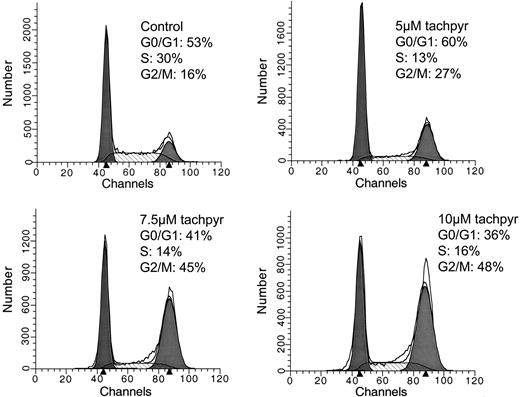
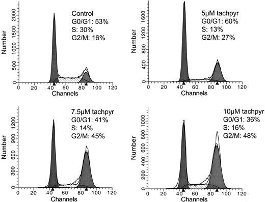
Tachpyridine induces G2/M arrest in HeLa cells. Cells were treated with 5, 7.5, and 10 μM tachpyridine for 24 hours, and cell-cycle distribution was determined by flow cytometry.
Tachpyridine induces G2/M arrest in HeLa cells. Cells were treated with 5, 7.5, and 10 μM tachpyridine for 24 hours, and cell-cycle distribution was determined by flow cytometry.
Results
Tachpyridine induces G2/M cell-cycle arrest
Tachpyridine is a metal chelator of potential clinical use as an anticancer agent.23,24 To probe the mechanism of action of this chelator, we assessed its effects on the cell cycle. As shown in Figure 1, treatment of HeLa human cervical cancer cells with 5, 7.5, and 10 μM tachpyridine for 24 hours induced a dose-dependent accumulation of cells in the G2/M phase of the cell cycle. The G2/M cell-cycle arrest induced by tachpyridine was achieved at concentrations and treatment durations that did not result in cytotoxicity, excluding the possibility that the accumulation of cells in G2/M was an artifact resulting from selective cytotoxicity of tachpyridine to cells in G0/G1 and S (data not shown).
Cell-cycle arrest requires metal binding activity
Our previous work has demonstrated that cytotoxic effects of tachpyridine are critically dependent on its metal binding activity, and that N-alkylated derivatives of tachpyridine that are unable to bind metals are nontoxic.23 To test whether tachpyridine's effects on the cell cycle were similarly dependent on its metal binding activity, we assessed the cell-cycle effect of N-methyl tachpyridine, an analog inhibited in its ability to bind iron because of steric constraints and an inability to undergo oxidative dehydrogenation.22 As shown in Table 1, N-methyl tachpyridine was unable to induce G2/M cell-cycle arrest. Thus, the ability of tachpyridine to induce G2/M arrest is linked to its metal binding activity.
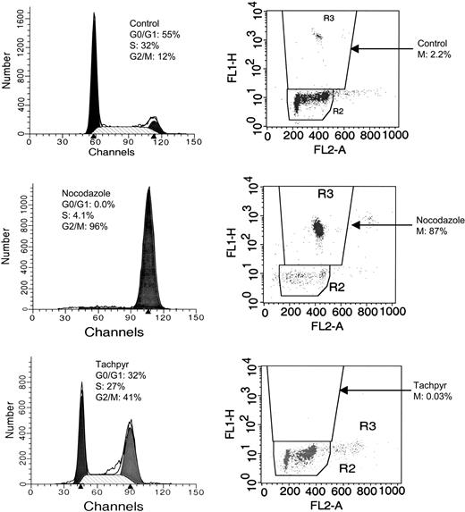
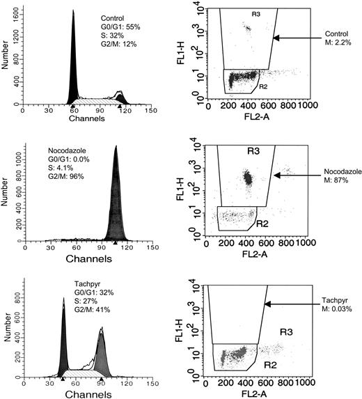
Tachpyridine induces cell-cycle arrest in the G2 phase of the cell cycle. HeLa cells treated with 7.5 μM tachpyridine for 18 hours were double-labeled for the M-phase marker phosphorylated H3 and for DNA content, followed by flow cytometry analysis. Untreated controls (top) show 12% of cells in G2/M by propidium iodide (PI) staining and 2.2% of those cells in mitosis. Cells treated with 50 ng/mL nocodazole, an M-phase arrestor, show 96% of cells in G2/M by PI staining and 87% of those cells in mitosis (middle). Tachpyridine-treated cells (bottom) display 41% of cells in G2/M by PI staining, and only 0.03% of cells in mitosis, consistent with the induction of a G2- and not M-phase arrest.
Tachpyridine induces cell-cycle arrest in the G2 phase of the cell cycle. HeLa cells treated with 7.5 μM tachpyridine for 18 hours were double-labeled for the M-phase marker phosphorylated H3 and for DNA content, followed by flow cytometry analysis. Untreated controls (top) show 12% of cells in G2/M by propidium iodide (PI) staining and 2.2% of those cells in mitosis. Cells treated with 50 ng/mL nocodazole, an M-phase arrestor, show 96% of cells in G2/M by PI staining and 87% of those cells in mitosis (middle). Tachpyridine-treated cells (bottom) display 41% of cells in G2/M by PI staining, and only 0.03% of cells in mitosis, consistent with the induction of a G2- and not M-phase arrest.
Tachpyridine arrests cells in G2 and not M phase
We next used labeling with anti-phospho histone H3 to determine whether cells treated with tachpyridine arrested in G2 or M. Phosphorylation of histone H3 occurs only when cells are in M phase. As shown in Figure 2, 2.2% of untreated control cells were in M phase as measured by staining with anti-phospho histone H3. The fraction of cells in M rose to 87% in cells treated with nocodazole, an agent known to arrest cells in M phase. In contrast, in cells treated with tachpyridine, only 0.03% of cells were stained with anti-phospho histone H3, despite an overall increase in cells in the G2/M peak from 12% to 41% (Figure 2). These results are consistent with the induction of a G2- and not M-phase arrest by tachpyridine.
To confirm this result, cells were treated with Colcemid following exposure to tachpyridine to trap cycling cells in mitosis. If cells are arrested in G2, they should be unable to progress into M, and therefore should not label with anti-phosphohistone H3 antibody after Colcemid treatment. These experiments revealed that, whereas treatment of control cells with Colcemid increased the fraction of cells labeled with phosphohistone H3 from 2% ± 0.9% SD (n = 3) to 73% ± 12% SD (n = 3), treatment of cells with tachpyridine prior to exposure to Colcemid blocked the increase in phosphohistone H3 staining: the number of phosphohistone H3-positive cells was 0.7% ± 0.3% SD (n = 3) in the presence of Colcemid and 0.8% ± 0.3% SD (n = 3) in its absence. Thus, evidence using both nocodazole and Colcemid indicates that tachpyridine induces a bona fide G2-phase arrest.
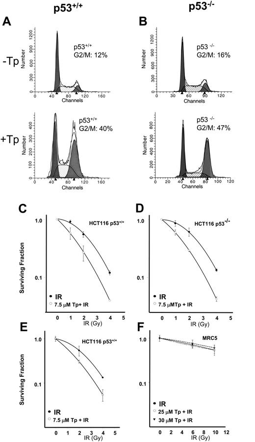
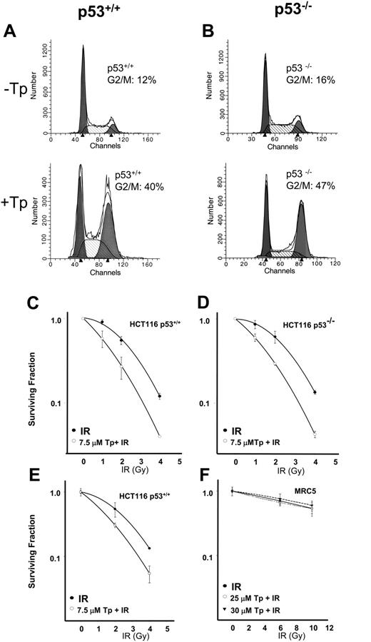
Tachpyridine induces G2/M arrest and radiosensitizes HCT 116 TP53+/+ and HCT 116 TP53-/- cells but does not radiosensitize normal diploid fibroblasts. (A) HCT 116 TP53+/+ or (B) HCT 116 TP53-/- cells were treated with 10 μM tachpyridine for 24 hours, and cell-cycle distribution was determined by flow cytometry. (C) HCT 116 TP53+/+ or (D) HCT 116 TP53-/- cells were pretreated with 7.5 μM tachpyridine for 12 hours, followed by irradiation at 1, 2, and 4 Gy, and clonogenic survival was determined as described in “Materials and methods.” (E) HCT 116 TP53+/+ cells were pretreated with 7.5 μM tachpyridine for 12 hours, followed by irradiation at 1, 2, and 4 Gy, and viability was determined by the MTT assay as described in “Materials and methods.” (F) MRC5 fibroblast cells were pretreated with 25 or 30 μM tachpyridine for 12 hours, followed by irradiation at 6 and 10 Gy, and viability was determined by the MTT assay. Data shown are the mean and SE of 3 independent experiments performed in triplicate.
Tachpyridine induces G2/M arrest and radiosensitizes HCT 116 TP53+/+ and HCT 116 TP53-/- cells but does not radiosensitize normal diploid fibroblasts. (A) HCT 116 TP53+/+ or (B) HCT 116 TP53-/- cells were treated with 10 μM tachpyridine for 24 hours, and cell-cycle distribution was determined by flow cytometry. (C) HCT 116 TP53+/+ or (D) HCT 116 TP53-/- cells were pretreated with 7.5 μM tachpyridine for 12 hours, followed by irradiation at 1, 2, and 4 Gy, and clonogenic survival was determined as described in “Materials and methods.” (E) HCT 116 TP53+/+ cells were pretreated with 7.5 μM tachpyridine for 12 hours, followed by irradiation at 1, 2, and 4 Gy, and viability was determined by the MTT assay as described in “Materials and methods.” (F) MRC5 fibroblast cells were pretreated with 25 or 30 μM tachpyridine for 12 hours, followed by irradiation at 6 and 10 Gy, and viability was determined by the MTT assay. Data shown are the mean and SE of 3 independent experiments performed in triplicate.
G2 arrest does not require p53
Our previous observations demonstrated that p53 is stabilized in cells treated with tachpyridine, although activation of p53 is not required for tachpyridine-mediated cell death.24 Because p53 can contribute to G2 cell-cycle arrest,36 we explored the role of p53 in tachpyridine-mediated G2 cell-cycle arrest. TP53 wild-type and TP53 knock-out HCT 116 human colon carcinoma cell lines were assessed for their response to tachpyridine. Cells were treated with 10 μM tachpyridine for 24 hours and analyzed by flow cytometry. As shown in Figure 3A-B, arrest in G2/M was observed and occurred to a similar extent in the presence and absence of p53. Analysis of p53 protein levels in the HCT 116 cell lines by Western blot analysis confirmed the lack of p53 protein expression in the knock-out cell line compared with the TP53 wild-type cell line (not shown).
Tachpyridine enhances the effect of ionizing radiation in cancer cells regardless of p53 status
G2/M is the most radiosensitive phase of the cell cycle,37 and agents that arrest cells in G2/M are known to be effective radiosensitization agents (reviewed in Wilson38 and Tenzer and Pruschy39 ). Because tachpyridine arrests cells in G2, experiments were performed to determine whether it acts as a radiosensitizer in cancer cells. We initially assessed radiosensitization in HCT 116 cells by clonogenic survival assays. As anticipated, exposure of HCT 116 cells to IR led to a dose-dependent decrease in cell survival compared with untreated cells (Figure 3C). We then tested whether pretreatment with tachpyridine would augment the sensitivity of these cells to IR. Cells were pretreated for 12 hours with 7.5 μM tachpyridine. This dose was selected because it only modestly decreased cell survival in response to tachpyridine alone (data not shown) and therefore facilitated assessment of the combined effects of tachpyridine and IR. After the 12 hours of pretreatment, cells were exposed to IR and allowed to recover for 1 hour. Tachpyridine was then removed, and surviving cells were allowed to form colonies. As shown in the clonogenic survival curves depicted in Figure 3C, tachpyridine increased the sensitivity of cells to the cytotoxic effect of ionizing radiation. The reduction in survival in cells pretreated with tachpyridine before exposure to IR was greater than that achieved with either tachpyridine or IR alone. Radioenhancement occurred at low levels of IR (1, 2, and 4 Gy), which represent clinically relevant doses of ionizing radiation exposure.
We also addressed the contribution of p53 to the radiation sensitization effect by using TP53 knock-out HCT 116 cells. As with the wild-type cell line, TP53 knock-out cells were also radioenhanced by tachpyridine (Figure 3D). Furthermore, these cells were sensitized to the same extent as p53 wild-type cells, indicating a p53-independent radiosensitization response.
To investigate whether the radioenhancing effect of tachpyridine was limited to HCT 116 cell lines, experiments were performed in RKO cells, another colon cancer cell line. Tachpyridine also enhanced the effect of IR in RKO cells as determined by clonogenic assays (data not shown).
Radiation sensitizers are clinically valuable because they enable a reduction in the dose of ionizing radiation. The efficacy of radiation sensitizers can be quantified by calculating a DER (dose enhancement ratio), which measures the fold increase in cytotoxic efficacy of ionizing radiation when delivered in the presence of the radiosensitizer. Dose enhancement ratios were calculated at 10% surviving fraction (SF) from radiation survival curves. For the HCT 116 TP53 wild-type cell line, a DER value at 10% SF of 1.39 ± 0.15 SEM (n = 3) was calculated (Figure 3C). The TP53 knock-out HCT 116 cell line produced a DER value at 10% SF of 1.35 ± 0.04 SEM (n = 3) (Figure 3D). These values are comparable to reported DER values of currently used radiosensitizers such as 5-fluoruracil (5-FU). For example, 5-FU results in DER values at 10% SF of 0.89 in HCT 116 TP53 wild-type cells, and 1.24 in HCT 116 TP53 knock-out cells.40 DER values of 1.70 and 2.05 in RKO cells were calculated at 10% SF for 30 μM and 45 μM tachpyridine, respectively.
Tachpyridine does not enhance the sensitivity of noncancer cells to ionizing radiation
The effect of radiation in normal tissue is an important consideration when studying radioenhancers. The desired effect is to achieve greater cytotoxicity in the tumor-cell environment with little effect in the surrounding normal tissue, thus preventing undesirable side effects. To assess potential effects of radiation on tachpyridine-treated normal cells, MRC5 human diploid fibroblasts were compared with HCT 116 cells. Because MRC5 human fibroblasts are poorly clonogenic, we used a modified MTT cell-survival assay to assess radiosensitivity (see “Materials and methods”), and compared this assay with the clonogenic assay we performed in HCT 116 cells. As shown in Figure 3E, the HCT 116 cancer cell line responded similarly in the MTT assay and clonogenic assay. In fact, 7.5 μM tachpyridine produced a nearly identical response curve in the MTT assay and the clonogenic assay across the same IR doses: DER values calculated at 50% surviving fraction were 1.46 for the MTT assay and 1.61 for the clonogenic assay. This demonstrates the validity of the MTT assay as a confirmatory technique for the clonogenic method.
From these results, the MTT assay was used to assess whether tachpyridine enhances radiation sensitivity in normal cells. As shown in Figure 3F, in contrast to the cancer cell lines we studied, tachpyridine does not enhance the effect of radiation in MRC5 cells. This observation is true even at 4 times the dose of tachpyridine and doses of IR as high as 10 Gy (Figure 3F).
Tachpyridine activates cell-cycle checkpoint kinases
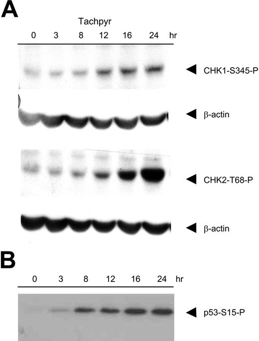
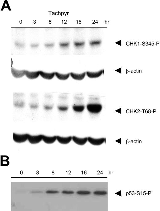
To explore the mechanism of tachpyridine-mediated G2 arrest, we tested the involvement of the checkpoint kinases. These kinases induce G2 arrest in response to genotoxic stress, at least in part through phosphorylation of Cdc25C.41,42 CHK1 is activated primarily in response to UV damage and replication arrest,34,43,44 whereas CHK2 is activated in response to ionizing radiation.45,46 However, recent work has suggested the existence of cross talk between these 2 pathways.47-50 To test whether tachpyridine activated the CHK kinases, HeLa cells were treated with 7.5 and 10 μM tachpyridine and analyzed by Western blotting using phosphospecific antibodies against activated CHK1 (S-345) and CHK2 (T-68). As shown in Figure 4A, tachpyridine induced a time-dependent activation of both kinases, with activation beginning at approximately 12 hours in both cases.
To test the relative contribution of each of these kinases to tachpyridine-mediated cell-cycle arrest we first examined the ability of tachpyridine to induce cell-cycle arrest in CHK2 knockout cells. We found that cell-cycle arrest by tachpyridine proceeded in CHK2 knock-out cells: the fraction of cells in G2/M rose from 17% ± 5% to 39% ± 8% (SD, n = 3) in cells treated with 7.5 or 10 μM tachpyridine, indicating that CHK2 is dispensible for tachpyridine-mediated cell-cycle arrest. We also examined the effect of UCN-01, a CHK1 kinase inhibitor, on cell-cycle arrest mediated by tachpyridine. As shown in Table 2, treatment of HCT 116 cells with 100 nM UCN-01 blocked the accumulation of tachpyridine-treated cells in G2. UCN-01 itself did not affect the fraction of cells in G2/M. These results point to an important role of CHK1 in tachpyridine-mediated G2 arrest.
Tachpyridine activates cell-cycle checkpoint kinases CHK1 and CHK2 and induces phosphorylation of p53 on serine-15. (A) HeLa cells were treated with 7.5 μM tachpyridine and analyzed by Western blotting using phosphospecific antibodies against activated CHK1 (S-345) and CHK2 (T-68). β-Actin was included as a loading control. (B) HeLa cells were treated with 10 μM tachpyridine and analyzed by Western blotting using a phosphospecific antibody against phosphorylated p53 (S-15). Equivalent loading of protein was confirmed by Ponceau S staining of the transfer membrane (not shown).
Tachpyridine activates cell-cycle checkpoint kinases CHK1 and CHK2 and induces phosphorylation of p53 on serine-15. (A) HeLa cells were treated with 7.5 μM tachpyridine and analyzed by Western blotting using phosphospecific antibodies against activated CHK1 (S-345) and CHK2 (T-68). β-Actin was included as a loading control. (B) HeLa cells were treated with 10 μM tachpyridine and analyzed by Western blotting using a phosphospecific antibody against phosphorylated p53 (S-15). Equivalent loading of protein was confirmed by Ponceau S staining of the transfer membrane (not shown).
Phosphorylation of p53 on serine 15 precedes activation of the checkpoint kinases
CHK1 and CHK2 are phosphorylated by ATM/ATR, members of the phosphoinositide 3-kinase-related kinase (PIKK) kinase family. To test whether activation of ATM/ATR might be responsible for phosphorylation of the CHK kinases in response to tachpyridine, we first examined the phosphorylation of p53 at serine 15, because p53ser15 is a known substrate of ATM/ATR.51-53 As shown in Figure 4B, tachpyridine induced a time-dependent increase in phosphorylation of p53 on serine 15 in HeLa cells. Phosphorylation was evident by 3 hours, a time point preceding the activation of the CHK kinases. This time course is consistent with early activation of ATM/ATR in tachpyridine-treated cells.
ATR and CHK1 play critical roles in tachpyridine-mediated G2 arrest
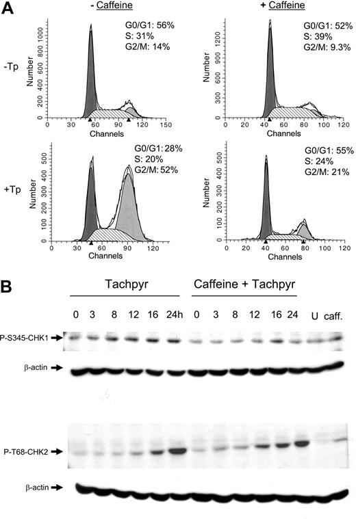
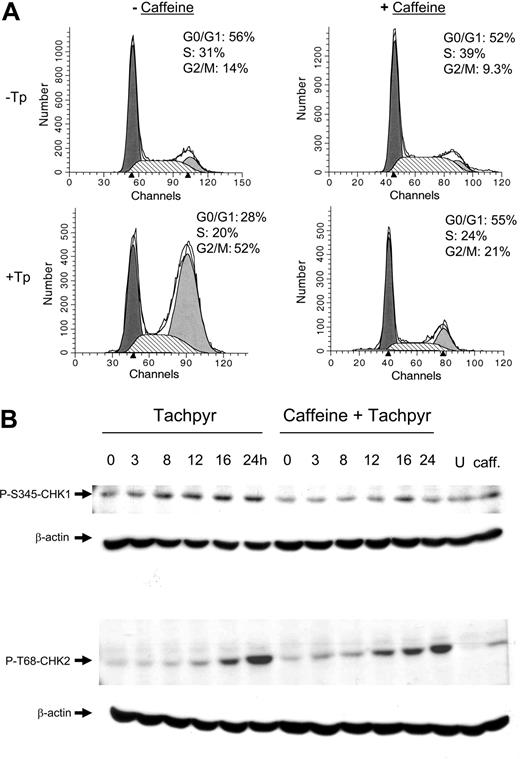
To further test the involvement of ATM/ATR in tachpyridine-mediated G2 arrest, HeLa cells were pretreated with caffeine, a PIKK kinase inhibitor that blocks activation of ATM/ATR,54,55 prior to exposure to tachpyridine. As shown in Figure 5A, pretreatment of HeLa cells with 5 mM caffeine abrogated tachpyridine-mediated G2 arrest, supporting the activation of ATM/ATR as an early event in tachpyridine-treated cells.
We also used caffeine to confirm the importance of CHK1 in triggering G2 arrest. As shown in Figure 5B, the inhibition of G2 arrest by caffeine was accompanied by a block in CHK1 phosphorylation, whereas CHK2 phosphorylation was not affected. These results are consistent with an important role for CHK1 in tachpyridine-mediated G2 arrest and suggest that the role of CHK2 may be less critical.
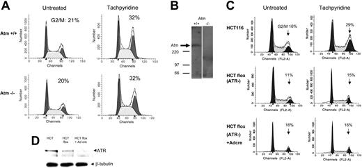
To probe whether ATM or ATR was the proximal kinase responsible for tachpyridine-mediated G2 arrest, we first tested the ability of tachpyridine to induce G2 arrest in isogenic ATM-deficient and -reconstituted cells.35 As shown in Figure 6A-B, tachpyridine induced an equivalent arrest regardless of the status of ATM, indicating that ATM is dispensable for tachpyridine-induced G2 arrest. To probe the role of ATR, we used HCT 116 cells containing a single functional ATR allele into which lox sites flanking exon 2 of the ATR gene have been introduced by homologous recombination.34 As previously observed, endogenous levels of ATR in this line, designated ATRflox/-, are reduced by approximately 80% compared with the parental HCT 116 cell line (Figure 6D). Furthermore, infection of ATRflox/- cells with a recombinant adenovirus expressing cre recombinase (Ad-cre) further reduces ATR levels. As shown in Figure 6C, tachpyridine-mediated G2 arrest was not observed in ATR-deficient cells in either the presence or absence of Ad-cre. Thus, a reduction in ATR levels abrogates tachpyridine-mediated G2 arrest, indicating the importance of ATR in activating G2 arrest in response to tachpyridine.
Caffeine inhibits tachpyridine-induced G2 arrest and activation of tachpyridine-induced CHK1 but not CHK2. (A) HeLa cells were either not pretreated or pretreated for 12 hours with 5 mM caffeine, followed by 18 hours of treatment with 7.5 μM tachpyridine, and analyzed by flow cytometry. (B) HeLa cells were either not pretreated or pretreated for 12 hours with 5 mM caffeine, followed by treatment with 7.5 μM tachpyridine over a time course of 3 to 24 hours, and analyzed by Western blotting (U indicates untreated control at 24 hours; caff, cells treated with caffeine alone for 24 hours).
Caffeine inhibits tachpyridine-induced G2 arrest and activation of tachpyridine-induced CHK1 but not CHK2. (A) HeLa cells were either not pretreated or pretreated for 12 hours with 5 mM caffeine, followed by 18 hours of treatment with 7.5 μM tachpyridine, and analyzed by flow cytometry. (B) HeLa cells were either not pretreated or pretreated for 12 hours with 5 mM caffeine, followed by treatment with 7.5 μM tachpyridine over a time course of 3 to 24 hours, and analyzed by Western blotting (U indicates untreated control at 24 hours; caff, cells treated with caffeine alone for 24 hours).
G2/M arrest proceeds in ATM-deficient cells but is inhibited in ATR-deficient cells. (A) Cells were treated with 10 μM tachpyridine for 24 hours and analyzed by flow cytometry. (B) Western blotting confirms absence of ATM in ATM-deficient cells. (C) ATRflox/- cells were treated with Adeno-Cre at a multiplicity of infection of 10 for 2 days to eliminate ATR expression. Cells were then treated with 10 μM tachpyridine for 24 hours and analyzed by flow cytometry. (D) Western blotting was used to assess ATR levels in HCT 116 parental cells, HCT 116 ATRflox-/- cells, and HCT 116 ATRflox-/- cells treated with Adeno-Cre.
G2/M arrest proceeds in ATM-deficient cells but is inhibited in ATR-deficient cells. (A) Cells were treated with 10 μM tachpyridine for 24 hours and analyzed by flow cytometry. (B) Western blotting confirms absence of ATM in ATM-deficient cells. (C) ATRflox/- cells were treated with Adeno-Cre at a multiplicity of infection of 10 for 2 days to eliminate ATR expression. Cells were then treated with 10 μM tachpyridine for 24 hours and analyzed by flow cytometry. (D) Western blotting was used to assess ATR levels in HCT 116 parental cells, HCT 116 ATRflox-/- cells, and HCT 116 ATRflox-/- cells treated with Adeno-Cre.
Discussion
The potential uses of iron chelators in the treatment of human disease, including neurodegenerative diseases, ischemia-reperfusion injury, iron overload, and cancer, are areas of active investigation. Several iron chelating agents are currently in cancer clinical15 and preclinical16,17 trials. An understanding of the intracellular mechanisms of action of iron chelators may be beneficial in optimizing their efficacy, as well as providing fundamental insights into the nature of intracellular targets of iron chelation. Here, we explore the mechanism of action of tachpyridine, a member of a chelator family currently in preclinical development with the NCI as a cancer therapeutic.
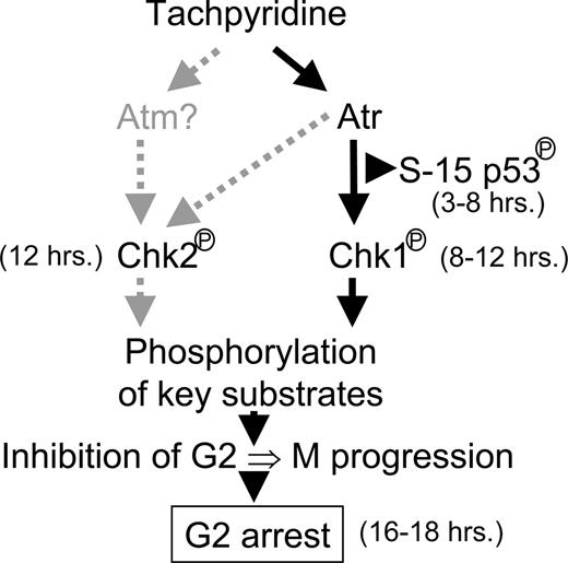
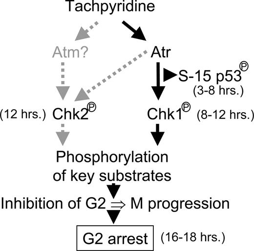
In this manuscript, we show that tachpyridine arrests cells in the G2 phase of the cell cycle. This result was unanticipated, because the majority of iron chelators arrest cells at the G1/S boundary.4,28-30,56-58 Although tachpyridine preferentially binds iron, it can also bind copper and zinc,27 and these activities may underlie its ability to arrest cells in G2. Alternatively or additionally, tachpyridine may induce a form of genotoxic stress different from that induced by other iron chelators, and this may trigger G2 arrest. Our results demonstrate that ATR and CHK1 are critical mediators of tachpyridine-induced G2 arrest. We also provide evidence that an important consequence of tachpyridine-mediated G2 arrest is the selective sensitization of cancer cells to ionizing radiation. A working model for the mechanism of G2 arrest induced by tachpyridine is shown in Figure 7.
Checkpoint kinases are activated in response to cellular stress, such as DNA damage. A major role of these kinases is the triggering of cell-cycle arrest to enable DNA repair. The activation of CHK1 or CHK2 is in part dependent on the type of DNA damage. For example, ionizing radiation primarily activates CHK2, whereas replicative stress and UV activate CHK1, although overlap between these pathways exists.47-50,59,60 Thus, DNA damage induced by exposure to alkylating agents activates both CHK1 and CHK2.50 Similarly, we found that tachpyridine activates both CHK1 and CHK2 (Figure 4A). The time course of activation of both kinases was comparable, suggesting that they are activated simultaneously.
Despite the activation of both CHK1 and CHK2, our experiments suggest that CHK1 may play the principal role in triggering G2 arrest in response to tachpyridine. First, inhibition of G2 arrest in cells treated with caffeine was associated with inhibition of tachpyridine-induced CHK1 phosphorylation (Figure 5). In contrast, under these same conditions, caffeine did not block phosphorylation of CHK2. Second, G2 arrest was not blocked in CHK2 knock-out cells. Finally, UCN-01, a CHK1 kinase inhibitor, blocked tachpyridine-mediated G2 arrest (Table 2). However, because neither caffeine nor UCN-01 are entirely selective in their inhibitory activity,34,61 and because CHK1 and CHK2 exhibit partial redundancy, we cannot completely exclude the possibility that under selected circumstances CHK2 can be recruited by tachpyridine and substitute for CHK1.
Working model of the mechanism of tachpyridine-induced G2 arrest. Tachpyridine activates ATR (and possibly ATM), which phosphorylates p53 on serine 15, followed by phosphorylation of CHK1 and CHK2. The CHK kinases then signal G2 arrest through phosphorylation of key substrates important for G2 to M progression. Signaling events not required for G2 arrest are indicated by gray dashed lines.
Working model of the mechanism of tachpyridine-induced G2 arrest. Tachpyridine activates ATR (and possibly ATM), which phosphorylates p53 on serine 15, followed by phosphorylation of CHK1 and CHK2. The CHK kinases then signal G2 arrest through phosphorylation of key substrates important for G2 to M progression. Signaling events not required for G2 arrest are indicated by gray dashed lines.
Known upstream activators of the CHK kinases include the ATM/ATR/ATX family of PIKK kinases. ATM and ATR participate in the response to DNA damage by recruiting multiprotein DNA repair complexes to sites of DNA damage. To test whether activation of these upstream kinases might be responsible for phosphorylation of the CHK kinases, we first examined the phosphorylation of p53 at serine 15 in cells treated with tachpyridine. p53ser15 is a known substrate of the ATM/ATR kinases.51-53 Although some studies suggest that DNA-PK (DNA-activated protein kinase) may also phosphorylate p53 at serine 15,62,63 this finding is controversial.62,64-68 We observed that p53 accumulated and was phosphorylated at serine 15 in a time-dependent manner, with phosphorylation observed as early as 3 hours (Figure 4B). This time point preceded activation of the CHK kinases (activated at 8-12 hours; Figures 4A,5B) and is consistent with the model shown in Figure 7, in which early activation of ATM/ATR triggers phosphorylation of p53 and the CHK kinases.
The second line of evidence supporting the involvement of ATM/ATR in tachpyridine-mediated cell-cycle arrest is our finding that caffeine, an inhibitor of the PIKK kinases, abrogated G2 arrest (Figure 5A). Further, ATR may be more critical than ATM in triggering the G2 response to tachpyridine. Thus, G2 arrest proceeded in cells genetically deficient in ATM (Figure 6A-B). In contrast, G2 arrest was not detected in HCT 116 cells containing reduced levels of ATR. Because knockout of ATR is lethal, these HCT 116 cells were designed to conditionally knock out ATR expression following introduction of cre recombinase.34 However, we and others34,59 have found that these cells express reduced levels of ATR even in the absence of cre. As shown in Figure 6C-D, HCT 116 ATR-deficient cells did not exhibit G2 arrest in response to tachpyridine, indicating that unlike ATM, ATR is required for G2 arrest.
Despite our observation that p53 accumulates and is phosphorylated in cells treated with tachpyridine (Figure 4B), our data suggest that p53 does not play an essential role in tachpyridine-mediated apoptosis24 or cell-cycle arrest (Figure 3). We have previously shown that p53 activated in response to tachpyridine is not fully transcriptionally competent, because p21, a transcriptional target of p53, is not induced in cells treated with tachpyridine.24 Although the nature of the block to p53-dependent transactivation in tachpyridine-treated cells remains unclear, this block may limit the contribution of p53 to both apoptosis and cell-cycle arrest, because transcriptional targets of p53 (eg, bax [bcl-2 (B-cell lymphoma-2) associated X protein], DR5 [death receptor-5], p21) contribute to both pathways. Ultimately, the ability of tachpyridine to enhance the response to ionizing radiation in a p53-independent manner may enhance its therapeutic utility, because of the wide-spread loss of the p53 pathway in human cancer.69,70
There are potentially important clinical ramifications for our findings. Because the G2/M phase of the cell cycle is the most sensitive to ionizing radiation,71-75 and we had observed striking G2 arrest with sublethal doses of tachpyridine, we explored the ability of tachpyridine to sensitize cells to the cytotoxic effects of ionizing radiation. We pretreated cells with tachpyridine prior to exposing them to ionizing radiation. Tachpyridine pretreatment enhanced the sensitivity of HCT 116 and RKO colon cancer cells to clinically relevant doses of ionizing radiation and did so via a p53-independent mechanism (Figure 3). Importantly, the sensitivity of noncancer MRC5 cells to ionizing radiation was not similarly enhanced (Figure 3F). In addition, the DER values derived from radiation survival curves with tachpyridine were comparable to those reported in the literature for clinically useful radiosensitizers, such as 5-FU.40 Notably, the concentrations of tachpyridine required to achieve radiosensitization are readily achievable in vivo (J. McCormack, R.P.P., and S.V.T., unpublished observations, March 1999). Our experiments therefore provide evidence that the iron chelator tachpyridine functions as a radioenhancer, an effect separable and distinct from our previous work that showed that tachpyridine functions as a cytotoxic agent.
Although the ability of other iron chelators to serve as radiosensitization agents has been observed (these include DFO, deferiprone, HBED [N,N′-bis(2-hydroxybenzyl)ethylenediamine-N,N′-diacetic acid], and mimosine),76 not all reports are consistent,77-79 and molecular mechanisms of chelator-mediated radiosensitization are poorly understood. However, one study using mimosine demonstrated that mimosine radiosensitized the human lung cancer cell lines H460 and H358,76 which differ in p53 status, consistent with our results that tachpyridine sensitizes cells to ionizing radiation via a p53-independent mechanism (Figure 3).
An additional implication of these experiments is that they suggest a point of convergence in the mechanism of action of different iron chelators. Unlike the G2 arrest reported here for tachpyridine, cell-cycle arrest induced by other iron chelators typically occurs at the G1/S boundary.4,28-30,56-58 This effect is generally attributed to inhibition of ribonucleotide reductase, an iron-dependent enzyme80,81 that catalyzes the rate-limiting step in DNA synthesis. For example, this enzyme is inhibited by desferrioxamine, a chelator in current clinical use in the treatment of iron overload, as well as by Triapine (Vion Pharmaceuticals, New Haven, CT), a chelator in clinical trials as an anticancer agent. Because replication arrest also represents a trigger for activation of a DNA damage cell-cycle checkpoint, activation of checkpoint kinases CHK1 and CHK2 may represent a point of convergence for iron chelators regardless of their proximal target; that is, the CHK kinases may be a focal point for a variety of iron chelators that target G1/S as well as those that target G2/M. Cell stresses that underlie the activation of CHK kinases by chelators remain to be elucidated, but they are likely to include metal depletion and/or genotoxic stress, which can be induced directly or indirectly by metal chelators.82 Further experiments will be required to fully define these mechanisms.
In summary, we have shown that tachpyridine triggers activation of the CHK kinases via an ATR-dependent mechanism, leading to cell-cycle arrest in G2. This is accompanied by a striking sensitization to ionizing radiation, which may have clinical implications. These studies suggest for the first time that ATR and the CHK kinases are targets of metal chelation.
Prepublished online as Blood First Edition Paper, July 12, 2005; DOI 10.1182/blood-2005-03-1263.
Supported by the National Institutes of Health (grant no. DK 57781) (S.V.T.); preclinical development of tachpyridine was supported by a grant from the National Cancer Institute (NCI) Rapid Access to Intervention Development (RAID) and the NCI Rapid Access to NCI Discovery Resources (RAND) programs of the National Institutes of Health (S.V.T.); and grant CA102289 (K.B.).
The publication costs of this article were defrayed in part by page charge payment. Therefore, and solely to indicate this fact, this article is hereby marked “advertisement” in accordance with 18 U.S.C. section 1734.
We thank Dr Ken Wheeler for thoughtful discussions and advice, and Christine Naczki and Dawn Eads for their vital technical assistance on clonogenic and MTT assays (Wake Forest University Health Sciences).

