FTY720, a potent immunomodulatory drug in phase 2/3 clinical trials, induces rapid and reversible sequestration of lymphocytes into secondary lymphoid organs, thereby preventing their migration to sites of inflammation. As prerequisite for its function, phosphorylation of FTY720 to yield a potent agonist of the sphingosine-1-phosphate receptor S1P1 is required in vivo, catalyzed by an as-yet-unknown kinase. Here, we report on the generation of sphingosine kinase 2 (SPHK2) knockout mice and demonstrate that this enzyme is essential for FTY720 phosphate formation in vivo. Consequently, administration of FTY720 does not induce lymphopenia in SPHK2-deficient mice. After direct dosage of FTY720 phosphate, lymphopenia is only transient in this strain, indicating that SPHK2 is constantly required to maintain FTY720 phosphate levels in vivo.
Introduction
Naive T cells regularly circulate between the bloodstream and lymphatic tissue in search for foreign antigen, as well as for tumor and autoantigen. Their activation in secondary lymphoid organs followed by regulated egress back into the circulation to reach sites of inflammation is a prerequisite for any adaptive immune response in the T-cell compartment. Recently, one of the G protein-coupled receptors for sphingosine-1-phosphate (S1P), namely the S1P1 receptor, was shown to be crucial for the tempo-spatial trafficking of T cells into and out of the secondary peripheral lymphoid organs.1 The importance of S1P1 in lymphocyte trafficking became clear through studies with FTY720, an analog of sphingosine. FTY720, after phosphorylation in vivo to FTY720 phosphate (FTY720-P), induces a reversible sequestration of lymphocytes into lymph nodes and Peyer patches.2,3 FTY720-P thereby acts as a functional antagonist of the S1P1 receptor, thus inducing aberrant internalization and consequently rendering T cells unresponsive to the obligatory egress signal provided by S1P.1,4-6
FTY720 has emerged as a potent immunomodulatory agent with usefulness in the control of organ transplant rejection and for treatment of autoimmune diseases. In animals, FTY720 is efficacious in prolonging graft survival, as well as in models of multiple sclerosis, acute lung injury, autoimmune diabetes, atherosclerosis, and renal ischemia-reperfusion injury.7 Promising results have been obtained from human trials on FTY720 for indications in renal transplantation and multiple sclerosis.7,8 Since FTY720 prodrug activation is essential for its action on T (and B) cells, understanding how the drug gets phosphorylated in vivo is of high interest, in particular for the design of novel analogs with altered pharmacologic properties.
FTY720 is known to be phosphorylated in vitro by the 2 mammalian sphingosine kinases SPHK1 and 2, with SPHK2 being considerably more efficient.9-11 As shown by a recent study in SPHK1-deficient mice,12 this enzyme appears to be dispensable for the action of FTY720 in vivo, as Sphk1-null mice are still rendered lymphopenic by the drug.
In this study, we describe the generation of a mouse strain deficient in SPHK2. These animals do not become lymphopenic upon administration of FTY720 due to drastic reduction in FTY720-P formation; this defect can be functionally rescued by dosage of FTY720-P. Thus, SPHK2 was identified as the enzyme essential for FTY720 prodrug activation in vivo.
Materials and methods
Generation of Sphk-/- mice
To generate Sphk1-/- mice, a targeting vector for homologous recombination was designed by cloning 1.3-kb genomic DNA containing Sphk1 exon 2 and 1.4-kb genomic DNA containing part of Sphk1 exon 6 into vector pRAY2 (Genbank accession no. U63120; http://www.ncbi.nlm.nih.gov/Genbank/GenbankOverview.html) harboring a neomycin expression cassette. For generation of Sphk2 knockout mice, a vector was designed by cloning 2.1-kb genomic DNA containing Sphk2 exons 1 and 2 and 1.6-kb genomic DNA containing Sphk2 exon 7 into vector pRAY2. After introduction into Balb/c embryonic stem cells, neomycin-resistant clones were screened by polymerase chain reaction (PCR) for homologous recombination. Correct targeting was confirmed by Southern blot analysis using gene-specific probes, while random integration was excluded likewise with a neomycin-specific probe. Selected cells were injected into C57BL/6 blastocysts and chimeric male mice were bred to Balb/c females, resulting in an F1 generation of heterozygous (+/-) inbred Balb/c mice, which allowed the generation of homozygous (-/-) mice. Genomic DNA was isolated from tail samples of the progeny. Primers used: Sphk2, exon 5, F: 5′AGGCATTGTCACTGTGTCTGG; Sphk2, exon 7, R: 5′AGGTCAACACCGACAACCTGCTC, neo F: 5′GGCCGGAGAACCTGC.
For experiments, Sphk2+/+ littermates of the Sphk2-/- (from breeding of Sphk2+/-) were used as wild-type (WT) controls. Mice were between 6 to 10 weeks old. The experimental procedures met all regulations and standards and were approved by the Austrian government.
mRNA expression analysis by real-time PCR
Total RNA was isolated using an Absolutely RNA Miniprep Kit (Stratagene, La Jolla, CA). For real-time PCR, an ABI PRISM 7900HT Sequence Detection System (Applied Biosystems, Weiterstadt, Germany) was used. Primers were designed using Primer Express 2.x software (Applied Biosystems). For relative quantification, data were analyzed using the ΔΔCT method as described previously.9 Expression levels of target genes in each sample were normalized to the average of housekeeping genes.
Analysis of SPHK activity in mouse tissues
Activity in the cytosolic fraction of mouse tissues and in whole blood was determined as described previously,9 using sphingosine or FTY720 and γ-[32P]ATP as substrates. Reaction buffers favored either SPHK1 activity (buffer 1; 50 mM HEPES, pH 7.4, 0.5% Triton, 15 mM MgCl2, 10% glycerol, 10 mM NaF, and 1.5 mM semicarbazide) or SPHK2 activity (buffer 2; 50 mM HEPES, pH 7.4, 15 mM MgCl2, 1 M KCl, 10% glycerol, 10 mM NaF, and 1.5 mM semicarbazide). Labeled lipids were extracted and separated by thin-layer chromatography (TLC); S1P and FTY720-P were quantified with a Storm 840 PhosphoImager (Molecular Dynamics, Sunnyvale, CA).9 Activity is reported as milliunits per gram (equal to nanomole of phosphorylated product formation per minute per gram of tissue).
Measurement of S1P, FTY720, and FTY720-P concentrations
Levels of S1P in serum, plasma, and tissues were determined by high-performance liquid chromatography (HPLC; Agilent, Palo Alto, CA) with mass detection. Samples were spiked with internal standard (C17-S1P; Avanti Polar Lipids, Alabaster, IL) and extracted with chloroform/methanol at acidic pH; extracts were subjected to acetylation with acetanhydride in pyridine (40°C, 20 minutes) as described.13 Following evaporation of solvents, samples were dissolved in methanol/0.2% formic acid and injected onto an Eclipse XDB C8 column (5μ, 4.6 × 150 mm; Agilent), which was eluted with a gradient (eluent A: 5 mM HCOONH4 + 0.1% HCOOH in MeOH/H2O (80/20); eluent B: 5 mM HCOONH4 + 0.1% HCOOH in MeOH/CH3CN/H2O (49/50/1); 70% to 100% B in 10 minutes) at a flow of 0.5 mL per minute at 40°C. Negative ion electrospray-ionization liquid chromatography with tandem mass spectroscopy (LC-MS/MS) was used for detection using an API 4000 QTrap instrument (MDS Sciex, Concord, Canada) as described.13 The optimal collision energy for derivatized S1P and C17-S1P was -28 and -26 V, respectively. The multiple reaction monitoring (MRM) transitions monitored were m/z 462/402 and m/z 448/388, respectively.
For determination of FTY720 and FTY720-P in plasma, samples were spiked with internal standard and extracted with chloroform/methanol at acidic pH; extracts were dried and reconstituted in methanol/0.2% formic acid. Samples were chromatographed on a Luna C8 column (3μ, 2 × 50 mm; Phenomenex, Torrence, CA), which was eluted with a gradient (eluent A: 10 mM ammonium acetate + 800 μL HCOOH in H2O; eluent B: 10 mM ammonium acetate + 800 μL HCOOH in MeOH; 50% to 98% B in 14 minutes) at a flow of 0.4 mL/min at 40°C. Analytes were detected by positive ion electrospray-ionization LC MS/MS analysis (API 4000 QTrap). The optimal collision energies for FTY720 and FTY720-P were 23 V and 25 V, respectively. The MRM transitions monitored for FTY720 and FTY720-P were m/z 308/255 and 388/255, respectively.
FTY720 or FTY720-P treatment
Mice were dosed with FTY720 (orally or intravenously) or with FTY720-P (intravenously) at 1 mg/kg. FTY720 was dissolved in distilled water (for oral dosing) or in 20% 2-hydroxypropyl-cyclodextrin (for intravenous dosing) at 0.2 mg/mL. In the case of FTY720-P, 1 mg compound was dissolved in 50 μL DMSO plus 2.6 μL 1 N HCl, followed by subsequent addition of 450 μL Cremophor EL (Fluka, Buchs, Switzerland) and 4 mL isotonic glucose in 50 mM HEPES, pH 7.3; absence of FTY720 as potential degradation product in this formulation prior to dosage was shown by LC/MS.
Lymphocyte counts
Heparinized blood samples were collected from the orbital sinus. After determination of the total number of leukocytes on a hemocytometer (Coulter AcT Diff; Coulter Corp, Miami, FL), differential (relative) cell counts of stained blood smears were performed based on morphology and staining characteristics. The absolute lymphocyte counts were calculated by multiplying these percentages by the total number of white blood cells.
Flow cytometry
Single-cell suspensions of peripheral lymph nodes and spleens were prepared in phosphate-buffered saline (PBS) by gently pressing the organs through a 70-μm nylon cell strainer (BD Falcon, Franklin Lakes, NJ). Flow cytometric analysis was done on a FACSCalibur (Becton Dickinson [BD], Mountain View, CA). For cell differentiation, cells were stained with PE-labeled anti-CD3 (Caltag, Hamburg, Germany), or FITC-labeled anti-CD19 (BD), or with 7-amino-actinomycin D (7-AAD; BD).
Migration of lymphocytes toward FTY720
WT and Sphk2-/- mice were treated with FTY720, FTY720-P (both 1 mg/kg), or vehicle. At 6 hours (FTY720-P) and 24 hours (FTY720, FTY720-P) after dosing, mice were killed and suspensions of excised peripheral lymph nodes were prepared. Lymphocytes of compound- or vehicle-treated animals (5 × 10⁵) were used for migration experiments using S1P in serial dilutions as chemoattractant in 5-μm pore size polycarbonate tissue culture inserts (Transwell; Corning-Costar, Corning, NY). After 2.5 hours at 37°C in a humidified incubator, the Transwell inserts were removed, and the cells accumulating at the bottom of the chamber were enumerated in a hemocytometer under an Axiovert 40 microscope (Zeiss, Vienna, Austria).
Results
Generation and characterization of Sphk2 knockout mice
To knock out Sphk2 expression in mice, a gene-targeting vector was prepared and transfected in Balb/c embryonic stem cells resulting in the replacement of Sphk2 exons 3 to 6 by a neomycin-resistant gene cassette (Figure 1A). Sphk2-/- mice were generated and correct targeting of the Sphk2 locus was confirmed by PCR on genomic DNA (Figure 1B) and verified by Southern blotting (data not shown). Consequently, in these animals Sphk2 mRNA expression was undetectable by quantitative real time PCR in kidney (Figure 1C), liver, and brain (data not shown). In contrast, expression of mRNA for Sphk1 and further genes encoding various enzymes involved in sphingolipid signaling, namely of sphingosine-1-phosphate phosphatases 1 and 2 (Sgpp1 and Sgpp2) sphingosine-1-phosphate lyase (Sgpl1), ceramide kinase (Cerk), and sphingomyelin phosphodiesterases (Smpd1 to 3) was not altered (Figure 1C). Homozygous Sphk2 knockout mice did not show an obvious phenotype; they were found to be viable and fertile, and did not exhibit histologic abnormalities in the major organs. Also, the percentages of CD3+ T cells and CD19+ B cells in the spleens and in the lymph nodes of WT and SPHK2-deficient mice were not different. In analogy, Sphk1-null mice, generated for the purpose of direct comparison on the same background (Balb/c), were also viable and fertile, as previously reported for Sphk1-null mice in the C57Bl/6 inbred strain,12 and also did not deviate from WT mice in the characteristics mentioned.
Disruption of the Sphk2 gene in the mouse genome by homologous recombination. (A) Schematic representation of the mouse Sphk2 locus before and after recombination. Homologous recombination removes exons 3 to 6. (B) Detection of knockout and WT alleles by PCR analysis of genomic DNA; bands of 511 and 733 bp are expected for +/+ and -/- animals, respectively, and both bands for heterozygotes. Locations of primers are indicated in panel A. (C) mRNA expression of Sphk1, Sphk2, Sgpp1, Sgpp2, Sgpl1, Cerk, Smpd1, Smpd2, and Smpd3 in kidneys of Sphk2-/- mice relative to WT as determined by quantitative real-time PCR. The data represent mean values ± SD, n = 3.
Disruption of the Sphk2 gene in the mouse genome by homologous recombination. (A) Schematic representation of the mouse Sphk2 locus before and after recombination. Homologous recombination removes exons 3 to 6. (B) Detection of knockout and WT alleles by PCR analysis of genomic DNA; bands of 511 and 733 bp are expected for +/+ and -/- animals, respectively, and both bands for heterozygotes. Locations of primers are indicated in panel A. (C) mRNA expression of Sphk1, Sphk2, Sgpp1, Sgpp2, Sgpl1, Cerk, Smpd1, Smpd2, and Smpd3 in kidneys of Sphk2-/- mice relative to WT as determined by quantitative real-time PCR. The data represent mean values ± SD, n = 3.
SPHK activity was determined in tissues from WT and Sphk2-null mice in reaction buffers favoring either SPHK1 activity (high Triton X-100 concentration) or SPHK2 activity (high salt).9 While under SPHK1-favoring conditions no difference between the WT and the Sphk2-/- strains was observed (Figure 2A), all tissues from Sphk2-null mice showed greatly reduced activity (by at least 90%) when using SPHK2-favoring buffer (Figure 2B). Using FTY720 instead of sphingosine as a substrate, we found equivalent but minimal phosphorylation in WT and Sphk2-/- mice in high-Triton buffer, but no detectable FTY-P formation in high-salt buffer for Sphk2-/- mice, in contrast to high levels in the WT animals (data not shown). Findings in Sphk1-/- mice—namely reduced S1P formation when using high Triton X-100 concentration, but unchanged FTY720 formation under any buffer condition compared with WT mice—were in line with observations by Allende et al.12 Levels of S1P in serum as measured by LC-MS/MS were found to be reduced by about 50% in SPHK1-deficient mice (WT: 2.0 ± 0.67 μM, Sphk1-/-: 0.93 ± 0.31 μM; n = 10), consistent with previous reported data.12 Such a reduction was not observed in the serum of Sphk2-/- mice (4.2 ± 0.4 μM, n = 10).
Lack of lymphopenic effect and reduced phosphorylation of FTY720 in Sphk2-/- mice
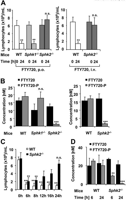
We treated SPHK1- and SPHK2-deficient mice and WT animals orally with FTY720 at a dose of 1 mg/kg. About 75% reduction in counts of peripheral lymphocytes was observed in WT, and, in line with Allende et al,12 also in Sphk1 knockout mice; in sharp contrast, Sphk2-/- mice were not rendered lymphopenic by FTY720 at all (Figure 3A, left). The same difference between WT and Sphk2-/- was also observed when the animals were dosed with FTY720 intravenously (Figure 3A, right). Corresponding analysis of plasma levels from the same individuals revealed that phosphorylation of FTY720 in the Sphk2-/- mice was reduced by at least 93% (Figure 3B), while there was no difference between WT and Sphk1-/- mice.
We asked whether FTY720-P would still function in the Sphk2 knockout mice to induce lymphopenia; the compound was dosed at 1 mg/kg by intravenous injection, since it is not orally bioavailable. In the WT mice, the phosphorylated drug-induced lymphopenia throughout the observation period up to 24 hours, comparable to the effect of dosing FTY720 itself. In the SPHK2-deficient animals, lymphopenia induced by FTY720-P was only transient: although at 6 hours the effect was comparable with that in the WT mice, it was completely lost at 24 hours (Figure 3C). The ratio of FTY720 to FTY720-P in the WT animals was approximately constant between 6 and 24 hours, while FTY720-P levels decreased in Sphk2 knockout mice (Figure 3D). This can be explained by the existence of phosphatase activity cleaving FTY720-P. While usually an equilibrium between FTY720 and FTY720-P is established by the opposing action of kinase and phosphatase activities, lack of SPHK2 in the mutant mice leads to decay of FTY720-P levels and hence to loss of lymphopenic effect. However, the fact that circumvention of the need for prodrug activation by applying the active form of the drug itself does lead to lymphopenia clearly points to the loss of SPHK2 activity as the only defect in these mice responsible for lack of effect of FTY720. These data establish that SPHK2 is critical in the phosphorylation of FTY720 in mice in vivo.
Migration to S1P is impaired for lymphocytes from FTY720-treated WT but not Sphk2-/- mice
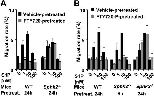
To assess the functional integrity of the lymphocytes from Sphk2-/- mice, we studied their in vitro migration to S1P with and without prior FTY720 or FTY720-P administration in vivo, both given intravenously for the sake of comparison. A typical bell-shaped dose-response curve was observed for cells from both WT and Sphk2-/- mice when isolated from animals that had received vehicle only (Figure 4). In agreement with the mode of action of FTY720, cells derived from WT animals treated with FTY720 for 24 hours in vivo failed to migrate, while cells from FTY720-treated Sphk2 knockout animals were not impaired in their migration toward S1P (Figure 4A). These experiments demonstrate the normal function of SPHK2-deficient lymphocytes in terms of migration before drug treatment. Since only the phosphorylated form of FTY720 has the capacity to down-regulate the S1P1 receptor,1,14 the unaffected migration of SPHK2-deficient lymphocytes in response to S1P is fully explained by the inability of the mutant mice to phosphorylate FTY720.
SPHK activity in tissues and blood from Sphk2-null mice. SPHK enzymatic activity in homogenates of tissues and in whole blood from Sphk2-null and WT mice. The assays were performed in buffers favoring SPHK1 activity (A) or SPHK2 activity (B). Values represent mean ± SD, with n = 3. Statistical significance was calculated using Student t test (**P < .01); no statistical significance was reached in panel A.
SPHK activity in tissues and blood from Sphk2-null mice. SPHK enzymatic activity in homogenates of tissues and in whole blood from Sphk2-null and WT mice. The assays were performed in buffers favoring SPHK1 activity (A) or SPHK2 activity (B). Values represent mean ± SD, with n = 3. Statistical significance was calculated using Student t test (**P < .01); no statistical significance was reached in panel A.
When animals were treated with FTY720-P intravenously, cells from WT animals taken at 6 hours (not shown) or 24 hours (Figure 4B, left) showed impaired migratory response to S1P. Sphk2-/- lymphocytes showed impaired migration at 6 hours, but the effect was lost after 24 hours (Figure 4B, middle and right panels). This result mirrors the transient lymphopenic effect of FTY720-P in the SPHK2-deficient mice (Figure 3C).
FTY720-P, but not FTY720 causes lymphopenia in Sphk2 knockout mice. (A) Lymphocyte counts in whole blood before and 24 hours after oral (left) or intravenous (right) treatment with 1 mg/kg FTY720. (B) FTY720 and FTY720-P plasma levels of the same individuals as in panel A at 24 hours. (C) Lymphocyte counts in whole blood before and after intravenous treatment with 1 mg/kg FTY720-P. (D) FTY720 and FTY720-P plasma levels 6 and 24 hours after dosage of FTY720-P. Results are expressed as the mean ± SD, n = 6. Statistical significance was calculated using Student t test (**P < .01; ***P < .001; n.s., not significant) in the case of panels A and C comparing lymphocyte counts before treatment (0 hours) to FTY720- or FTY720-P-treated groups, or for panels B and D comparing Sphk-null mice versus WT mice.
FTY720-P, but not FTY720 causes lymphopenia in Sphk2 knockout mice. (A) Lymphocyte counts in whole blood before and 24 hours after oral (left) or intravenous (right) treatment with 1 mg/kg FTY720. (B) FTY720 and FTY720-P plasma levels of the same individuals as in panel A at 24 hours. (C) Lymphocyte counts in whole blood before and after intravenous treatment with 1 mg/kg FTY720-P. (D) FTY720 and FTY720-P plasma levels 6 and 24 hours after dosage of FTY720-P. Results are expressed as the mean ± SD, n = 6. Statistical significance was calculated using Student t test (**P < .01; ***P < .001; n.s., not significant) in the case of panels A and C comparing lymphocyte counts before treatment (0 hours) to FTY720- or FTY720-P-treated groups, or for panels B and D comparing Sphk-null mice versus WT mice.
Migratory behavior of lymphocytes to different concentrations of S1P. Ex vivo chemotaxis assay using unsorted T and B cells from WT or Sphk2 knockout mice treated for 24 hours with FTY720 (A) or for 6 or 24 hours with FTY720-P (B) (1 mg/kg intravenously, each). The percentage of the input cell population that responded to S1P was determined (mean values ± SD, n = 6).
Migratory behavior of lymphocytes to different concentrations of S1P. Ex vivo chemotaxis assay using unsorted T and B cells from WT or Sphk2 knockout mice treated for 24 hours with FTY720 (A) or for 6 or 24 hours with FTY720-P (B) (1 mg/kg intravenously, each). The percentage of the input cell population that responded to S1P was determined (mean values ± SD, n = 6).
Discussion
We report here on the generation of SPHK2-deficient mice, a strain that we found to be viable and without obvious phenotype. Deficiency of the enzyme was proven on the genomic and mRNA level. Consistently, on the protein level, we found more than 90% reduction of SPHK activity in a high-salt buffer favoring SPHK2 activity. In part, the low residual S1P generation in this buffer can be attributed to SPHK1, which has minimal activity under high-salt conditions.9 Furthermore, based on data from Igarashi and coworkers,15 who demonstrated the existence of SPHK activity not attributable to the types 1 and 2 enzymes, additional enzyme activity may have to be considered.
As had previously been suggested by in vitro studies using recombinant enzymes,9-11 SPHK2 was here identified as the enzyme that is primarily responsible for phosphorylation of FTY720 in vivo (Figure 3B). At 24 hours after oral dosage of FTY720, concentrations of the drug are similar in the WT and the Sphk2 knockout mice, while doubling might have been expected in the knockout mice due to absence of the phosphorylated form. However, there is preliminary evidence that oxidative metabolism occurs only on the parent compound, not on the phosphorylated form (J. Kovarik, Novartis; personal written communication, August 2005); therefore, faster elimination of FTY720 in the knockout strain is conceivable.
As a consequence of the largely abolished phosphorylation of FTY720 in the Sphk2-/- mice, the drug does not induce lymphopenia in this mouse strain (Figure 3A), and fails to impair migratory capacity of lymphocytes toward S1P (Figure 4). These experiments highlight that the phosphorylated form of FTY720 is essential for both lymphodepletion and abrogation of signaling through the S1P1 receptor. The low residual FTY720-P levels encountered in Sphk2-/- mice, contributed by SPHK1, are pharmacologically not relevant in the test systems studied here. This makes Sphk2-/- mice an important tool to dissect between actions of FTY720 dependent on its phosphorylated form and potential effects that do not require formation of FTY70-P.
Prepublished online as Blood First Edition Paper, October 13, 2005; DOI 10.1182/blood-2005-07-2628.
All of the authors are employed by Novartis Pharma, whose potential product FTY720 was studied in the present work.
B.Z. designed and performed the lymphodepletion and migration studies; B.K. designed and constructed the vectors for targeted disruption of the Sphk genes; M.M. planned and performed the ES-cell transfection, blastocyst injection, and breeding of the KO mice; R.R. established and performed LC/MS analytics; D.M. designed and performed RT-PCR measurements; N.U. performed genotyping of the mice and assisted in the conduction of lymphodepletion and migration studies; F.B. provided guidance for the study design and took part in the data analysis; T.B. provided guidance for the study design, took part in the data analysis, and edited the manuscript; and A.B. measured SPHK activity, provided guidance for the study design, analyzed data, and edited the manuscript.
The publication costs of this article were defrayed in part by page charge payment. Therefore, and solely to indicate this fact, this article is hereby marked “advertisement” in accordance with 18 U.S.C. section 1734.
We thank Drs P. Ettmayer and P. Nussbaumer and R. Csonga, E. Dobrowolski, T. Doll, M. Haffner, M. Hahn, E. Haupt, M. Lemaistre, L. Malacek, E. Pursch, R. Kutil, W. Mayer-Granitzer, V. Schuler, and A. Wlachos for discussion and technical assistance.


