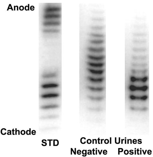
The brief report by Beullens et al1 is misleading regarding the urine test that the World Anti-Doping Agency (WADA) uses to detect recombinant human erythropoietin (rhEpo). The WADA-recommended test is based on immunoelectorphoresis and double blotting (IEF/DB), and was developed by Lasne and de Ceaurriz in 2000.2
Our WADA-accredited laboratory has performed the IEF/DB test for rhEpo on more than 6800 urine samples, including more than 2600 doping control samples from athletes. Of the latter, we have reported 9 positive cases for rhEpo: 3 of these have publicly confessed to using rhEpo, 3 have accepted penalties, the physician of a seventh has been indicted for distribution of rhEpo, and 2 maintained their innocence but lost on appeal.
We take issue with Beullens's use of the term “false positive” because, as the authors emphasize, the compound they are discussing is not rhEpo. If the compound detected can be identified as not rhEpo, then it cannot cause a false positive. This term sensationalizes an otherwise interesting case report that could in due course contribute to the body of science.
The criteria used by the WADA laboratories are well known and readily available.3 Beullens et al do not state the criteria they used to make the “false-positive” claim. Using the WADA criteria, the “false-positive” electropherogram1 (Fig1A) is clearly negative. Moreover, they do not include a negative or a positive urine quality-control sample. Forensic testing results are normally accompanied by a comprehensive documentation package that supports the conclusion. A report such as this that raises a profound issue (false accusations against an athlete) at a minimum requires far more documentation.
Another important but unexplained issue is the nature of the compound that appears to migrate in the same general region as rhEpo and is characterized by bands. The pH range of the ampholytes is needed in order to fully interpret the data. In the left panel of their Figure 1A, the epoietin-β lane shows 3 faint bands and possibly a very weak fourth band. The bands in the darbepoetin lane are overly dense. Knowing the pH range of the ampholytes might explain why the darbepoetin region seems closer to the rHuEPO region than we customarily observe (compare with Figure 1 here).
The right panel of their Figure 1A shows an apparent protein with bands, but it does not look like a typical rhEpo positive (Figure 1 here). Further, 1 and maybe 2 of the bands migrate more basically than the most basic epoietin-β band. Under the WADA rules, the identification criteria are not met.
Electropherogram showing a darbepoetin alfa/rhEpo standard (left lane) and human quality control urines. The darbepoetin alfa/rhEpo standard is on the left, the negative human quality control urine (from a known rhEpo-free donor) is in the middle, and the positive human quality control urine (pooled from known donors on rhEpo) is on the right.
Electropherogram showing a darbepoetin alfa/rhEpo standard (left lane) and human quality control urines. The darbepoetin alfa/rhEpo standard is on the left, the negative human quality control urine (from a known rhEpo-free donor) is in the middle, and the positive human quality control urine (pooled from known donors on rhEpo) is on the right.
Finally, the athlete has a puzzling renal disease characterized by a concentrating defect and an excessive number of casts that apparently does not interfere with his athletic prowess. He should have a full nephrology evaluation.


