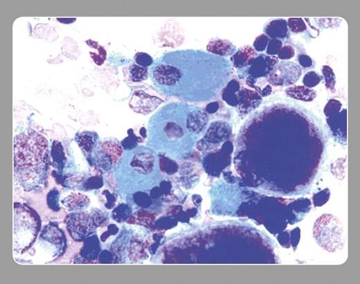
29-year-old woman found unconscious was brought to the emergency room. Her past history included seizures, emotional disorders, and morbid obesity that had been treated with gastric bypass. She was found to have mild pancytopenia and slight elevations of alkaline phosphatase, aspartate aminotransferase, and alanine aminotransferase. Her drugs were amitriptyline, alprazolam, venlafaxine, quetiapine, baclofen, vicoprofen, and valproic acid.
A bone marrow examination showed mild hypercellularity with myeloid hyperplasia and megaloblastic changes of the red cells. “Sea-blue histiocytes” were very prominently seen as well.
Sea-blue histiocytes, hepatosplenomegaly, and thrombocytopenia were reported in 1970 (Silverstein et al, N Engl J Med. 282:1-4). Similar histiocytes were reported in the liver, spleen, lymph nodes, and tonsils. Sea-blue histiocytes are also noted as a secondary phenomena of myeloproliferative disease, hyperlipidemia, lysosomal storage disease (Gaucher and Niemann-Pick), lymphoma, myelodysplastic disorders, with total parenteral nutrition, idiopathic thrombocytopenic purpura, and viral disorders.
The patient's medications were continued except for valproate. Shortly thereafter, her complete blood count normalized. One month later the marrow was repeated, and sea-blue histiocytes had disappeared. An association between sea-blue histiocytes and a drug, such as valproic acid, has not yet been reported.
Many Blood Work images are provided by the ASH IMAGE BANK, a reference and teaching tool that is continually updated with new atlas images and images of case studies. For more information or to contribute to the Image Bank, visit www.ashimagebank.org.


