Abstract
The blast colony-forming cell (BL-CFC) was identified as an equivalent to the hemangioblast during in vitro embryonic stem (ES) cell differentiation. However, the molecular mechanisms underlying the generation of the BL-CFC remain largely unknown. Here we report the isolation of mouse lysocardiolipin acyltransferase (Lycat) based on homology to zebrafish lycat, a candidate gene for the cloche locus. Mouse Lycat is expressed in hematopoietic organs and is enriched in the Lin−C-Kit+Sca-1+ hematopoietic stem cells in bone marrow and in the Flk1+/hCD4+(Scl+) hemangioblast population in embryoid bodies. The forced Lycat transgene leads to increased messenger RNA expression of hematopoietic and endothelial genes as well as increased blast colonies and their progenies, endothelial and hematopoietic lineages. The Lycat small interfering RNA transgene leads to a decrease expression of hematopoietic and endothelial genes. An unbiased genomewide microarray analysis further substantiates that the forced Lycat transgene specifically up-regulates a set of genes related to hemangioblasts and hematopoietic and endothelial lineages. Therefore, mouse Lycat plays an important role in the early specification of hematopoietic and endothelial cells, probably acting at the level of the hemangioblast.
Introduction
It was proposed nearly a century ago that a common progenitor generates both the hematopoietic and endothelial lineages.1,2 Using in vitro mouse embryonic stem (ES) cell differentiation, the blast colony-forming cell (BL-CFC) that clonally generates both endothelial and hematopoietic cells in the presence of vascular endothelial growth factor (VEGF) was characterized.3-5 The BL-CFC was later isolated in vivo from the posterior primitive streak of mid-gastrulation mouse embryos.6 Flk1+Scl+ and Brachyury+Flk1+ cells are enriched for the hemangioblast.6,7 Fate mapping in the zebrafish gastrula suggests that hemangioblasts are interspersed with cells that only give rise to either blood cells or endothelial cells in the ventral mesoderm.8,9 However, the molecular identity and plasticity of the hemangioblast remain largely unknown
The in vitro differentiation of mouse ES cells, along with genetically modified ES cells, has proved to be valuable in deciphering the underlying signaling pathways in hemangioblast development.10,11 Several pathways are revealed to participate in hemangioblast development, including the Bmp4-Gata2 signaling in embryoid bodies (EBs), the VEGF-Flk1-Plcg1 signaling in mice and EBs, and the transcription factors Scl, Runx1, Mixl1, and Hex in EBs.7,12-25 In zebrafish, the cloche (clo) mutant provides genetic evidence that a single gene is required for both endothelial and hematopoietic lineages.26,27 cloche acts upstream of all known hematopoietic (scl, gata1, gata2) and endothelial (flk1, etsrp, fli1) genes.26,28-34 We have recently cloned the zebrafish lysocardiolipin acyltransferase (lycat) gene from the cloche genetic interval. lycat is required for the generation of both endothelial and hematopoietic lineages and acts upstream of scl and etsrp in zebrafish embryos (J.-W.X., Qingming Yu, Jiaojiao Zhang, and John D. Mably, “An acyltransferase controls the generation of hematopoietic and endothelial lineages in zebrafish,” manuscript submitted July 2007). Interestingly, we found that mouse Lycat messenger RNA (mRNA) could partially rescue the cloche mutant phenotype. Therefore the mouse Lycat gene may also play an important role in hemangioblast, endothelial, and hematopoietic cell development.
Here we report the isolation of a mouse orthologue of zebrafish lycat and the characterization of mouse Lycat role in the generation of hemangioblasts and endothelial and hematopoietic lineages during in vitro ES cell differentiation. Mouse Lycat was reported to be part of the Cardiolipin remodeling pathway but its function in hemangioblast development is not known.35 Our data show that Lycat mRNA is enriched in the Flk1+Scl+ hemangioblast population, and is essential for the formation of both endothelial and hematopoietic lineages during in vitro ES cell differentiation. To our knowledge, Lycat is the first acyltransferase essential for hematopoietic and endothelial development in mouse ES cells.
Materials and methods
ES cell culture and differentiation
The 129/Sv ES cell lines R1 (a gift from Dr Andras Nagy, Samuel Lunenfeld Research Institute, Mount Sinai Hospital, Toronto, ON) and Scl+/hCD4 R1 (kindly provided by Dr Kyunghee Choi, Washington University School of Medicine, St Louis, MO) were cultured on mouse embryonic fibroblast (MEF) feeder cells pretreated with Mitomycin-C in ES cell medium containing Dulbecco modified Eagle medium (DMEM; Gibco/BRL, Carlsbad, CA), 1000 U/mL leukemia inhibitory factor (LIF; Chemicon International, Temecula, CA), 15% fetal bovine serum (FBS; Gibco/BRL), 2 mM glutamine (Gibco/BRL), 0.1 mM nonessential amino acids (Gibco/BRL), 100 μM monothioglycerol (MTG; Sigma, St Louis, MO), 50 U/mL penicillin, and 50 μg/mL streptomycin.
ES cell differentiation into EBs was carried out as described.3,36 Briefly, ES cells were dissociated into single cells with 0.25% Trypsin/EDTA (Gibco/BRL). After MEF feeder cells were depleted by incubating cell suspensions for 10 minutes at 37°C, ES cells were suspended and counted by Trypan Blue staining (Bioss, Beijing, China). Approximately 3000 to 5000 cells were transferred into 6 cm Petri dishes containing 5 mL of the EB induction medium that includes DMEM, 15% FBS, 5% protein free hybridoma medium (PFHM-II; Gibco/BRL), 0.5 mM ascorbic acid (Sigma), 2 mM glutamine, 0.1 mM nonessential amino acid, 450 μM MTG, 50 U/mL penicillin, and 50 μg/mL streptomycin. EBs were cultured at 37°C and EB culture medium was changed daily.
Generation of stable Lycat transgenic ES cell lines
R1 ES cells were electroporated with 15 μg linearized plasmid DNA of VC-Flk1 (vector control contains the mouse Flk1 promoter but without Lycat cDNA, and a PGK-neo cassette), Flk1-Lycat (Flk1:Lycat;PGK:neo contains Lycat cDNA driven by the Flk1 promoter and a PGK-Neo cassette), VC-β-actin (vector control contains the β-actin promoter but without Lycat cDNA, and a PGK-neo cassette), or β-actin-Lycat (β-actin:Lycat;PGK:neo contains Lycat cDNA driven by the β-actin promoter and a PGK-neo cassette) as described.36 Transfected ES cells were placed on neomycin-resistant SNL (STO [mouse embryonic fibroblast cell line] that produces LIF) feeder cells (a gift from Dr K. Choi) in the ES cell medium. After 24 hours, ES cells were selected in the ES medium supplemented with 0.25 mg/mL G418 (Geneticin Selective Antibiotic; Invitrogen, Carlsbad, CA) for 10 days. Single colonies were picked up and expanded. ES cell genomic DNA and RNA were purified as previously described.36 The ES clones containing each transgene were identified by polymerase chain reaction (PCR) with transgene-specific primers. To identify high levels of expression of the Lycat transgene in transgenic ES clones, we examined Lycat mRNA expression in all transgene-containing ES cell clones by reverse transcriptase (RT)–PCR and quantitative RT-PCR.
Generation of stable Lycat siRNA ES cell lines
Two mouse Lycat small interfering RNA (siRNA) retroviral clones (V2MM_232680 and V2MM_103078), containing short-hairpin RNAs (shRNAs) targeting Lycat, were obtained from Open Biosystems (Huntsville, AL).37 The retrovirus vector pBabe-puro was used as the vector control (gift from Dr Dan Littman, New York University, New York). Retroviral supernatants were produced in 293GP cells by transient transfection with calcium phosphate.38 Briefly, 293GP cells were grown to 60% confluence in DMEM/10% FBS. pVSV-G and retroviral DNA (Lycat siRNA or vector control DNA) were mixed with 2.5 mM CaCl2 and sterilized water in a final volume of 0.5 mL, and the mixture was slowly added into 2× HEPES (N-2-hydroxyethylpiperazine-N-2-ethanesulfonic acid)–buffered saline with gentle vortexing. The resulting 1-mL mixture was added into 293GP cells. The transfection medium was replaced by fresh medium after 16 hours. Retroviral supernatants were harvested at 48 hours after transfection and centrifuged at 1400g to remove cell debris. Single ES cells were resuspended in 1 mL retroviral supernatants with 4 μg/mL polybrene at 37°C for 30 minutes, and were then placed on puromycin-resistant SNP feeder cells (a gift from Dr K. Choi). After infection for 48 hours, the infected cells were cultured and selected with the ES medium supplemented with 3 μg/mL puromycin for 7 days. Single puromycin-resistant clones were picked and expanded. The Lycat siRNA ES cell clones were identified by PCR-based genotyping, and knockdown of Lycat mRNA was confirmed by semiquantitative and quantitative RT-PCR.
Blast and hematopoietic colony assays
To assess BL-CFCs, we collected and dissociated EBs at day 4 (D4) into single cells by trypsin treatment. Viable cells were counted and 2 × 10⁴ cells/mL were replated in 1% methylcellulose matrix in the presence of Iscove modified Dulbecco medium (IMDM; Gibco/BRL), 15% FBS, 2 mM glutamine, 450 μM MTG, 25 μg/mL ascorbic acid, 20% BIT9500 (StemCell Technologies, Vancouver, BC, Canada), 5 ng/mL human vascular endothelial growth factor (hVEGF), 50 ng/mL stem cell factor (SCF), 10 ng/mL human fibroblast growth factor 2 (hFGF2), and 4 U/mL human erythropoietin (hEPO; Kirin Brewery, Tokyo, Japan). After post-replating culture for 4 days, blast colonies were recognized and quantified.
To determine primitive erythroid progenitors (Eryp), we dissociated D6 EBs into single cells by trypsinization. Cells (2 × 10⁴) were replated in 1% methylcellulose medium (M3333; StemCell Technologies). Eryp colonies could be identified as small bright red colonies and were scored 5 to 7 days after post-replating cultures.3
To determine definitive hematopoietic progenitors, we dissociated day 9 to 12 EBs into single cells. Cells (2 × 10⁴) were replated in 1% methylcellulose medium (M3434, StemCell Technologies) supplemented with an additional 100 ng/mL SCF, 20 ng/mL granulocyte colony-stimulating factor (G-CSF), 5 ng/mL macrophage colony-stimulating factor (M-CSF), 5 ng/mL thrombopoietin (TPO), and 3 ng/mL GM-CSF (PeproTech, London, United Kingdom). Hematopoietic colonies were counted 10 to 14 days after re-plating.
Endothelial cell sprouting assay
ES cell differentiation into endothelial cells was carried out as described.39 Fifty day 11 EBs of each experimental group were used and replated on 3.5-cm culture dishes with 1.5 mL collagen gel medium supplemented with IMDM, 1.25 mg/mL rat tail collagen, type I (BD Biosciences, San Jose, CA), 15% FBS, 450 μM MTG, 10 μg/mL insulin (Invitrogen), 50 U/mL penicillin, and 50 μg/mL streptomycin. EB cultures were incubated at 37°C for 3 to 5 days. The vascular endothelial spindlelike EBs were classified into 3 groups based on numbers of endothelial cell sprouting and numbers of EBs in each group after replating for 3 days.
Fluorescence-activated cell sorting
EB cells were dissociated into single cells with 0.25% Trypsin-EDTA for 3 minutes. Cells were centrifuged, washed with staining/wash buffer (5% fetal calf serum in phosphate-buffered saline [PBS]) twice, and stained with Fc blocking antibody (anti-CD16/CD32, FcγIII/II receptor) on ice in the dark for 5 minutes. For single-color staining for Flk1, CD41, C-Kit, or CD31, cells were stained with primary antibodies (1:200) on ice in the dark for 15 to 30 minutes. For CD45 analysis, cells were stained first with biotinylated anti-CD45 for 15 minutes, washed with PBS 3 times, and followed by staining with streptavidin-Cy5 (Sav-Cy5). For hCD4/Flk1 double staining, cells were stained first with biotinylated antihuman CD4 for 15 minutes and followed by staining with streptavidin-phycoerythrin-Cy5 (Sav-PE-Cy5) and phycoerythrin (PE)–conjugated anti-Flk1. After staining with the secondary antibody, cells were washed with PBS 3 times and resuspended in 500 μL PBS. Stained cells were analyzed on a fluorescence-activated cell sorting (FACS) Caliber (Becton Dickinson, San Jose, CA), and data were analyzed using CellQuest Software (Becton Dickinson). PE-anti-Flk1, fluorescein isothiocyanate (FITC)–conjugated anti-CD41, allophycocyanin (APC)–conjugated anti-C-Kit, biotin-anti-CD45, biotin-anti-hCD4, Sav-Cy5, and Sav-PE-Cy5 were purchased from BD Bioscience Pharmingen (San Diego, CA).
DNA microarray analysis
For microarray analyses, the Flk1:Lycat FC5 clone and vector control clone were examined in 2 independent experiments. Total RNA was isolated from day 4 EBs using TRIzol (Invitrogen). cDNA synthesis and labeling were performed as described in the Affymetrix user's manual (Santa Clara, CA). SmartArray (CapitalBio, Beijing, China) chips containing about 25 000 mouse genes were hybridized with labeled cDNA probes on a GeneChip system. All arrays were globally scaled to a target value of 150 using the average signal from all gene features. Scanned chip images were analyzed using the Microarray Suite software version 5.0 (Affymetrix). Data analysis was further carried out using Microsoft Excel. More than 1.5-fold changes in gene expression were considered to be significant. In this report, we have chosen to study the top 200 affected genes that are affected with more than 2-fold, except HoxB1 with a 1.5-fold decrease (data not shown and Table S1, available on the Blood website; see the Supplemental Materials link at the top of the online article). Fold changes in affected genes were averaged from 2 independent array experiments.
Semiquantitative RT-PCR and quantitative RT-PCR
Total RNA from cultured cells was prepared using TRIzol (Invitrogen) and total RNA from sorted cells was extracted with a microscale RNA isolation kit (RNAqueous-Micro, Ambion, TX). Trace amounts of DNA in RNA samples were removed using DNase I, and DNase I was then inactivated by DNase Inactivation Reagent (Ambion). Reverse transcription was carried out according to the manufacturer's instruction (Promega, Madison, WI). RNA expression levels were quantified using semiquantitative RT-PCR and quantitative RT-PCR (Q-PCR). Q-PCR was performed using SYBR Green (Invitrogen). The PCR reactions included 2 mM MgCl2, 0.4 mM dNTP, 8% glycerol, 3% DMSO, 150 nM of each primer, 0.75 μL of 1:1000 dilution of reference dye, and 2.5 μL of a 1:2000 dilution of SYBR Green. RNA levels were normalized using GAPDH as an internal control. Primer sequences and PCR length are listed in Table S2.
Annexin-V assay
Apoptosis was detected by double staining for Annexin-V–FITC and propodium iodide (PI; Baosai Reagent, Beijing, China). Briefly, single cells were washed in cold PBS twice, resuspended with 200 μL binding buffer, and stained with Annexin V–FITC on ice in the dark for 15 minutes. The stained cells were then mixed with 300 μL binding buffer and 5 μL of 2.5 μg/mL PI. Stained cells were analyzed on a FACS Caliber within 30 minutes, and FACS data were analyzed using Cell Quest software version 7.5.3 (Becton Dickinson).
Results
Mouse Lycat is the only highly conserved homologue of zebrafish lycat in the mouse genome
We identified the lysocardiolipin acyltransferase gene from the zebrafish clochem39 deletion interval by positional cloning (J.-W.X., Qingming Yu, Jiaojiao Zhang, and John D. Mably, “An acyltransferase controls the generation of hematopoietic and endothelial lineages in zebrafish,” manuscript submitted July 2007). Morpholino knockdown of lycat in wild-type zebrafish embryos led to cloche mutant phenotypes, whereas overexpression of lycat mRNA could partially rescue cloche mutant phenotypes. To further investigate lycat gene function in hematopoietic and endothelial development, we isolated mouse Lycat by homology to zebrafish lycat and the synteny between mouse and zebrafish lycat neighboring genes. CGI127 (AF172940), Lycat, and Lbh (NM_030915) are tightly linked in zebrafish chromosome 13, mouse chromosome 17, and human chromosome 2. Mouse Lycat protein is 47% identical to human LYCAT, 50% identical to zebrafish Lycat, and 65% identical to Tetraodon nigroviridis Lycat (Figure S1). The N-terminal acyltransferase domain is highly conserved and the H(X)4D and EGTD catalytic domains are identical among Lycat proteins. Mouse Lycat is expressed in the heart and localized in the endoplasmic reticulum.35 However, it has not been addressed if Lycat is essential for the development of hematopoietic and endothelial lineages. Therefore, we embarked on characterization of the Lycat role in hematopoietic and endothelial lineages using an in vitro ES cell differentiation system.
Mouse Lycat mRNA is enriched in the Flk1+ mesodermal and hemangioblast populations in EBs and the Lin−Sca1+C-Kit+ hematopoietic stem cells in bone marrow
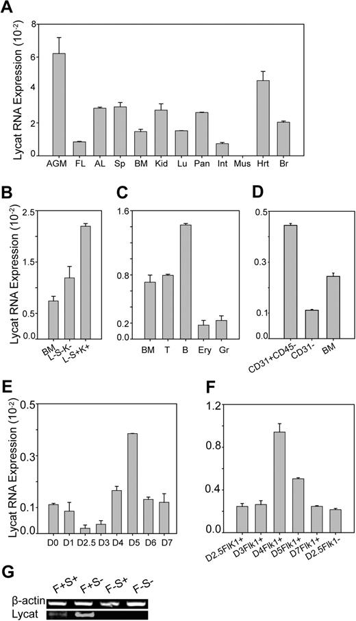
We found that Lycat mRNA is weakly expressed in most adult and embryonic tissues tested by quantitative RT-PCR (Figure 1A). Lycat is expressed at higher levels in the E12.5 aorta-gonad-mesonephros (AGM), an intraembryonic hematopoietic site,40-42 but is expressed at lower levels in the E16 fetal liver (FL) and the adult bone marrow (BM), the definitive hematopoietic sites supporting hematopoietic expansion and differentiation during fetal and adult life.43 In addition, Lycat expression is relatively high in the heart, which supports its potential function in the cardiovascular system, but is not detectable in skeletal muscle.35 To further determine Lycat expression in the bone marrow, we sorted out different cell populations using flow cytometry with a variety of stem cell, endothelial, and hematopoietic markers. We found that Lycat is enriched in the Lin−Sca1+C-Kit+ HSCs, compared with whole BM and Lin−Sca-1−C-Kit− cells (Figure 1B) and is enriched in the CD31+CD45− endothelial cells, compared with CD31− and the whole BM (Figure 1D). In addition, Lycat has higher expression in B cells (B), compared with whole BM, T cells (T), erythrocytes (Ery), and granulocytes (Gr; Figure 1C). During in vitro differentiation of mouse ES cells into EBs, we found that Lycat has dynamic low-level expression in embryoid bodies from day 0 to day 7, peaks at day 5 (Figure 1E), and is enriched in the Flk1+ cells in day 4 and 5 embryoid bodies (Figure 1F). The Flk1+hCD4+/Scl+ cells in embryoid bodies were reported to have high hemangioblast activities using blast colony assays.7 We found that Lycat is expressed in the Flk1+Scl+ and Flk1+Scl− but not in Flk1− cell populations in day 4 EBs, supporting that Lycat may function in generation of Flk1+ mesodermal cells and hemangioblasts (Figure 1G). Therefore, Lycat mRNA is enriched in the Flk1+ mesodermal cells and hemangioblasts in embryoid bodies as well as in the Lin−Sca-1+C-Kit+ HSCs and the CD31+CD45− endothelial cells in bone marrow.
Distribution of mouse Lycat mRNA in tissues and cell populations detected by RT-PCR. (A) Lycat mRNA had highest expression in the AGM and heart among multiple mouse tissues including E12.5 AGM, E16 fetal liver (FL), adult liver (AL), spleen (Sp), bone marrow (BM), kidney (Kid), lung (Lu), pancreas (Pan), intestine (Int), muscle (Mus), heart (Hrt), and brain (Br). (B) Lycat mRNA was enriched in the Lin−Sca-1+C-Kit+ HSCs in BM. Cells were sorted by flow cytometry with lineage markers, Sca-1, and C-Kit for hematopoietic stem cells. Lineage−Sca-1−C-Kit−, L−S−K−; Lineage−Sca-1+C-Kit+, L−S+K+ (Figure S2A). (C) Lycat mRNA expression was higher in B cells. Cells were sorted by flow cytometry with mature hematopoietic lineage markers including T4 and T8a for T lymphocytes (T), B220 for B lymphocytes (B), TER119 for erythrocytes (Ery), and Gr-1 for granulocytes (Gr). (D) Lycat mRNA was enriched in the CD31+CD45− endothelial cells in adult mouse BM. Cells were sorted from BM by flow cytometry with anti-CD31 and anti-CD45. (E) Lycat mRNA could be detected in embryoid bodies from day 0 to day 7 (D0 to D7). The morphology of EBs is presented in Figure S3. (F) Lycat mRNA was 2-fold more enriched in the Flk1+ cells in embryoid bodies from day 4 than those from day 5. Flk1+ cells were sorted from staged embryoid bodies by anti-Flk1. (G) Lycat mRNA was enriched in the Flk1+ cell population in embryoid bodies. Lycat mRNA could be detected in Flk1+hCD4+(Scl+) (F+S+) and Flk1+hCD4−(Scl−) (F+S−) but not in the Flk1−hCD4+(Scl+) (F−S+) and Flk1−hCD4−(Scl−) (F−S+) cells, which were sorted by flow cytometry with anti-Flk1 and anti-hCD4 (Figure S2B). The RNA levels were determined and normalized by GAPDH using quantitative RT-PCR (A-F) and using semiquantitative RT-PCR by β-actin as an internal RNA control (G). Error bars in panels A-F represent standard deviations.
Distribution of mouse Lycat mRNA in tissues and cell populations detected by RT-PCR. (A) Lycat mRNA had highest expression in the AGM and heart among multiple mouse tissues including E12.5 AGM, E16 fetal liver (FL), adult liver (AL), spleen (Sp), bone marrow (BM), kidney (Kid), lung (Lu), pancreas (Pan), intestine (Int), muscle (Mus), heart (Hrt), and brain (Br). (B) Lycat mRNA was enriched in the Lin−Sca-1+C-Kit+ HSCs in BM. Cells were sorted by flow cytometry with lineage markers, Sca-1, and C-Kit for hematopoietic stem cells. Lineage−Sca-1−C-Kit−, L−S−K−; Lineage−Sca-1+C-Kit+, L−S+K+ (Figure S2A). (C) Lycat mRNA expression was higher in B cells. Cells were sorted by flow cytometry with mature hematopoietic lineage markers including T4 and T8a for T lymphocytes (T), B220 for B lymphocytes (B), TER119 for erythrocytes (Ery), and Gr-1 for granulocytes (Gr). (D) Lycat mRNA was enriched in the CD31+CD45− endothelial cells in adult mouse BM. Cells were sorted from BM by flow cytometry with anti-CD31 and anti-CD45. (E) Lycat mRNA could be detected in embryoid bodies from day 0 to day 7 (D0 to D7). The morphology of EBs is presented in Figure S3. (F) Lycat mRNA was 2-fold more enriched in the Flk1+ cells in embryoid bodies from day 4 than those from day 5. Flk1+ cells were sorted from staged embryoid bodies by anti-Flk1. (G) Lycat mRNA was enriched in the Flk1+ cell population in embryoid bodies. Lycat mRNA could be detected in Flk1+hCD4+(Scl+) (F+S+) and Flk1+hCD4−(Scl−) (F+S−) but not in the Flk1−hCD4+(Scl+) (F−S+) and Flk1−hCD4−(Scl−) (F−S+) cells, which were sorted by flow cytometry with anti-Flk1 and anti-hCD4 (Figure S2B). The RNA levels were determined and normalized by GAPDH using quantitative RT-PCR (A-F) and using semiquantitative RT-PCR by β-actin as an internal RNA control (G). Error bars in panels A-F represent standard deviations.
Overexpression of Lycat increased hematopoietic and endothelial gene expression
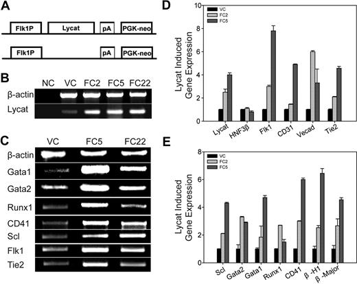
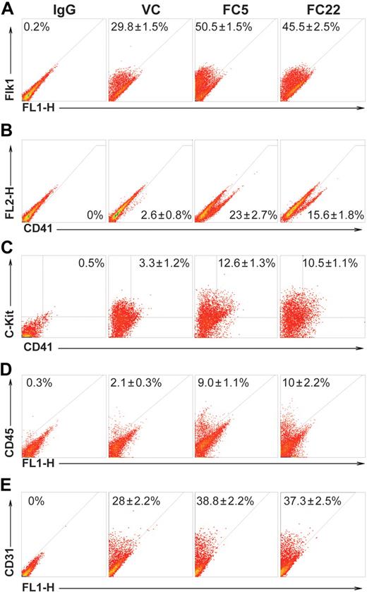
To test if Lycat is necessary and sufficient to direct cell fate toward the hematopoietic and endothelial lineages, we first evaluated Lycat gene function in EBs using a gain-of-function assay, in which Lycat expression is driven by the mouse Flk1 promoter or β-actin promoter (Figure 2A; Figure S4A).44,45 Stable R1 ES cell clones were obtained by electroporation of the vector control (VC) or Lycat expression construct DNA, followed by G418 selection. After selection, stable transgenic ES clones were identified using PCR with transgene-specific primers (not shown). High levels of Lycat transgene expression were verified using semiquantitative RT-PCR. In day 4 EBs, Lycat mRNA was 5-fold higher in 3 ES cell clones (FC2, FC5, FC22) compared with the VC ES clone (Figure 2B). Lycat transgene expression increased hematopoietic (Gata1, Gata2, Runx1, CD41, Scl) and endothelial (Flk1, Tie2) gene expression in day 4 EBs by semiquantitative RT-PCR analysis (Figure 2C). This result was further confirmed by quantitative RT-PCR (Figure 2D,E). mRNA levels of endothelial (Flk1, CD31, VE-Cadherin, Tie1; Figure 2D), as well as primitive hematopoietic (Scl, Gata2, Gata1, β-H1 hemoglobin) and definitive hematopoietic (Runx1, CD41, β-major hemoglobin; Figure 2E) genes were elevated in the Lycat transgenic ES–derived EBs at day 4. However, the Lycat transgene did not affect the endodermal gene HNF3β expression, suggesting its specific function in endothelial and hematopoietic lineages (Figure 2D). In addition, we found that the Lycat transgene–induced gene expression was similar in EBs from ES cell clones using the Lycat transgene driven by the cell-specific Flk1 promoter (Figure 2) or ubiquitous β-actin promoter (Figure S4; not shown). This indicates that Lycat may play a permissive role in the generation of these lineages. Furthermore, the Lycat transgene could also increase the hematopoietic and endothelial cell populations in day 4 EBs detected by flow cytometry (Figure 3). The Lycat transgene increased Flk1+ cells 1.6-fold, CD41+ cells 7.4-fold; CD41+/C-Kit+ putative definitive HSCs 3.5-fold, CD45+ panhematopoietic cells 4.5-fold, and CD31+ endothelial cells 1.4-fold in day 4 EBs. Therefore, the Lycat transgene can sufficiently drive the development of endothelial and hematopoietic lineages in EBs.
Overexpression of the Lycat transgene increased hematopoietic and endothelial cell gene expression in embryoid bodies. (A) Schematic representation of the Flk1:Lycat expression construct in which Lycat expression was driven by the mouse Flk1 promoter (Flk1P) and a PGK-neo cassette was inserted (top), and the vector control (VC) without Lycat cDNA (bottom). (B) Lycat mRNA was significantly increased in D4 EBs from transgenic ES clones of FC2, FC5, and FC22 as determined by semiquantitative RT-PCR. NC indicates negative control without input of DNA templates in the PCR reaction; β-actin, an internal RNA control. (C) The forced Lycat transgene increased hematopoietic and endothelial gene expression in D4 EBs from FC5 and FC22 ES clones detected by RT-PCR. Hematopoietic genes Gata1, Gata2, Runx1, CD41, Scl; endothelial genes Tie2 and Flk1; and β-actin as an internal RNA control. (D,E) The forced Lycat transgene increased mRNA expression of endothelial (panel D) and hematopoietic (panel E) but not endodermal (HNF3β; panel D) genes in D4 EBs from FC2 and FC5 ES clones detected by quantitative RT-PCR. Endothelial genes VE-Cadherin (Vecad), Flk1, CD31, Tie2; endodermal gene, HNF3β; hematopoietic genes Scl, Gata1, Gata2, Runx1, CD41, β-H1 hemoglobin (β-H1), and β-major hemoglobin (β-Major). The mRNA levels were normalized by GAPDH. (D,E) Error bars represent standard deviations.
Overexpression of the Lycat transgene increased hematopoietic and endothelial cell gene expression in embryoid bodies. (A) Schematic representation of the Flk1:Lycat expression construct in which Lycat expression was driven by the mouse Flk1 promoter (Flk1P) and a PGK-neo cassette was inserted (top), and the vector control (VC) without Lycat cDNA (bottom). (B) Lycat mRNA was significantly increased in D4 EBs from transgenic ES clones of FC2, FC5, and FC22 as determined by semiquantitative RT-PCR. NC indicates negative control without input of DNA templates in the PCR reaction; β-actin, an internal RNA control. (C) The forced Lycat transgene increased hematopoietic and endothelial gene expression in D4 EBs from FC5 and FC22 ES clones detected by RT-PCR. Hematopoietic genes Gata1, Gata2, Runx1, CD41, Scl; endothelial genes Tie2 and Flk1; and β-actin as an internal RNA control. (D,E) The forced Lycat transgene increased mRNA expression of endothelial (panel D) and hematopoietic (panel E) but not endodermal (HNF3β; panel D) genes in D4 EBs from FC2 and FC5 ES clones detected by quantitative RT-PCR. Endothelial genes VE-Cadherin (Vecad), Flk1, CD31, Tie2; endodermal gene, HNF3β; hematopoietic genes Scl, Gata1, Gata2, Runx1, CD41, β-H1 hemoglobin (β-H1), and β-major hemoglobin (β-Major). The mRNA levels were normalized by GAPDH. (D,E) Error bars represent standard deviations.
The Lycat transgene increased protein expression of hematopoietic and endothelial cells, as detected by flow cytometry. The Lycat transgenic and vector control ES cell clones were induced into EBs at day 4 to day 6. Expression of Flk1 (A), CD41 (B), CD41 and C-Kit (C), and CD31 (E) was increased in D4 EBs and expression of CD45 (D) was increased in D6 EBs from transgenic ES cells (FC5 and FC22). IgG indicates negative controls of FACS without input of the primary antibody. These quantitative effects were calculated from 3 independent experiments. Numbers within quadrants indicate percentages of positive cells.
The Lycat transgene increased protein expression of hematopoietic and endothelial cells, as detected by flow cytometry. The Lycat transgenic and vector control ES cell clones were induced into EBs at day 4 to day 6. Expression of Flk1 (A), CD41 (B), CD41 and C-Kit (C), and CD31 (E) was increased in D4 EBs and expression of CD45 (D) was increased in D6 EBs from transgenic ES cells (FC5 and FC22). IgG indicates negative controls of FACS without input of the primary antibody. These quantitative effects were calculated from 3 independent experiments. Numbers within quadrants indicate percentages of positive cells.
Overexpression of Lycat led to the increased formation of both hematopoietic and endothelial cells
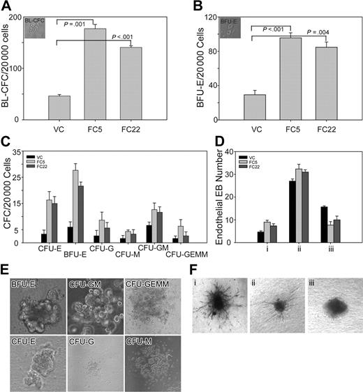
We then explored how overexpression of Lycat influenced the formation of BL-CFCs and their progenies, including endothelial cell and hematopoietic colonies using semisolid methylcellulose or collagen matrix.19,39 We observed that the Lycat transgene increased BL-CFCs 3-fold (an equivalent to the hemangioblast; Figure 4A) and we confirmed that these blast colonies could generate suspended hematopoietic cells and adherent endothelial cells (not shown). We also observed that Lycat overexpression increased primitive erythroid progenitors (BFU-Es) 3-fold in day 6 EBs (Figure 4B). For definitive hematopoietic lineages, we found that the Lycat transgene in FC5 and FC22 led to a 4-fold increase of CFU-Es, 5-fold of BFU-Es, 2-fold of CFU-Gs, 2-fold of CFU-Ms, 2-fold of CFU-GMs, and 2-fold of CFU-GEMMs in day 9 EBs (Figure 4C). The morphology of representative hematopoietic colonies are shown in Figure 4E. To examine the effect of Lycat on endothelial cell differentiation, primary day 11 EBs were dissociated and cultured in collagen matrix with growth factors for endothelial cell differentiation for 4 days.39,46 We found that the Lycat transgene increased endothelial tube sprouting 2-fold in FC5 and FC22 clones grown on collagen matrix (Figure 4Di). The sprouting endothelial tubes were CD31+ (not shown). The morphology of endothelial sprouting of EBs is shown in Figure 4F. Thus, our in vitro functional assays further substantiate that the Lycat transgene sufficiently increases the derivation of hemangioblasts and endothelial and hematopoietic lineages in EBs.
Overexpression of the Lycat transgene increased the formation of BL-CFCs and hematopoietic and endothelial cells by in vitro function assay. (A) D4 EBs from FC5 and FC22 were dissociated and determined to have 3-fold more potency than D4 EBs from VC to generate BL-CFCs in methylcellulose cultures. Top left, morphology of a representative BL-CFC (×100) by 10× objective lens. (B) D6 EBs from FC5 and FC22 were dissociated and determined to have 3-fold more potency than D6 EBs from VC to generate primitive erythroids in methylcellulose cultures in the presence of hEPO. Top left, morphology of 2 representative BFU-E colonies (×100) by 10× objective lens. (C) D9 EBs from VC, FC5, and FC22 were dissociated and assayed for multilineage hematopoietic colony formation. The Lycat transgene in FC5 and FC22 increased the formation of CFU-Es (4-fold), BFU-Es (5-fold), CFU-Gs (2-fold), CFU-Ms (2-fold), CFU-GMs (2-fold) and CFU-GEMMs (2-fold). (D) D11 EBs from FC5 and FC22 had better potential than those from VC to form long endothelial sprouting. (i) The EBs producing much long endothelial sprouting as shown in panel Fi (×100) by 10× objective lens. (ii) The EBs producing only some endothelial sprouting as shown in panel Fii (×40) by 4× objective lens. (iii) The EBs producing no endothelial sprouting as shown in panel Fiii (×100) by 10× objective lens. (E) Representative colonies of BFU-Es (×200), CFU-Es (×200), CFU-GMs (×200) by 20× objective lens, CFU-Gs (×40) by 4× objective lens, CFU-Ms (×100) by 10× objective lens, and CFU-GEMMs (×40) by 4× objective lens. Live images were taken under a Nikon ECLIPSE TE2000-U fluorescence microscope (Nikon, Yokohama, Japan). Images were acquired with a SPOT charge-coupled device camera (Diagnostic Instruments, Sterling Heights, MI), imported with SPOT software version 4.6.4.4, and prepared with Adobe Photoshop 7.0 (Adobe, San Jose, CA). Results are presented as the mean plus or minus the standard error of the mean (SEM). Statistical significance in Figure 4A,B was determined using an unpaired Student t test. P values were calculated from quantitative effects in 3 independent experiments, compared with those of FC5 and FC22 with those of VC, respectively. (A-D) Error bars represent standard deviations.
Overexpression of the Lycat transgene increased the formation of BL-CFCs and hematopoietic and endothelial cells by in vitro function assay. (A) D4 EBs from FC5 and FC22 were dissociated and determined to have 3-fold more potency than D4 EBs from VC to generate BL-CFCs in methylcellulose cultures. Top left, morphology of a representative BL-CFC (×100) by 10× objective lens. (B) D6 EBs from FC5 and FC22 were dissociated and determined to have 3-fold more potency than D6 EBs from VC to generate primitive erythroids in methylcellulose cultures in the presence of hEPO. Top left, morphology of 2 representative BFU-E colonies (×100) by 10× objective lens. (C) D9 EBs from VC, FC5, and FC22 were dissociated and assayed for multilineage hematopoietic colony formation. The Lycat transgene in FC5 and FC22 increased the formation of CFU-Es (4-fold), BFU-Es (5-fold), CFU-Gs (2-fold), CFU-Ms (2-fold), CFU-GMs (2-fold) and CFU-GEMMs (2-fold). (D) D11 EBs from FC5 and FC22 had better potential than those from VC to form long endothelial sprouting. (i) The EBs producing much long endothelial sprouting as shown in panel Fi (×100) by 10× objective lens. (ii) The EBs producing only some endothelial sprouting as shown in panel Fii (×40) by 4× objective lens. (iii) The EBs producing no endothelial sprouting as shown in panel Fiii (×100) by 10× objective lens. (E) Representative colonies of BFU-Es (×200), CFU-Es (×200), CFU-GMs (×200) by 20× objective lens, CFU-Gs (×40) by 4× objective lens, CFU-Ms (×100) by 10× objective lens, and CFU-GEMMs (×40) by 4× objective lens. Live images were taken under a Nikon ECLIPSE TE2000-U fluorescence microscope (Nikon, Yokohama, Japan). Images were acquired with a SPOT charge-coupled device camera (Diagnostic Instruments, Sterling Heights, MI), imported with SPOT software version 4.6.4.4, and prepared with Adobe Photoshop 7.0 (Adobe, San Jose, CA). Results are presented as the mean plus or minus the standard error of the mean (SEM). Statistical significance in Figure 4A,B was determined using an unpaired Student t test. P values were calculated from quantitative effects in 3 independent experiments, compared with those of FC5 and FC22 with those of VC, respectively. (A-D) Error bars represent standard deviations.
The Lycat siRNA decreased endothelial and hematopoietic gene expression
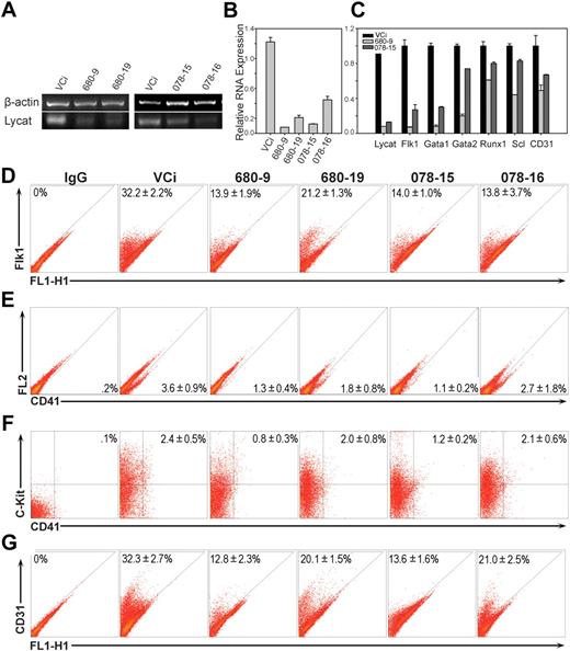
Seeing that overexpression of Lycat leads to expansion of hematopoietic and endothelial progenitors, we determined if siRNA-mediated gene knockdown influenced hematopoietic and endothelial lineages.37 We generated stable ES cell clones containing the retroviral vector pBabe-puro control (VCi), ES cell clones 680-9 and 680-19 containing the Lycat siRNA transgene V2MM_232680 (Open Biosystems, Huntsville, AL), and ES cell clones 078-15 and 078-16 containing the Lycat siRNA transgene V2MM_103078 (Open Biosystems). We found that Lycat mRNA levels were significantly knocked down in day 5 EBs from all 4 Lycat siRNA ES cell clones by semiquantitative RT-PCR (Figure 5A). This knockdown result was confirmed by quantitative RT-PCR (Figure 5B). Furthermore, the Lycat siRNA inhibited mRNA expression of hematopoietic (Gata1, Gata2, Runx1, Scl) and endothelial (Flk1, CD31) genes in day 5 EBs from both 680-9 and 078-15 clones. In addition, the Lycat siRNA V2MM_232680, from which the clone 680-9 was derived, had more potent knockdown of Lycat mRNA and so had more pronounced reduction of endothelial and hematopoietic genes expression (Figure 5C). By flow cytometric analysis, we observed that the Lycat siRNA generated percentages showing a reduction in Flk1+ cells (Figure 5D) and CD41+ cells in clones 680-9, 680-19 and 078-15 but not 078-16 (Figure 5E); of C-Kit+CD41+ cells (Figure 5F); and of CD31+ cells (Figure 5G). In addition, efficiency of Lycat mRNA knockdown correlates very well with its effects on protein expression of these marker genes in EBs (Figure 5B, D-G). Therefore Lycat is found to influence both endothelial and hematopoietic cell gene and protein expression in EBs.
Lycat siRNA reduced hematopoietic and endothelial gene expression during ES cell differentiation into EBs. (A) Lycat mRNA knockdown was detected in day 4 EBs from siRNA ES cell clones by semiquantitative RT-PCR. VCi, retroviral vector control ES cell clone; 680-9 and 680-19, 2 independent Lycat siRNA ES cell clones derived from V2MM_232680 (Open Biosystems); 078-15 and 078-16, 2 independent Lycat siRNA ES cell clones from V2MM_103078 (Open Biosystems). (B) The Lycat siRNA knockdown in panel A was confirmed by Q-PCR. (C) The Lycat siRNA reduced expression of endothelial (Flk1 and CD31) and hematopoietic (Gata1, Gata2, Runx1, and Scl) genes using Q-PCR. (D) The Lycat siRNA reduced the formation of Flk1+, C-Kit+, C-Kit+ and CD41+, and CD31+ cells in day 4 EBs from all 4 siRNA ES cell clones by flow cytometry. These were calculated from 3 independent experiments except those of CD31+, which was calculated from 2 independent experiments. (B,C) Error bars represent standard deviations. (D-G) Numbers within quadrants indicate percentages of positive cells.
Lycat siRNA reduced hematopoietic and endothelial gene expression during ES cell differentiation into EBs. (A) Lycat mRNA knockdown was detected in day 4 EBs from siRNA ES cell clones by semiquantitative RT-PCR. VCi, retroviral vector control ES cell clone; 680-9 and 680-19, 2 independent Lycat siRNA ES cell clones derived from V2MM_232680 (Open Biosystems); 078-15 and 078-16, 2 independent Lycat siRNA ES cell clones from V2MM_103078 (Open Biosystems). (B) The Lycat siRNA knockdown in panel A was confirmed by Q-PCR. (C) The Lycat siRNA reduced expression of endothelial (Flk1 and CD31) and hematopoietic (Gata1, Gata2, Runx1, and Scl) genes using Q-PCR. (D) The Lycat siRNA reduced the formation of Flk1+, C-Kit+, C-Kit+ and CD41+, and CD31+ cells in day 4 EBs from all 4 siRNA ES cell clones by flow cytometry. These were calculated from 3 independent experiments except those of CD31+, which was calculated from 2 independent experiments. (B,C) Error bars represent standard deviations. (D-G) Numbers within quadrants indicate percentages of positive cells.
We wondered if overexpression of Lycat and Lycat siRNA interfered with normal ES cell differentiation and apoptosis. We found that Flk1 had similar dynamic expression in EBs from the vector control (VC) ES clone and Lycat transgenic ES clone (FC5) from day 2.75 to 4 (Figure S5A). We observed relatively normal apoptosis by double staining of PI and Annexin-V–FITC in day 4 EBs from VC as well as transgenic FC2, FC5, and FC22 ES clones (Figure S5B). This suggests that overexpression of Lycat did not affect overall ES cell differentiation and cell death. We also observed that Lycat siRNA did not affect apoptosis by double-staining PI and Annexin-V–FITC in day 4 EBs from transgenic Lycat siRNA ES cell clones (Figure S5C). These data suggest that the stable transgenic ES cell clones we used in this study have the typical characteristics of normal cell differentiation and death.
Lycat specifically regulated hematopoietic and endothelial gene expression using an unbiased genomewide microarray analysis
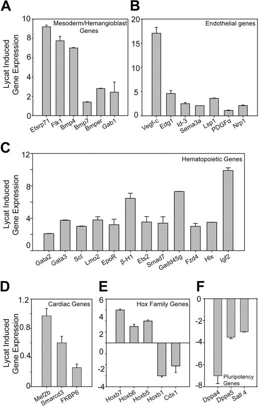
We have shown that overexpression of Lycat could increase hematopoietic and endothelial progenitor frequency, whereas Lycat siRNA reduced their frequency in EBs (Figures 2,3,5; Figure S4). We further explored if Lycat specifically regulated endothelial and hematopoietic gene expression using microarray analysis of about 25 000 mouse genes, by comparing differential gene expression in day 4 EBs in VC and Lycat transgenic clone FC5. We observed that 65 of 200 (32.5%) affected genes with more than 2-fold increased signals were related to endothelial and hematopoietic cell development or signaling (Table S1 and data not shown). Using quantitative RT-PCR, we confirmed the increased expression for 35 of the 65 genes and HoxB1 (with 1.5-fold decrease) in EBs from FC5 and VC (Figure 6; Table S1). Lycat induced several mesoderm/hemangioblast genes (Figure 6A), including Etsrp71 (a potential orthologue of zebrafish etsrp),32,33 Flk1, Bmp4, Bmp7, Bmper,47 and Gab148 ; a panel of hematopoietic genes (Figure 6C), including Gata2, Gata3, Scl, Lmo2, EpoR, β-H1 hemoglobin, Ets2,49 Smad7,50 Gadd45g,51 Fzd4,52 Hlx,53 and Igf254 ; and several endothelial genes (Figure 6B), including Vegf-C, Edg1,55 Id-3,56 Sema3a,57 Lsp1,58 PDGFα, and Nrp159 (Figure 6A-C). The Lycat transgene had little effect on cardiac genes including Mef2b, Smarcd3,60 and FKBP661 (Figure 6D). Furthermore, the Lycat transgene increased mRNA expression of several important Hox family members, including HoxB5, HoxB6, and HoxB7 but reduced mRNA expression of HoxB1 and Cdx1, supporting the role of Lycat in hematopoiesis.62,63 In addition, the Lycat transgene reduced, to some extent, mRNA expression of several ES cell pluripotency genes including Dppa4, Dppa5, and Sall4.64,65 In summary, our data strongly argue that Lycat specifically influences the development of endothelial and hematopoietic lineages during ES cell differentiation.
Overexpression of the Lycat transgene specifically increased hematopoietic and endothelial gene expression in EBs using a microarray analysis. We identified differentially expressed genes between VC and FC5 ES cell-derived D4 EBs using SmartArray (CapitalBio, Beijing, China) chips containing about 25 000 mouse genes. The top 200 affected genes were shown to be up- and down-regulated from 2- to 37-fold (averaged from 2 independent experiments) in FC5 EBs, of which 65 genes are related to hemangioblastic, hematopoietic, and endothelial lineages. Thirty-five of these genes and HoxB1 (1.5-fold decrease) were chosen and verified by Q-PCR. These genes were clustered into mesoderm/hemangioblast genes (A), endothelial genes (B), hematopoietic genes (C), cardiac genes (D), Hox family genes (E), and pluripotency genes (F). Q-PCR was carried out using SYBR Green, and RNA levels were normalized by GAPDH. Error bars represent standard deviations
Overexpression of the Lycat transgene specifically increased hematopoietic and endothelial gene expression in EBs using a microarray analysis. We identified differentially expressed genes between VC and FC5 ES cell-derived D4 EBs using SmartArray (CapitalBio, Beijing, China) chips containing about 25 000 mouse genes. The top 200 affected genes were shown to be up- and down-regulated from 2- to 37-fold (averaged from 2 independent experiments) in FC5 EBs, of which 65 genes are related to hemangioblastic, hematopoietic, and endothelial lineages. Thirty-five of these genes and HoxB1 (1.5-fold decrease) were chosen and verified by Q-PCR. These genes were clustered into mesoderm/hemangioblast genes (A), endothelial genes (B), hematopoietic genes (C), cardiac genes (D), Hox family genes (E), and pluripotency genes (F). Q-PCR was carried out using SYBR Green, and RNA levels were normalized by GAPDH. Error bars represent standard deviations
Discussion
Here we present the first characterization of the mouse Lycat gene in hemangioblast development using in vitro ES cell differentiation. We provide evidence that mouse Lycat mRNA is expressed in several hematopoietic and endothelial organs in fetal and adult mice and in the Flk1+/Scl+ hemangioblast cell population in EBs (Figure 1). Overexpression of Lycat increased mRNA and protein expression of both endothelial and hematopoietic markers, as well as increased hematopoietic CFC frequency and endothelial cell sprouting (Figures 2-4). Lycat siRNA reduced hematopoietic and endothelial gene expression (Figure 5). The stable ES cell clones containing the Lycat or Lycat siRNA did not impair ES cell differentiation or cell death program (Figure S5). Lycat specifically regulates endothelial and hematopoietic gene expression in EBs evaluated by a microarray analysis (Figure 6). All of our data support a notion that Lycat is the first acyltransferase gene found to influence both endothelial and hematopoietic lineage development in EBs.
Palmitate modification of proteins by acyltransferases has emerged as an important mechanism for regulating protein trafficking, sorting, and development. Acyltransferases are a family of integral membrane proteins that modify cytoplasmic and secreted signaling molecules.66-69 Two Drosophila acyltransferases, Rasp and Porc, modify Hedgehog, Wingless, and Spitz to generate fully functional ligands.70-75 Among the ligands, Hedgehogs and Wnts are involved in hematopoiesis.52,68,76 Mouse Lycat was shown to have acyltransferase activity for cardiolipin and may modify additional proteins.35 Unlike Porc and Rasp, Lycat is distinct from the membrane-bound O-acyltransferases (MBOAT) class of acyltransferases and its protein target(s) remain to be identified.77
In mouse embryos and EBs, several signaling pathways are essential for the formation of BL-CFCs. Bmp4-Smad1-Gata2 signaling is important for the derivation of Flk1+ cells and the BL-CFCs from mesodermal cells in embryoid bodies.7,12,78 Phenotypical analyses of mouse mutants in VEGF, Flk1, and Plcg1 suggest that the VEGF-Flk1 signaling pathway is essential for generation of both endothelial and hematopoietic lineages.13-16 In vitro differentiation of Flk1-/- ES cells does give rise to both hematopoietic and endothelial lineages but at greatly reduced blast colony numbers, suggesting that Flk1 is not absolutely required for the formation of hemangioblasts but may be required for their subsequent migration and expansion.17,18 Scl is essential for the development of all hematopoietic lineages and for vascular remodeling in mice.79-81 In addition, Scl is required for the formation of BL-CFC in EBs.19,20 Runx1 is required for definitive but not primitive hematopoieis in mice.82 Careful studies show that Runx1 is also involved in BL-CFC development in EBs in a dose-dependent manner.21,22 The homeobox gene Hex functions as a negative regulator of the hemangioblast and the endothelial lineage in EBs.25 The double Mixl1+Flk1+ population in day 4 EBs is enriched for BL-CFCs, and Mixl1−/− ES cells show reduced and delayed Flk1 expression and decreased BL-CFCs in EBs, whereas conditional activation of Mixl1 leads to increased numbers of mesodermal, hemangioblastic, and hematopoietic progenitors.23,24 It is possible that Lycat could act on one or several components of these known or other unknown pathways that control the specification of hematopoietic and endothelial lineages in EBs. Mouse Lycat mRNA is enriched in the Flk1+Scl− and Flk1+/Scl+ hemangioblastic and mesodermal cell populations, and is for the generation of the hemangioblast and its progenies. Lycat may introduce another level of control on ligands involved in mesoderm to hematopoietic/endothelial specification. The resulting effect could be analogous to the lipid modification of Shh, Wntless, and Spitz and their roles in the embryonic patterning in Drosophila.66,83 Future studies are required to determine if Lycat is directly modifying important known molecules, such as the Hedgehogs, Wnts, BMPs, Etsrp71/Etsrp, Scl, Runx1, Lmo2, Mixl1, Hex, and VEGF pathway components.10,11,32,33,52,68,76,84 It also remains of great interest to discover novel Lycat targets that play an important role in hemangioblast development using an unbiased genomewide proteomics approach.85
Mouse ES cell–derived HSCs have been extensively studied and were found to have limited engrafting efficiency in mice.10,86 HSCs derived from ES cells transduced with HoxB4 or HoxB4 and Cdx4 have been shown to have characteristics of multipotency, engraftment, and long-term population in lethally irradiated mice.63,87 Our data show that mouse Lycat has the highest expression in the AGM region, a definitive hematopoietic site.40-42 More interestingly, Lycat mRNA is also enriched in the Lin−Sca1+C-Kit+ hematopoietic stem cells (HSCs). These data suggest a role for mouse Lycat in adult HSC function. Overexpression of Lycat can increase expression of both primitive (Gata1, Gata2, Scl, β-H1 hemoglobin, etc) and definitive hematopoietic (Runx1, HoxB4, CD41, β-major hemoglobin, etc) genes (Figures 2, 3, 6), and expand primitive and definitive hematopoietic lineages from ES cells (Figure 4B,C). A microarray analysis reveals specific roles of Lycat in controlling the expression of many hematopoietic genes (Figure 6C). It will be worth exploring if Lycat can increase the derivation of HSCs from mouse and human ES cells, and if the Lycat-transduced ES cells can increase their engraftment efficiency in mice and humans. The outcome of such future studies will help evaluate the potential of the Lycat gene and protein in regenerative medicine.
An Inside Blood analysis of this article appears at the front of this issue.
The online version of this article contains a data supplement.
The publication costs of this article were defrayed in part by page charge payment. Therefore, and solely to indicate this fact, this article is hereby marked “advertisement” in accordance with 18 USC section 1734.
Acknowledgments
J.-W.X. acknowledges Dr Mark C. Fishman's advice, encouragement, and enthusiastic support on the zebrafish cloche project in his laboratory at the Cardiovascular Research Center, Massachusetts General Hospital (Boston). We are grateful to Dr Kyunghee Choi (Washington University, St Louis, MO) for providing the scl:hCD4 knock-in R1 ES line and valuable cell culture reagents; Dr Cam Patterson (University of North Carolina-Chapel Hill) for the mouse Flk1 promoter and enhancer clone; Dr Andras Nagy (Mount Sinai Hospital, Toronto, ON) for the chicken β-actin promoter clone; Dr Xiangen Li (Massachusetts General Hospital) for sharing protocols for hematopoietic colony assays; Liyin Du (Peking University, Beijing, China) for helping flow cytometry analysis; and Yizhe Zhang (Peking University) for technical support on real-time PCR. We also thank Yan Shi, Zhenchuan Miao, Yao Zhao, Xiuxia Qu, Qin Han, Yanxia Liu, Jun Cai, Chen Yu, Haisheng Zhou, Jiefang You (Peking University), and Qingming Yu and Jiaojiao Zhang (Massachusetts General Hospital) for valuable suggestions and assistance in our laboratories. We acknowledge Dr Amin Arnaout (Massachusetts General Hospital) for his valuable comments on the manuscript.
This project was supported by National Basic Research Program for China (973 program, 2005CB522504 and 2007CB947900), the National Nature Science Foundation of China for Creative Research Groups (5051003), the Beijing Nature Science Foundation (30421004), and the Bill and Melinda Gates Foundation (37 871); as well as funding to J.-W.X. from the National Institute on Aging (K01 AG19676) and the Massachusetts General Hospital Nephrology Division.
Authorship
Contribution: C.W. designed and performed experiments, analyzed data, and wrote the paper; P.W.F. designed and performed experiments; Z.T., Y.L., P.Z., and Y.G. performed experiments; H.D. conceived and designed experiments, analyzed data, and wrote the paper; and J.-W.X. conceived and designed experiments, analyzed data, and wrote the paper.
Conflict-of-interest disclosure: The authors declare no competing financial interests.
Correspondence: Jing-Wei Xiong, Nephrology Division, Massachusetts General Hospital, Harvard Medical School, 149 13th St, Rm 8216, Charlestown, MA 02129; e-mail:xiong@cvrc.mgh.harvard.edu; or Hongkui Deng, Key Laboratory of Cell Proliferation and Differentiation of the Ministry of Education, College of Life Sciences, Peking University, Yiheyuan Rd 5, Beijing 100871, China; e-mail: hongkui_deng@pku.edu.cn.

