Abstract
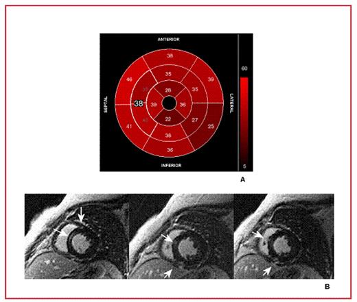
Little is known about cardiac involvement in Thalassemia Intermedia (TI) using magnetic resonance imaging (MRI). Thus, we investigated myocardial and liver iron overload, myocardial fibrosis, biventricular function parameters and their inter-relationship in 60 TI using MRI. Then, we compared the data of 35 selected TI with age < 39 years with that of 60 Thalassemia Major (TM) patients matched for age. Myocardial Iron Overload (MIO) was assessed using a multislice multiecho T2* approach. Biventricular function parameters were measured quantitatively by cine images. Myocardial fibrosis was evaluated by late gadolinium-enhanced. MIO was present in the 22% of TI patients, more frequently (92%) with an heterogeneous distribution. TI patients with myocardial fibrosis (32%) showed significantly reduced left ventricular ejection fraction (LVEF) (P=0.01). No significant correlation was found between myocardial fibrosis and MIO. Figure shows a TI patient with no myocardial iron overload (all 16 segments and the mid-ventricular septum with T2* values > 20 ms) (A) and late enhancement (white arrows) in the antero-septal junction, in the infero-septal junction and in the mid-ventricular septum (B). In TI patients MIO was significantly lower respect to TM patients; conversely volumes, cardiac indexes, LVEF, bi-atrial areas, and liver iron overload were significantly higher. Myocardial fibrosis was comparable between the 2 groups. TI patients showed lower myocardial iron burden and more pronounced high cardiac output findings with myocardial fibrosis significantly associated to impaired heart function. These data seem to suggest using MRI also for TI patients and could place the basis to reconsider the current cardiological and haematological management in this population.
Disclosures: No relevant conflicts of interest to declare.
Author notes
Corresponding author


