E2-2 belongs to the basic helix-loop-helix (bHLH) family of transcription factors. E2-2 associates with inhibitor of DNA binding (Id) 1, which is involved in angiogenesis. In this paper, we demonstrate that E2-2 interacts with Id1 and provide evidence that this interaction potentiates angiogenesis. Mutational analysis revealed that the HLH domain of E2-2 is required for the interaction with Id1 and vice versa. In addition, Id1 interfered with E2-2–mediated effects on luciferase reporter activities. Interestingly, injection of E2-2–expressing adenoviruses into Matrigel plugs implanted under the skin blocked in vivo angiogenesis. In contrast, the injection of Id1-expressing adenoviruses rescued E2-2–mediated inhibition of in vivo angiogenic reaction. Consistent with the results of the Matrigel plug assay, E2-2 could inhibit endothelial cell (EC) migration, network formation, and proliferation. On the other hand, knockdown of E2-2 in ECs increased EC migration. The blockade of EC migration by E2-2 was relieved by exogenous expression of Id1. We also demonstrated that E2-2 can perturb VEGFR2 expression via inhibition of VEGFR2 promoter activity. This study suggests that E2-2 can maintain EC quiescence and that Id1 can counter this effect.
Introduction
Angiogenesis is the formation of new vessels from preexisting ones by sprouting or by intussusceptive microvascular growth. Angiogenesis takes place throughout development as well as in adulthood. Although the vasculature in adults is generally quiescent, angiogenesis occurs to ensure physiologic homeostasis and integrity after wound healing, inflammation, ischemia, and during the female reproductive cycle.1 Angiogenesis encompasses 2 phases: activation and resolution. The activation phase is initiated by growth factor signals (eg, vascular endothelial growth factor [VEGF], fibroblast growth factor-2 [FGF-2]) or by hypoxia. In this process, endothelial cells (ECs) proliferate, vascular permeability increases, and extracellular matrix components are degraded. These serial events allow ECs to migrate and form new capillary sprouts. During the resolution stage, ECs cease proliferation and migration, the basement membrane is reconstituted, and the vessels mature. The transition from the activation phase to the resolution phase, and vice versa, is referred to as the “angiogenic switch” and is determined by a tightly regulated balance between angiogenic inducers and inhibitors.2
The inhibitor of the DNA-binding (Id) family of proteins, consisting of Id1, Id2, Id3, and Id4, belongs to the helix-loop-helix (HLH) family of transcription factors. The basic HLH (bHLH) family of transcription factors regulates transcription by binding to DNA as either homodimers or heterodimers. Id proteins, which lack a DNA-binding domain, interact with bHLH proteins to prevent dimer formation and/or DNA binding.3 In addition, Id1 can associate with members of the Ets protein family and Rb.4,5 Several observations support a crucial role for Id proteins in development, differentiation, and proliferation of cells, and in tumorigenesis. For example, it has been demonstrated that Id1 can block MyoD-mediated myogenic responses.6,–8 Id1 and Id3, identified as direct targets of bone morphogenetic protein (BMP) signaling,9,,,,,–15 are abundantly expressed during blood vessel formation. Id1/Id3 double-knockout mice display abnormal angiogenesis characterized by enlarged, dilated blood vessels.16 Introduction of Id1 into ECs induces EC proliferation and migration.17,18 Because knockdown of Id1 in ECs treated with BMP perturbs EC activation, Id1 is considered to be essential for BMP-induced EC activation.18 Loss of Id1 in tumor ECs leads to down-regulation of integrin α6, integrin β4, FGFR1, MMP2, and laminin5.19 Thus, Id1 positively regulates proangiogenic transcripts even though it cannot bind directly to DNA. Although many bHLH transcription factors have been implicated in EC angiogenic activities, which proteins Id1 regulates remains unclear.
We have reported that Hey1/Herp2/Hesr1, one of the bHLH proteins induced by Notch signaling, antagonizes activated BMP receptor-induced EC migration. This antagonism is caused by the degradation of Id1 interacting with Hey1/Herp2/Hesr1 in ECs. Thus, Id1-induced EC migration is blocked by Hey1/Herp2/Hesr1.20 However, the mechanism by which Id1 triggers EC angiogenic activation is still poorly understood.
To gain more insight into the molecular mechanisms by which Id1 positively regulates EC activation, we searched for Id1 interaction partners using a yeast 2-hybrid system and identified E2-2. E2-2 (also known as ITF2, TCF4, SEF2, and SEF2-1B) is classified into the E-protein family (or the class A type of bHLH transcription factors), whose expression is virtually ubiquitous. In addition to E2-2, E2A and HEB also belong to the E-protein family. This family of transcription factors recognizes a consensus DNA sequence known as the E-box (CANNTG) in dimer form, whereas monomeric forms have no discernible DNA-binding activity.21,22 The E-protein family of proteins is known to regulate lymphocyte development,23 neural differentiation,24 and myogenesis.25 Our previous study demonstrated that E2-2 can inhibit the activity of VEGFR2 luciferase reporters in ECs.26 However, how E2-2 counteracts the activity of the VEGFR2 promoter and whether E2-2 influences the actions of ECs remain veiled. In this study, we explored the role of E2-2 in angiogenesis both in vitro and in vivo. We found that E2-2 represses VEGFR2 promoter activity to inhibit angiogenesis and that E2-2–mediated EC inactivation can be alleviated through interaction with Id1.
Methods
Plasmids and adenoviruses
cDNAs for human and mouse E2-2 as well as mouse LMO2 were cloned by reverse-transcription polymerase chain reaction (RT-PCR). Each cDNA was sequenced before use. E2-2ΔHLH and Id1ΔHLH were generated by Pfx DNA polymerase (Invitrogen) using human E2-2 or mouse Id1 as templates, respectively. Mouse stem cell leukemia hematopoietic transcription factor (SCL) was a kind gift from Dr M. Ema (University of Tsukuba, Japan).27 cDNAs were inserted into Flag-pcDNA3 or Myc-pcDNA328 and subsequently ligated into the pcDEF3 vector.29 DEF3-Flag-Id1, DEF3-Myc-Id1, and DEF3-Id1 have been previously described.20 MCKpfos-luc and pGL2b-VEGFR2-luc (−166 bp/267 bp) were generously provided by Drs M. Sigvardsson (Lund University, Sweden) and C. C. W. Hughes (University of California, Irvine), respectively.26,30,31 VEGFR2-luc mutant constructs were also generated by PCR. After verification of each sequence, reporter constructs were used for each experiment. Adenoviruses expressing Myc-E2-2 were generated using the pAdTrack-CMV vector.32 After recombination of pAdTrack-CMV-Myc-E2-2 with pAdEasy-1,32 the resulting plasmid was transfected into 293T cells, and adenoviruses were amplified. Adenoviruses expressing Flag-Id1 have been reported.20
Cell culture
COS7 cells and mouse embryonic endothelial cells (MEECs)17 were maintained in Dulbecco modified Eagle medium (DMEM; Invitrogen) containing 10% fetal calf serum (FCS; Invitrogen), minimum essential medium nonessential amino acids (Invitrogen), and 100 U/mL penicillin/streptomycin (Wako). Calf pulmonary aortic endothelial cells (CPAEs)33 were cultured in DMEM with 10% FCS, 20mM N-2-hydroxyethylpiperazine-N′-2-ethanesulfonic acid (Wako), and 100 U/mL penicillin/streptomycin. Primary human umbilical vein endothelial cells (HUVECs) were cultured in endothelial basal medium (Lonza Walkersville) supplemented with 2% FCS. MEECs and HUVECs were grown on 0.1% gelatin-coated dishes.
Adenoviral infections
Adenoviruses were incubated in DMEM containing polybrene (Sigma-Aldrich; 80 μg/mL) for 2 hours and added to the dishes. Two hours after infection, cells were washed and allowed to recover 24 hours before the experiments. If necessary, cells were starved by removal from the FCS overnight and then stimulated with 50 ng/mL recombinant human VEGF (Wako).
Immunoprecipitation and Western blotting
To detect interactions among the proteins, plasmids were transfected into COS7 cells (5 × 10⁵ cells/6-cm dish) using FuGENE6 (Roche Diagnostics). Forty hours after transfection, the cells were lysed in 500 μL of TNE buffer (10mM Tris [pH 7.4], 150mM NaCl, 1mM ethylenediamine-N′, N′, N′, N′-tetraaccetic acid, 1% NP-40, 1mM phenymethylsulfonyl-l-fluoride, 5 μg/mL leupeptin, 100 U/mL aprotinin, 2mM sodium vanadate, 40mM NaF, and 20mM β-glycerophosphate). Cell lysates were precleared with protein G-Sepharose beads (GE Healthcare) for 30 minutes at 4°C and then incubated with anti-Flag M5 antibody (Sigma-Aldrich) for 2 hours at 4°C. Protein complexes were immunoprecipitated by incubation with protein G-Sepharose beads for 30 minutes at 4°C followed by 3 washes with TNE buffer. Immunoprecipitated proteins and aliquots of total cell lysates were boiled for 5 minutes in sample buffer, separated by sodium dodecyl sulfate-polyacrylamide gel electrophoresis, and transferred to Hybond-C Extra membranes (GE Healthcare). The membranes were probed with anti-Myc 9E10 antibody (Santa Cruz Biotechnology). Primary antibodies were detected using horseradish peroxidase-conjugated goat anti–mouse antibody (GE Healthcare) and chemiluminescent substrate (Thermo Electron). Protein expression in total cell lysates was evaluated by Western blotting using anti-Flag M5 or anti-Myc 9E10 antibody. To detect the endogenous interaction between Id1 and E2-2, either an anti–E2-2 monoclonal antibody (anti-TCF4 M03; Abnova) or an anti-Id1 polyclonal antibody (Santa Cruz Biotechnology) was used.
RNA isolation and RT-PCR
Total RNA was isolated using the RNeasy kit (QIAGEN). Reverse transcription was carried out using a First-Strand cDNA Synthesis Kit (Takara). PCR was performed using Taq polymerase (Invitrogen) as directed by the manufacturer. Primer sets used are shown in supplemental Tables 1 and 2 (available on the Blood Web site; see the Supplemental Materials link at the top of the online article).
Transcriptional reporter assay
MEECs were seeded at 5 × 10⁴ cells/well in 12-well plates 1 day before transfection.
Cells were transfected using Lipofectamine (Invitrogen) and Plus Reagent (Invitrogen). After 40 hours of transfection, lysates were prepared and luciferase activity was measured using a luciferase assay system (Promega). Results were corrected by measuring β-galactosidase activity (pCH110; GE Healthcare). Each experiment was carried out in triplicate and repeated at least twice. Values represent the mean plus or minus SD (n = 3).
Immunofluorescence
Immunofluorescence assay was performed as previously described.34 Briefly, MEECs grown on the cover glass were stimulated with 25 ng/mL BMP6 to induce Id1. After treatment, the glasses were washed once with phosphate-buffered saline (PBS), fixed for 10 minutes with 4% paraformaldehyde (PFA; Wako), washed 3 times with PBS, subsequently permeabilized with 0.5% Triton X-100 in PBS for 5 minutes, and washed again 3 times with PBS. Glasses were blocked with 5% normal swine serum (Dako Denmark) in PBS at 37°C for 1 hour and incubated with 5% normal swine serum (in PBS) containing mouse monoclonal anti–E2-2 (anti-TCF4 M03) and rabbit polyclonal Id1 antibodies at 4°C overnight. The glasses were then washed 3 times with PBS, incubated with 5% normal swine serum (in PBS) including both fluorescein isothiocyanate-conjugated goat anti–mouse IgG antibody (diluted 1:250; Invitrogen) and Texas red-conjugated goat anti–rabbit IgG antibody (diluted 1: 250; Invitrogen) at room temperature for 1 hour, and washed 3 times with PBS. To visualize the fluorescence, an immunofluorescence microscope (Axiovert 200M; Carl Zeiss) was used.
Migration assay
Cell migration assays were performed using a Boyden chamber. Costar nucleopore filters (8-μm pore diameter) were coated with 10 μg/mL fibronectin (Sigma-Aldrich) overnight at 4°C. The chambers were washed 3 times with PBS. Adenovirus-infected HUVECs starved for 12 hours without FCS were added to the top of each migration chamber at a density of 1.5 × 10⁴ cells/chamber in 150 μL of endothelial basal medium without FCS. Cells were allowed to migrate to the underside of the chamber in the presence or absence of 50 ng/mL VEGF in the lower chamber. After 6 hours, cells were fixed in 4% PFA and stained with 0.5% crystal violet (dissolved in 25% methanol; Wako). The upper surface was wiped with cotton swabs to remove nonmigrating cells. Cells present on the lower surface were counted. Each experiment was carried out in triplicate and repeated several times. Values represent the mean plus or minus SD (n = 3).
Network formation assay
HUVECs (2 × 10⁴ cells/well in an 8-well Lab-Tek chamber; Thermo Electron) infected with adenoviruses were seeded on growth factor-reduced Matrigel (BD Biosciences). Ninety minutes later, images were captured every 15 minutes for 4 hours using a time-lapse microscope (Axiovert 200M; Carl Zeiss) to monitor cell behavior.
Erk phosphorylation
HUVECs were seeded at 1 × 10⁵ cells/well in 12-well plates. Twenty-four hours after HUVECs were infected with the E2-2–expressing adenoviruses, HUVECs were starved without FCS for 12 hours. Then, HUVECs were stimulated with 50 ng/mL VEGF for the indicated times. Phospho-Erk1/2, Erk2, Myc-E2-2, and β-actin were analyzed by Western blots of total cell lysates using antiphospho-Erk(p44/p42) (Cell Signaling), anti-Erk2 (Santa Cruz Biotechnology), anti-Myc9E10 (Santa Cruz Biotechnology), and anti–β-actin (Sigma-Aldrich) antibodies, respectively.
Cell proliferation
Proliferation was measured by direct counting of cultures in 12-well plates. CPAEs were seeded at 1 × 10⁴ cells/well in 12-well plates 1 day before adenoviral infection. We began counting cells 1 day after adenoviral infection.
Mouse angiogenesis assay
The formation of new vessels in vivo was evaluated by Matrigel plug assay with some modifications to the previously described method.35 Adenoviruses expressing 1 × 10⁹ pfu of green fluorescent protein (GFP), 1 × 10⁹ pfu of E2-2, and/or 2.5 × 10⁸ pfu of Id1 were mixed in the Matrigel solution at 4°C with 200 ng/mL VEGF-A (Wako), 1 μg/mL bFGF (R&D Systems), and 100 μg/mL heparin (Wako). A total of 500 μL of Matrigel-containing adenoviruses was injected subcutaneously into the abdomen of male ICR mice. The mice were killed 7 days after the injection. The Matrigel plugs with adjacent subcutaneous tissues were recovered by en bloc resection, and images were then taken using a stereomicroscope (Leica). Thereafter, each sample was embedded in the optimal cutting temperature (OCT) compound and quickly frozen in liquid nitrogen to be sectioned at a thickness of 5 μm at −18°C. After being fixed with 4% PFA/PBS, anti–rat monoclonal PECAM-1 antibody (1:200 dilution) was used as a primary antibody. Then, the sections were incubated with Alexa568-conjugated goat anti–rat IgG antibody (1:200 dilution; Invitrogen). Images were taken using a fluorescence microscope (Carl Zeiss). Mice were housed in the animal facilities of Laboratory Animal Resource Center in University of Tsukuba under specific pathogen-free conditions with constant temperature and humidity and fed a standard diet. Treatment of mice was approved by the Animal Care and Use Program at the University of Tsukuba.
Knock-down of E2-2
The pLVTHM lentiviral vector for shRNA was purchased from Addgene. Double-strand DNAs for 5′-cgcgtccccAGAGCTGAGTGATTTACTGttcaagagaCAGTAAATCACTCAGCTCTtttttggaaat/3′-aggggTCTCGACTCACTAAATGACaagttctctGTCATTTAGTGAGTCGAGAaaaaacctttagc-5′ (shE2-2#1) and 5′-cgcgtccccGAAATTAGATGACGACAAGttcaagaga-CTTGTCGTCATCTAATTTCtttttggaaat-3′/3′-aggggCTTTAATCTACTGCTGTTCaagttctctGAACAGCAGTAGATTAAAGaaaaacctttagc-5′ (shE2-2#4) were inserted into pLVTHM digested with both MluI and ClaI. Lentiviral vectors expressing shE2-2 were transfected into 293T cells together with psPAX2 and pMD2.G. The lentiviruses were incubated for 2 hours in DMEM containing polybrene (80 μg/mL) and then added to the dishes. Two hours after being infected, the cells were washed and cultured in medium. Infected HUVECs, which became GFP-positive, were isolated by fluorescence-activated cell sorting and used for the experiments.
Results
Identification of Id1-interacting proteins
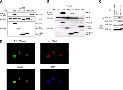
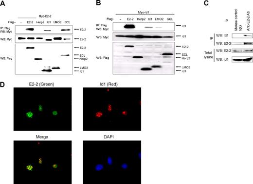
To obtain insight into how Id1 activates angiogenesis, we isolated proteins that interact with Id1 using a yeast 2-hybrid system. We used Id1 as bait to screen an amplified human aorta cDNA library (with an original complexity of 3.5 × 10⁶ independent cDNA clones). Among the positive clones identified, the cDNA encoding E2-2 was isolated most frequently. We then investigated whether E2-2 interacts with Id1 in mammalian cells. We also tested the interaction of E2-2 with Herp2, LMO2, and SCL, which are known to modulate EC activation.20,36,–38 As shown in Figure 1A, E2-2 interacted with Id1, SCL,26 and itself, but not with Herp2 or LMO2. In the reciprocal experiment, Id1 bound strongly with E2-2 and marginally with LMO2 and itself (Figure 1B). When the membrane was exposed for a longer time, an interaction between Id1 and Herp2, which we have described previously,20 was observed (data not shown). These results indicated that the interaction between Id1 and E2-2 is the most prominent of the combinations tested.
Interaction between E2-2 and Id1. (A) Interaction of Myc-E2-2 with Flag-Id1. Myc-E2-2 was cotransfected with Flag-E2-2, Flag-Herp2, Flag-Id1, Flag-LMO2, or Flag-SCL. Immunoprecipitations were carried out using anti-Flag M5 antibody, and coimmunoprecipitated E2-2 was detected by Western blotting using anti-Myc 9E10 antibody (top panel). The expression of Myc-E2-2 and proteins conjugated with Flag at the N-terminus was evaluated using anti-Myc 9E10 (middle panel) and anti-Flag M5 antibodies (bottom panel), respectively. (B) Interaction of Myc-Id1 with Flag-E2-2. The experiment was performed in a manner similar to that described in panel A. Interaction of Myc-Id1 with Flag-tagged proteins (top panel). Expression of Myc-Id1 and Flag-tagged proteins was checked using anti-Myc 9E10 antibodies (middle panel) and anti-Flag M5 antibodies (bottom panel), respectively. (C) Endogenous interaction between Id1 and E2-2. MEECs were stimulated with BMP6 for 3 hours. Cell lysates were immunoprecipitated with a mouse anti–E2-2 monoclonal antibody, followed by Western blotting with a rabbit anti-Id1 polyclonal antibody (top panel). Expression of E2-2 in immunoprecipitates was checked using an anti–E2-2 monoclonal antibody (second panel). To show expression of E2-2 and Id1 in total lysates, an anti–E2-2 monoclonal antibody (third panel) and an anti-Id1 polyclonal antibody (bottom panel) were used. As a negative control, mouse control IgGs were used for immunoprecipitation. (D) Colocalization of E2-2 with Id1 in MEECs. MEECs were stained with a mouse anti–E2-2 monoclonal antibody (green) or a rabbit anti-Id1 polyclonal antibody (red). Nuclei were visualized using 4′,6-diamidino-2-phenylindole. After samples were mounted with Fluorescent Mounting Medium (Dako Denmark), they were visualized using an immunofluorescence microscope (Axiovert 200M; Carl Zeiss) with a 63×/1.4 oil objective lenses (Carl Zeiss). Images were acquired with AxioCam MRm 60-C1 (Carl Zeiss) and processed with the AxioVision Rel 4.4 (Carl Zeiss) and Adobe Photoshop 7.0.1 software (Adobe).
Interaction between E2-2 and Id1. (A) Interaction of Myc-E2-2 with Flag-Id1. Myc-E2-2 was cotransfected with Flag-E2-2, Flag-Herp2, Flag-Id1, Flag-LMO2, or Flag-SCL. Immunoprecipitations were carried out using anti-Flag M5 antibody, and coimmunoprecipitated E2-2 was detected by Western blotting using anti-Myc 9E10 antibody (top panel). The expression of Myc-E2-2 and proteins conjugated with Flag at the N-terminus was evaluated using anti-Myc 9E10 (middle panel) and anti-Flag M5 antibodies (bottom panel), respectively. (B) Interaction of Myc-Id1 with Flag-E2-2. The experiment was performed in a manner similar to that described in panel A. Interaction of Myc-Id1 with Flag-tagged proteins (top panel). Expression of Myc-Id1 and Flag-tagged proteins was checked using anti-Myc 9E10 antibodies (middle panel) and anti-Flag M5 antibodies (bottom panel), respectively. (C) Endogenous interaction between Id1 and E2-2. MEECs were stimulated with BMP6 for 3 hours. Cell lysates were immunoprecipitated with a mouse anti–E2-2 monoclonal antibody, followed by Western blotting with a rabbit anti-Id1 polyclonal antibody (top panel). Expression of E2-2 in immunoprecipitates was checked using an anti–E2-2 monoclonal antibody (second panel). To show expression of E2-2 and Id1 in total lysates, an anti–E2-2 monoclonal antibody (third panel) and an anti-Id1 polyclonal antibody (bottom panel) were used. As a negative control, mouse control IgGs were used for immunoprecipitation. (D) Colocalization of E2-2 with Id1 in MEECs. MEECs were stained with a mouse anti–E2-2 monoclonal antibody (green) or a rabbit anti-Id1 polyclonal antibody (red). Nuclei were visualized using 4′,6-diamidino-2-phenylindole. After samples were mounted with Fluorescent Mounting Medium (Dako Denmark), they were visualized using an immunofluorescence microscope (Axiovert 200M; Carl Zeiss) with a 63×/1.4 oil objective lenses (Carl Zeiss). Images were acquired with AxioCam MRm 60-C1 (Carl Zeiss) and processed with the AxioVision Rel 4.4 (Carl Zeiss) and Adobe Photoshop 7.0.1 software (Adobe).
To confirm that the interaction between E2-2 and Id1 is physiologically significant, we investigated the interaction between endogenous proteins in MEECs stimulated with BMP6 for 3 hours because Id1 is known to be induced by BMPs.39 After cell lysis, immunoprecipitation was performed using an anti–E2-2 antibody. Subsequently, immunoprecipitates were blotted with an anti-Id1 antibody. As shown in Figure 1C, endogenous Id1 formed a complex with endogenous E2-2 in cells. Conversely, the specific band corresponding to E2-2 could be detected (arrow) when cell lysates were immunoprecipitated with an anti-Id1 antibody (supplemental Figure 1). The interaction between E2-2 and Id1 led us to investigate whether E2-2 colocalizes with Id1. To show colocalization of E2-2 with Id1 in ECs, subcellular localization was determined with a fluorescence microscopy by staining E2-2 with fluorescein isothiocyanate-conjugated goat anti–mouse IgG and Id1 with Texas Red-conjugated goat anti–rabbit IgG. E2-2 and Id1 colocalized in nucleus (Figure 1D), which is consistent with an interaction between the 2 proteins. In addition, we used the aorta ring assay to visualize the expressions of E2-2 and Id1 with fluorescent imaging. When both E2-2 and Id1 were costained with fluorescence probes, we recognized that both proteins in sprouting ECs could colocalize in the nucleus (data not shown).
HLH domains play an important role in protein-protein interactions. To examine the contribution of HLH domains to the E2-2/Id1 interaction, we made E2-2 and Id1 mutants lacking the respective HLH domains (supplemental Figure 2A). As shown in supplemental Figure 2B and C, E2-2 did not interact with E2-2ΔHLH or Id1ΔHLH. Similarly, Id1 did not interact with E2-2ΔHLH (supplemental Figure 2D). To further confirm that the HLH domain of E2-2 is enough for E2-2 to interact with Id1, we made an expression construct for GFP, which was fused with the HLH domain derived from E2-2 (GFP-HLH). When GFP-HLH was cotransfected with Id1 in COS7 cells, we could observe the interaction between GFP-HLH and Id1 (supplemental Figure 2E). However, its interaction seemed to be weak. Thus, the other domain(s) of E2-2, except for its HLH domain, might contribute to the adequate interaction between E2-2 and Id1. Taken together, these results indicated that E2-2 heterodimerizes with Id1 and homodimerizes through its HLH domain. In addition, Id1 requires its HLH domain to associate with E2-2.
Inhibition of E2-2–mediated transcription by Id1
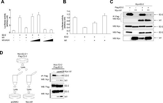
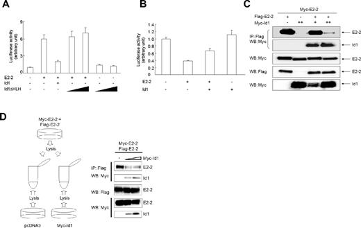
The MCKpfos-luc reporter construct consisting of 4 E-box elements has been used to investigate the function of E-proteins.26,30 E2-2 enhanced the activity of this reporter in a dose-dependent manner (supplemental Figure 3A), but E2-2ΔHLH was unable to potentiate reporter activity (data not shown). Id proteins have been reported to interact with bHLH transcription factors and prevent them from binding to DNA or forming active heterodimers (or homodimers).3 Although we did not know whether E2-2 forms a homodimer or a heterodimer with other bHLH proteins in MEECs to activate this reporter, Id1 blocked E2-2–induced reporter activity as anticipated (Figure 2A; supplemental Figure 3B). Consistent with the undetectable interaction between E2-2 and Id1ΔHLH, E2-2–induced reporter activity was not reduced by Id1ΔHLH (Figure 2A).
Id1 counteracts E2-2–mediated luciferase activity. (A) Id1ΔHLH does not perturb E2-2–induced MCKpfos-luc activity. MEECs were transfected with MCKpfos-luc, E2-2, and either Id1 or different amounts of Id1ΔHLH. (B) Id1 relieves the inhibition of pGL2b-VEGFR2-luc (−166 bp/267 bp) activity by E2-2. MEECs were transfected with pGL2b-VEGFR2-luc (−166 bp/267 bp), Id1, and E2-2. (C) Id1 disrupts E2-2 homodimer formation. The experiment was performed as described in Figure 1A. E2-2 homodimer formation (top panel) and E2-2/Id1 heterodimer formation (second panel) are shown. Expressions of Myc-E2-2 (third panel) and Myc-Id1 (bottom panel) were evaluated using an anti-Myc 9E10 antibody, and the expression of Flag-E2-2 was shown using an anti-Flag M5 antibody (fourth panel). (D) Id1 disturbs the preexisting E2-2 homodimer formation. Left panel: Illustration of how cell lysates were prepared from each dish in which indicated plasmids were transfected in COS7 cells. Right panel: After each cell lysate was mixed, the experiment was performed as described in Figure 1A. E2-2 homodimer formation (top panel) and E2-2/Id1 heterodimer formation (second panel) are shown. The expression of Flag-E2-2 was shown using an anti-Flag M5 antibody (third panel). Expressions of Myc-E2-2 (fourth panel) and Myc-Id1 (bottom panel) were evaluated using an anti-Myc 9E10 antibody.
Id1 counteracts E2-2–mediated luciferase activity. (A) Id1ΔHLH does not perturb E2-2–induced MCKpfos-luc activity. MEECs were transfected with MCKpfos-luc, E2-2, and either Id1 or different amounts of Id1ΔHLH. (B) Id1 relieves the inhibition of pGL2b-VEGFR2-luc (−166 bp/267 bp) activity by E2-2. MEECs were transfected with pGL2b-VEGFR2-luc (−166 bp/267 bp), Id1, and E2-2. (C) Id1 disrupts E2-2 homodimer formation. The experiment was performed as described in Figure 1A. E2-2 homodimer formation (top panel) and E2-2/Id1 heterodimer formation (second panel) are shown. Expressions of Myc-E2-2 (third panel) and Myc-Id1 (bottom panel) were evaluated using an anti-Myc 9E10 antibody, and the expression of Flag-E2-2 was shown using an anti-Flag M5 antibody (fourth panel). (D) Id1 disturbs the preexisting E2-2 homodimer formation. Left panel: Illustration of how cell lysates were prepared from each dish in which indicated plasmids were transfected in COS7 cells. Right panel: After each cell lysate was mixed, the experiment was performed as described in Figure 1A. E2-2 homodimer formation (top panel) and E2-2/Id1 heterodimer formation (second panel) are shown. The expression of Flag-E2-2 was shown using an anti-Flag M5 antibody (third panel). Expressions of Myc-E2-2 (fourth panel) and Myc-Id1 (bottom panel) were evaluated using an anti-Myc 9E10 antibody.
As we have reported,26 E2-2 inhibited VEGFR2 reporter activity in MEECs in a dose-dependent manner (supplemental Figure 3C), whereas the introduction of Id1 into MEECs relieved the E2-2–mediated repression (Figure 2B). To show that Id1 indeed hampers E2-2 dimer formation, COS7 cells were cotransfected with Myc-E2-2 and Flag-E2-2 in the absence or presence of Myc-Id1. Immunoprecipitation of COS7 cell extracts revealed that Id1 blocked E2-2 homodimer formation (Figure 2C). We also investigated whether Id1 can disrupt the preexisting E2-2 homodimer. We prepared lysates from cells transfected with either Myc-Id1 or pcDNA3. Subsequently, each lysate was mixed with lysate prepared from cells transfected with both Myc-E2-2 and Flag-E2-2, immunoprecipitated with anti-Flag antibody, and then analyzed by Western blotting with anti-Myc antibody. As seen in Figure 2D, Id1 could efficiently make the preexisting E2-2 homodimer dissociated.
Inhibition of EC activation by E2-2
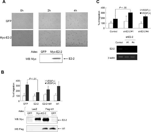
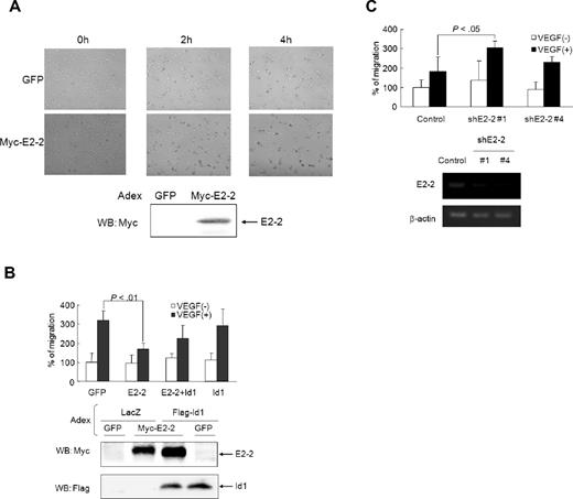
We speculated that E2-2 would have an opposite effect on EC activation compared with Id1 because Id1 antagonized E2-2–mediated reporter activity. To investigate the effect of E2-2 on network formation, we infected HUVECs with either GFP- or E2-2–expressing adenoviruses before seeding the cells on Matrigel. We then observed the formation of cord-like structures by time-lapse microscopy (Figure 3A). As seen in video (supplemental Figure 4A-B), HUVECs expressing E2-2 did not form cord-like structures as quickly as HUVECs expressing GFP as a control did. We also investigated the effect of E2-2 on cell migration in the presence of VEGF. HUVECs challenged with VEGF after infection with E2-2–expressing adenoviruses exhibited decreased migration compared with cells infected with GFP-expressing adenoviruses (Figure 3B). Because Id1 perturbs the function of E2-2, we examined the possibility that Id1 would relieve the E2-2–mediated repression of the VEGF-induced chemotactic response. Indeed, Id1 marginally rescued the inhibitory effect of E2-2 on VEGF-induced cell migration (Figure 3B). Importantly, VEGF-induced HUVEC migration was also increased when E2-2 levels were decreased via lentiviral expression of human shE2-2s but not in response to control lentiviruses (Figure 3C).
E2-2 blocks EC activation. (A) E2-2 inhibits the formation of cord-like structures on the Matrigel. Forty hours after adenoviral infection, HUVECs were seeded on the Matrigel. Ninety minutes later, images were recorded every 15 minutes by time-lapse microscopy (supplemental Figure 4A-B). The images at 0-, 2-, and 4-hour time points are shown. Bottom panel: Expression of Myc-E2-2. Samples were visualized using a conventional microscope (Axiovert 200M; Carl Zeiss) with a 5×/0.12 dry objective lenses (Carl Zeiss). Images were acquired with AxioCam MRm 60-C1 (Carl Zeiss) and processed with the AxioVision Rel 4.4 (Carl Zeiss) and Adobe Photoshop 7.0.1 software (Adobe). (B) Id1 rescues E2-2–mediated inhibition of cell migration. After adenoviral infection, HUVECs were seeded on the upper membrane of the Boyden chamber. VEGF (50 ng/mL) was added to the lower chamber. After 6 hours, cells were stained with crystal violet, and the number of transmigrated cells was counted. Adenoviruses expressing LacZ or GFP were used as controls. Values represent the mean plus or minus SD (n = 3). Bottom panels: Myc-E2-2 and Flag-Id1 expression levels. Significant difference was calculated by the Student t test. (C) shE2-2 enhances VEGF-induced effect on HUVEC migration. Lentiviruses expressing GFP alone, shE2-2#1, or shE2-2#4 were infected in HUVECs. After sorting lentivirus-infected cells using GFP as a marker, sorted HUVECs were used for migration assay as described in panel B. Bottom panels: Expressions of E2-2 and β-actin by RT-PCR. Values represent the mean plus or minus SD (n = 3). Significant difference was calculated by the Student t test.
E2-2 blocks EC activation. (A) E2-2 inhibits the formation of cord-like structures on the Matrigel. Forty hours after adenoviral infection, HUVECs were seeded on the Matrigel. Ninety minutes later, images were recorded every 15 minutes by time-lapse microscopy (supplemental Figure 4A-B). The images at 0-, 2-, and 4-hour time points are shown. Bottom panel: Expression of Myc-E2-2. Samples were visualized using a conventional microscope (Axiovert 200M; Carl Zeiss) with a 5×/0.12 dry objective lenses (Carl Zeiss). Images were acquired with AxioCam MRm 60-C1 (Carl Zeiss) and processed with the AxioVision Rel 4.4 (Carl Zeiss) and Adobe Photoshop 7.0.1 software (Adobe). (B) Id1 rescues E2-2–mediated inhibition of cell migration. After adenoviral infection, HUVECs were seeded on the upper membrane of the Boyden chamber. VEGF (50 ng/mL) was added to the lower chamber. After 6 hours, cells were stained with crystal violet, and the number of transmigrated cells was counted. Adenoviruses expressing LacZ or GFP were used as controls. Values represent the mean plus or minus SD (n = 3). Bottom panels: Myc-E2-2 and Flag-Id1 expression levels. Significant difference was calculated by the Student t test. (C) shE2-2 enhances VEGF-induced effect on HUVEC migration. Lentiviruses expressing GFP alone, shE2-2#1, or shE2-2#4 were infected in HUVECs. After sorting lentivirus-infected cells using GFP as a marker, sorted HUVECs were used for migration assay as described in panel B. Bottom panels: Expressions of E2-2 and β-actin by RT-PCR. Values represent the mean plus or minus SD (n = 3). Significant difference was calculated by the Student t test.
Impairment of in vivo angiogenesis by E2-2
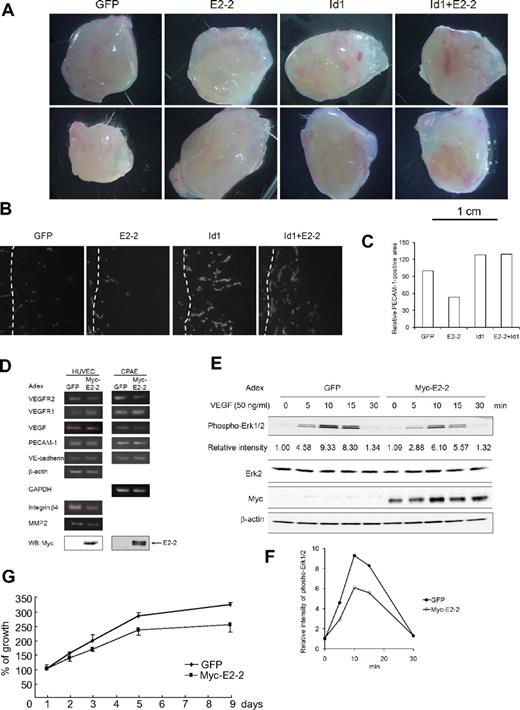
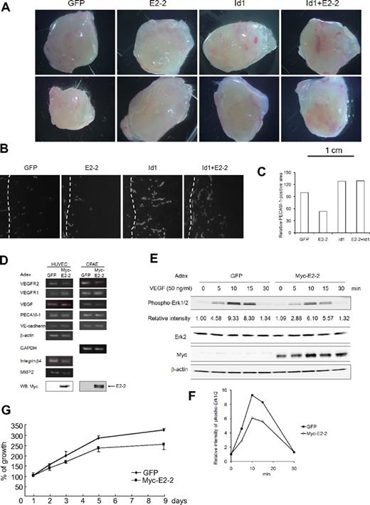
Because E2-2 inhibits EC activation in vitro, we tried to examine whether in vivo angiogenesis would be affected by overexpression of E2-2. To elucidate this possibility, we used the Matrigel plug assay by which the angiogenic (or antiangiogenic) ability of proteins, cytokines, or compounds can be assigned. To overexpress E2-2 and/or Id1, we subcutaneously injected Matrigel mixed with E2-2– and/or Id1-expressing adenoviruses.35,40 Seven days after implantation of the Matrigel plugs together with adenoviruses expressing E2-2 and/or Id1, the Matrigels were removed. When we investigated whether the protein expression by the method of adenoviral gene transfer was kept for 7 days, GFP-positive cells were detected in the Matrigel plugs (supplemental Figure 5). As seen in Figure 4A, there were fewer blood vessels observed in the Matrigel plugs containing E2-2–expressing adenoviruses than in those containing control adenoviruses. Because Id1 influenced the E2-2–mediated inhibition of EC activation, we mixed the Id1-expressing adenoviruses with E2-2–expressing adenoviruses and injected them into the Matrigel plugs implanted under the skin. As expected, the injection of Id1-expressing adenoviruses restored in vivo angiogenesis suppressed by E2-2. When the cells expressing PECAM-1 were visualized using the frozen sections, there were fewer PECAM-1–positive cells seen in the Matrigel plugs injected with adenoviruses expressing E2-2 than in those injected with control adenoviruses or a combination of Id1-expressing adenoviruses with E2-2–expressing adenoviruses (Figure 4B-C). These results indicated that, in contrast to Id1, E2-2 is an antagonistic molecule for angiogenic reaction.
Suppression of angiogenesis by E2-2. (A) Photographs for en bloc resection, including Matrigel plugs with adjacent subcutaneous tissues. Samples were visualized using a stereomicroscope (S8APO; Leica). Images were acquired with EC3 (Leica) and processed with the LAS EZ (Leica) and Adobe Photoshop 7.0.1 software (Adobe). (B) PECAM-1 staining of frozen sections. Matrigels are located to the right side of broken lines, whereas there are mouse subcutaneous tissues to the left side of broken lines. PECAM1-positive areas are shown as white. After samples were mounted with Fluorescent Mounting Medium (Dako Denmark), they were visualized using an immunofluorescence microscope (Axiovert 200M; Carl Zeiss) with a 63×/1.4 oil objective lenses (Carl Zeiss). Images were acquired with AxioCam MRm 60-C1 (Carl Zeiss) and processed with the AxioVision Rel 4.4 (Carl Zeiss) and Adobe Photoshop 7.0.1 software (Adobe). (C) Relative PECAM-1-positive area in Matrigel plugs. Five fields were randomly chosen in each condition from panel B, and PECAM-1-positive areas were scored using the imaging software. PECAM-1-positive areas were normalized with the areas of the field. Each relative PECAM-1-positive area was calculated by the comparison of the score in Matrigel plugs, including GFP. (D) Effect of E2-2 on the expression of mRNAs implicated in EC activation. mRNAs involved in EC activation were analyzed by RT-PCR. HUVECs or CPAEs were infected with adenoviruses expressing E2-2 or GFP as a negative control. Gene transcript names are indicated to the left of the figure. mRNAs for β-actin and GAPDH were included as internal controls. The expression of Myc-E2-2 was evaluated in total cell lysates using an anti-Myc 9E10 antibody (bottom panel). Adex indicates adenovirus. To show that EC markers are expressed in CPAEs, RT-PCR was carried out (supplemental Figure 6A). (E) E2-2 inhibits VEGF-induced Erk phosphorylation. Phospho-Erk1/2 (top panel), Erk2 (second panel), Myc-E2-2 (third panel), and β-actin (bottom panel) levels were analyzed by Western blots of total cell lysates. Adenoviruses expressing GFP were used as a negative control. The expression for phosphorylation of Erk1/2 was normalized using the intensity of the band corresponding to Erk2. Each relative intensity was calculated by the comparison of the value for cells infected with GFP-expressing adenoviruses in the absence of VEGF. (F) Graphical presentation for relative intensity of phospho-Erk1/2 levels in panel E: ● represents GFP-expressing adenoviruses; ○, Myc-E2-2–expressing adenovirusues. (G) E2-2 perturbs EC proliferation. Each experiment was performed in triplicate and repeated a few times. Values represent the mean plus or minus SD (n = 3).
Suppression of angiogenesis by E2-2. (A) Photographs for en bloc resection, including Matrigel plugs with adjacent subcutaneous tissues. Samples were visualized using a stereomicroscope (S8APO; Leica). Images were acquired with EC3 (Leica) and processed with the LAS EZ (Leica) and Adobe Photoshop 7.0.1 software (Adobe). (B) PECAM-1 staining of frozen sections. Matrigels are located to the right side of broken lines, whereas there are mouse subcutaneous tissues to the left side of broken lines. PECAM1-positive areas are shown as white. After samples were mounted with Fluorescent Mounting Medium (Dako Denmark), they were visualized using an immunofluorescence microscope (Axiovert 200M; Carl Zeiss) with a 63×/1.4 oil objective lenses (Carl Zeiss). Images were acquired with AxioCam MRm 60-C1 (Carl Zeiss) and processed with the AxioVision Rel 4.4 (Carl Zeiss) and Adobe Photoshop 7.0.1 software (Adobe). (C) Relative PECAM-1-positive area in Matrigel plugs. Five fields were randomly chosen in each condition from panel B, and PECAM-1-positive areas were scored using the imaging software. PECAM-1-positive areas were normalized with the areas of the field. Each relative PECAM-1-positive area was calculated by the comparison of the score in Matrigel plugs, including GFP. (D) Effect of E2-2 on the expression of mRNAs implicated in EC activation. mRNAs involved in EC activation were analyzed by RT-PCR. HUVECs or CPAEs were infected with adenoviruses expressing E2-2 or GFP as a negative control. Gene transcript names are indicated to the left of the figure. mRNAs for β-actin and GAPDH were included as internal controls. The expression of Myc-E2-2 was evaluated in total cell lysates using an anti-Myc 9E10 antibody (bottom panel). Adex indicates adenovirus. To show that EC markers are expressed in CPAEs, RT-PCR was carried out (supplemental Figure 6A). (E) E2-2 inhibits VEGF-induced Erk phosphorylation. Phospho-Erk1/2 (top panel), Erk2 (second panel), Myc-E2-2 (third panel), and β-actin (bottom panel) levels were analyzed by Western blots of total cell lysates. Adenoviruses expressing GFP were used as a negative control. The expression for phosphorylation of Erk1/2 was normalized using the intensity of the band corresponding to Erk2. Each relative intensity was calculated by the comparison of the value for cells infected with GFP-expressing adenoviruses in the absence of VEGF. (F) Graphical presentation for relative intensity of phospho-Erk1/2 levels in panel E: ● represents GFP-expressing adenoviruses; ○, Myc-E2-2–expressing adenovirusues. (G) E2-2 perturbs EC proliferation. Each experiment was performed in triplicate and repeated a few times. Values represent the mean plus or minus SD (n = 3).
To investigate the mechanism by which E2-2 inhibits angiogenic reaction both in vitro and in vivo, we examined the expression of several genes known to be implicated in angiogenesis (eg, VEGFR1, VEGFR2, VEGF, PECAM-1, and VE-cadherin). Of the genes examined, VEGFR2 mRNA was considerably reduced in both human and bovine ECs after infection with E2-2–expressing adenoviruses (Figure 4D); the expression of VEGF was marginally decreased by E2-2. VEGFR1, PECAM-1, and VE-cadherin mRNA levels, however, did not change in response to E2-2 overexpression (Figure 4D). Because VEGFR2 mRNA was prominently decreased by E2-2, we were prompted to check its protein expression after the infection of E2-2 to HUVECs. Consistent with the result of VEGFR2 mRNA expression, E2-2 slightly inhibited the expression of VEGFR2 protein, whereas Id1 partially rescued decrease of VEGFR2 protein mediated by E2-2 (supplemental Figure 6B).
VEGF signaling through the activation of the VEGFR2 receptor elicits multiple effects on ECs, such as migration, proliferation, survival, and differentiation.41 One of the signaling pathways downstream of VEGF/VEGFR2 is the MAP kinase pathway. When HUVECs were stimulated with VEGF, Erk was transiently phosphorylated (Figure 4E). Its phosphorylation peaked at 10 minutes after VEGF stimulation and declined rapidly thereafter. When E2-2 was overexpressed in HUVECs by adenoviral gene transfer, VEGF induced phosphorylation of Erk with similar kinetics to those of HUVECs infected with GFP-expressing adenoviruses; however, the extent of Erk phosphorylation was diminished (Figure 4E-F). Consistent with the inhibition of VEGF-induced Erk phosphorylation by E2-2, CPAEs and HUVECs expressing E2-2 proliferated more slowly than those expressing GFP (Figure 4G; supplemental Figure 6C). Taken together, these observations support the idea that E2-2 has the ability to inhibit EC activation by antagonizing VEGF signaling.
Id1 has been implicated in the regulation of the expression of integrin α6, integrin β4, FGFR1, MMP2, and laminin5; loss of Id1 results in decreased protein levels.19 Because Id1 antagonizes the function of E2-2, we hypothesized that genes suppressed transcriptionally by the loss of Id1 might be negatively regulated by E2-2. Transcript levels of MMP2 and integrin β4, but not of other genes, in ECs were reduced by overexpression of E2-2 (Figure 4D; and data not shown). Thus, E2-2 may also block EC activation through transcriptional repression of other proangiogenic genes.
Suppression of VEGFR2 promoter by E2-2
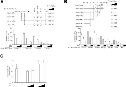
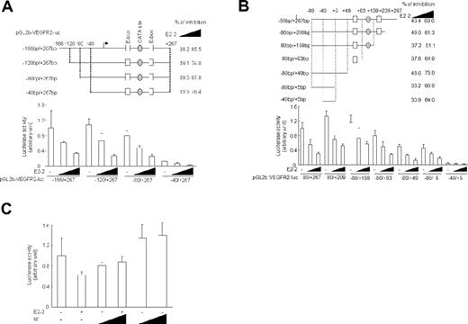
Because ectopic E2-2 reduced VEGFR2 mRNA levels in ECs (Figure 4D), we tried to determine which regions of the VEGFR2 promoter could be affected by E2-2. We tested several VEGFR2 promoter constructs truncated downstream of position −166 for E2-2 responsiveness. Basal reporter activity was greatly reduced on removal of the sequence between −80 and −40. However, all of the constructs were strongly inhibited by E2-2 in a dose-dependent manner (Figure 5A). Two typical E-boxes located in the 5′ noncoding region of the VEGFR2 gene were predicted to be involved in the inhibition of VEGFR2 promoter activity by E2-2. To test this hypothesis, we made 7 3′-truncation mutants of the VEGFR2 promoter (Figure 5B). Basal reporter activity gradually decreased as the 5′ noncoding region was truncated. Unexpectedly, E2-2 could still attenuate the luciferase activity even when both E-boxes were deleted. Furthermore, the activity of pGL2b-VEGFR2-luc (−40 bp/5 bp) was also inhibited by E2-2 despite very low basal luciferase activity. GATA-1 and/or GATA-2 are known to positively regulate VEGFR2 promoter activity by binding to the 5′ noncoding region (98-122 bp).42 However, the basal activity of the reporter lacking the GATA binding site (pGL2b-VEGFR2-luc [-80 bp/93 bp]) remained at 60% of the reporter activity with the binding site included (pGL2b-VEGFR2-luc, −80 bp/159 bp). Thus, it appears that neither GATA sequences nor E-boxes are required for E2-2 to repress the activity of the VEGFR2 promoter.
Analysis of the VEGFR2 promoter. (A) Effect of E2-2 on 5′ deletion mutants of the VEGFR2 promoter. MEECs were transfected with 1 of the indicated reporters with different amounts of E2-2. The inhibition percentage of reporter activities by E2-2 is indicated to the right of the top panel. (B) Effect of E2-2 on 3′ deletion mutants of the VEGFR2 promoter. The experiment was carried out as described in panel A. (C) Effect of Id1 on E2-2–mediated suppression of VEGFR2 promoter activity. Different amounts of Id1 (0.1 and 0.5 pg) were transfected together with E2-2. The experiment was carried out as described in panel A.
Analysis of the VEGFR2 promoter. (A) Effect of E2-2 on 5′ deletion mutants of the VEGFR2 promoter. MEECs were transfected with 1 of the indicated reporters with different amounts of E2-2. The inhibition percentage of reporter activities by E2-2 is indicated to the right of the top panel. (B) Effect of E2-2 on 3′ deletion mutants of the VEGFR2 promoter. The experiment was carried out as described in panel A. (C) Effect of Id1 on E2-2–mediated suppression of VEGFR2 promoter activity. Different amounts of Id1 (0.1 and 0.5 pg) were transfected together with E2-2. The experiment was carried out as described in panel A.
We next explored the effect of Id1 on the E2-2–mediated repression of VEGFR2 promoter activity. For this purpose, we used pGL2b-VEGFR2-luc (−80 bp/5 bp) instead of pGL2b-VEGFR2-luc (−40 bp/5 bp) because the basal activity of pGL2b-VEGFR2-luc (−40 bp/5 bp) was quite low. Similar to the effect of Id1 on the pGL2b-VEGFR2-luc (−166 bp/267 bp) reporter (Figure 2B), Id1 relieved the E2-2–mediated repression of pGL2b-VEGFR2-luc (−80 bp/5 bp) activity (Figure 5C), but Id1ΔHLH did not (data not shown). This evidence further convinced us that E2-2 negatively regulates the VEGFR2 promoter. Because the HLH domain of E2-2 is required to activate the MCKpfos-luc reporter (data not shown), we also tested the effect of E2-2ΔHLH on the activity of the VEGFR2 promoter. As expected, E2-2ΔHLH was not capable of inhibiting VEGFR2 promoter activity (data not shown). To test the possibility that E2-2 can bind to the VEGFR2 promoter (from −40 to 5), we performed electrophoresis mobility shift assay using glutathione S-transferase (GST) fusion proteins. GST or GST-Id1 showed no specific binding to the VEGFR2 promoter. However, a specific shifted band appeared when GST-E2-2 was mixed with the probe. Interestingly, the shifted complex decreased when GST-Id1 was added to the mixture containing GST-E2-2 and the probe (supplemental Figure 7A). To further confirm that Id1 promotes the dissociation of E2-2 from the VEGFR2 promoter, we carried out chromatin immunoprecipitation analysis using HUVECs infected with E2-2– and/or Id1-expressing adenoviruses. In the presence of Id1, the VEGFR2 promoter showed decrease in E2-2 binding (supplemental Figure 7B). These results support the data that Id1 rescues E2-2–mediated repression of VEGFR2 promoter activity.
Discussion
It is widely accepted that ubiquitous bHLH proteins, such as E2-2 and E2A, interact with tissue-restricted bHLH proteins to bind consensus E-box sequences and activate transcription of target genes. E-proteins are pivotal in the regulation of cell proliferation and differentiation. For example, E2-2 and its family member E2A play critical roles in B-cell development and perturbed T-cell development.43,–45 In the present study, we found that E2-2 could inhibit angiogenesis in Matrigel plugs implanted under the skin, whereas Id1 overcame E2-2–mediated antagonism of angiogenesis in vivo.
We showed that E2-2 associated with Id1 in MEECs (Figure 1C; supplemental Figure 1A). Consistent with this interaction, Id1 blocked the E2-2–induced activity of the artificial E-box–containing reporter (MCKpfos-luc) in a manner dependent on the HLH domains of Id1 (Figure 2A; supplemental Figure 3B). On the other hand, E2-2 blocked the VEGFR2 reporter activity in ECs independently of typical E-box sequences (Figure 5B).26 Although E2-2 generally activates the transcription of target genes by forming dimers with other bHLH proteins, a few reports have shown that E2-2 can interfere with transcription.46,47 In the present study, we clarified that E2-2 negatively regulated the activity of the VEGFR2 promoter, which does not possess typical E-box sequences. However, the VEGFR2 promoter from −40 to 5 contains one imperfect E-box sequence (CANNTC), which has been identified as one of the binding sites of E2-2.46 It is possible that E2-2 negatively regulates the transcription of the VEGFR2 gene through its binding to the imperfect E-box sequence because GST-E2-2 could make a specific complex with this promoter region. Excepting that possibility, E2-2 might displace TFII-I from the initiator (inr) sequences or interact with TFII-I on inr sequences to prevent TFII-I from activating transcription because TFII-I was recently found to enhance VEGFR2 transcription in an inr-dependent fashion.48 Further investigation is needed to examine in detail how E2-2 inhibits VEGFR2 promoter activity. In an analogous fashion to E2-2, we found that E2A potentiated and inhibited MCKpfos-luc activity and VEGFR2-luc activity, respectively (supplemental Figure 8A-B), and also interacted with Id1 (supplemental Figure 8C). Furthermore, Id1 perturbed E2A-induced MCKpfos-luc activity (supplemental Figure 8A). These observations provide further evidence that E-protein family members interfere with EC activation. The mechanism by which Id1 potentiates EC activation has not been fully explored. Our study demonstrates one possible mechanism by which Id1 enhances EC activation: by antagonizing the inhibitory function of E2-2 on VEGFR2 promoter activity (supplemental Figure 9).
A number of transcription factors, such as SCL, Ets, Sp1, and NF-κB, have been identified as positive regulators of the VEGFR2 promoter.49,,–52 E2-2 may also modulate the activity of these transcription factors, although the sequences between −40 and 5 of the VEGFR2 promoter are critical for the ability of E2-2 to negatively regulate VEGFR2 promoter activity.
The mRNA expressions of integrin β4 and MMP2, both of which are thought to be involved in EC activation, are reduced on ablation of the Id1 gene in cells.19 As expected, overexpression of E2-2 in ECs down-regulated the transcripts of integrin β4 and MMP2 (Figure 4D). Hey1/Herp2/Hesr1 has been shown to inhibit the activity of both the MMP2 and the VEGFR2 promoters via the inr element.31 The inhibition of MMP2 transcription by E2-2 might also occur via inr sequences in the MMP2 promoter. Genes other than VEGFR2, integrin β4, and MMP2 that contribute to EC activation may also be negatively regulated by E2-2. We are currently searching for such genes by DNA microarray or chromatin immunoprecipitation (ChIP-on-ChIP) analysis.
The present study demonstrated that E2-2 affects EC activation through suppression of the VEGFR2 transcript as one of its inhibitory actions. The antagonistic actions of E2-2 on EC activation were not so strong that we could exclude other possible explanations for why E2-2 blocks in vivo angiogenesis. Thus, it is possible that E2-2 also possesses the ability to influence vascular smooth muscle cells in addition to ECs to perturb angiogenic reaction in vivo. Therefore, we need to further investigate the action of E2-2 on vascular smooth muscle cells.
The addition of Id1 restored E2-2–mediated inhibition of angiogenesis. E2-2 is ubiquitously expressed, whereas Id1 is induced by a number of proangiogenic factors.3 The function of Id1 can also be regulated by the subcellular localization during angiogenesis.53 Thus, the balance between E2-2 and Id1 in the nucleus might determine either the activation or the resolution phase in ECs. It is possible that a compound that enhances the expression of E2-2 in ECs might provide an effective drug to inhibit tumor angiogenesis.
Recently, Deleuze et al reported that E2A (also known as E12/E47) together with SCL and LMO2 could up-regulate VE-cadherin expression to potentiate EC activation.38 The E2-2–mediated EC suppression observed in our present study conflicts with that report. However, in their report, E2A alone did not enhance VE-cadherin expression, although the knock-down of E2A did inhibit VE-cadherin expression.38 Furthermore, there is no evidence that E2A overexpression can activate ECs. We have presented evidence clearly demonstrating that E2-2 overexpression inactivates ECs. In addition, E2A alone, like E2-2, blocked VEGFR2-luc reporter activity (supplemental Figure 8B). It is possible that the bilateral activation of ECs by E-proteins is dependent on the presence of other cofactors, such as SCL and LMO2.
Mice lacking the E2-2 gene are initially viable but die within 1 week of birth. No vascular abnormality in E2-2–deficient embryos is observed.54 It is possible that related genes (E2A and HEB) might compensate for E2-2 gene deficiency during development. If EC-specific knockout mice bearing triple deletions of the E2-2, E2A, and HEB genes can be established, vascular defect(s) might then be seen.
In conclusion, we discovered a novel mechanism by which E2-2 impairs VEGFR2 transcription in ECs. Id1 can overcome the inhibitory effect of E2-2 on VEGFR2 transcription to potentiate EC activation. The identification of compounds that bolster E2-2 activity in an EC-specific manner may provide an exciting opportunity to modulate tumor angiogenesis for therapeutic intervention.
The online version of this article contains a data supplement.
The publication costs of this article were defrayed in part by page charge payment. Therefore, and solely to indicate this fact, this article is hereby marked “advertisement” in accordance with 18 USC section 1734.
Acknowledgments
The authors thank Dr M. Ema for mouse SCL cDNA, Dr K. Sampath for BMP-6, and Ms F. Miyamasu for excellent English revision.
This work was supported by Grants-in-aid for Scientific Research (17390073, 20012007, 21590328; M.K., S.I.); a Genome Network Project grant (M.K.) from the Japanese Ministry of Education, Culture, Sports, Science and Technology; a Grant-in-Aid for Fellows from the Japan Society for the Promotion of Science (F.I.); the University of Tsukuba Research Project (M.K.);an AstraZeneca Research Grant 2004 (S.I.); the Kowa Life Science Foundation (S.I.); and the Kato Memorial Bioscience Foundation (S.I.).
Authorship
Contribution: A.T. performed most of the research and analyzed the data; F.I. performed the yeast 2-hybrid experiment and critically reviewed the manuscript; K.N. carried out aorta ring assay, interpreted the data, and critically reviewed the manuscript; T.T. contributed to the network formation assay; S.I. designed most aspects of the research, interpreted the data, and drafted the manuscript; and H. K. and M.K. designed parts of the research, interpreted the data, and critically reviewed the manuscript.
Conflict-of-interest disclosure: The authors declare no competing financial interests.
Correspondence: Susumu Itoh, Department of Biochemistry, Showa Pharmaceutical University, 3-3165, Higashi-Tamagawa Gakuen, Machida, Tokyo 194-8543, Japan; e-mail: sitoh@ac.shoyaku.ac.jp.

