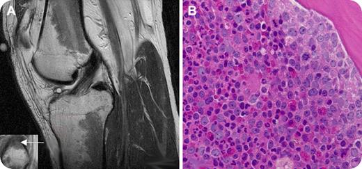
A 27-year-old man presented with a complaint of several months of progressive left knee pain with ambulation. An MRI scan of the left knee revealed extensive intermediate-low-signal intensity within the bone marrow in the diametaphyseal regions of the distal femur and proximal tibia (panel A, sagittal T2-weighted image of knee joint) and the epiphysis of the proximal fibula (panel A inset, coronal T1-weighted detail of fibular epiphysis; the arrow shows the bone marrow abnormality). The patient denied a history of knee trauma and was otherwise healthy. He had no knee abnormality on examination. There was no enlargement of lymph nodes, liver, or spleen.
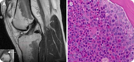
Laboratory tests showed a white blood cell count of 25.3 × 10⁹/L with 55% neutrophils, 8% bands, 5% myelocytes, 6% metamyelocytes, 21% lymphocytes, 4% monocytes, and 1% basophils; hematocrit of 43.7%; and platelet count of 203 × 109/L. A bone marrow aspirate and biopsy from the left posterior-superior iliac crest revealed hypercellularity with granulocytic predominance (panel B). Karyotypic analysis revealed a Philadelphia chromosome (t (9;22)). The patient was diagnosed with chronic myelogenous leukemia (CML) and therapy with imatinib mesylate was initiated.
The abnormalities on the MRI scan, when viewed in the absence of known hematologic abnormalities, were initially attributed to red marrow reconversion, which may be secondary to athletic activity. However, similar findings can indeed be seen in myeloproliferative disorders and marrow replacement disorders. Clinicians should be aware of unusual presentations of hematologic disorders in seemingly healthy patients with abnormal bone marrow signal on MRI scans.
Many Blood Work images are provided by the ASH IMAGE BANK, a reference and teaching tool that is continually updated with new atlas images and images of case studies. For more information or to contribute to the Image Bank, visit www.ashimagebank.org.


