Abstract
Abstract 4268
Heart disease remains the main cause of mortality in thalassemia major patients. Female patients with thalassemia major have a proved lower prevalence of cardiac complications than males and survive longer. It has been suggested that females have a better compliance than males, and therefore accumulate less iron in crucial organs like the heart (Borgna-Pignatti C et al, Haematologica 2004). The aim of our study was to verify if the decreased prevalence of cardiac disease in females could be attributed to lesser iron accumulation in their hearts as measured by multislice multiecho T2* Magnetic Resonance Imaging (MRI) technique.
We performed a retrospective review of the MRI results and of clinical data about the thalassemia major patients enrolled in the Myocardial Iron Overload in Thalassemia (MIOT) project. The MIOT is a network where MRI is performed using standardized and validated procedures and the MRI and thalassemia centers are linked by a web-based network, configured to collect patients' clinical and diagnostic data (Meloni A et al, Int J Med Inform 2009). Myocardial iron concentrations were measured by T2* multislice multiecho technique (Pepe A et al, JMRI 2006).Biventricular function parameters were quantitatively evaluated by cine images.
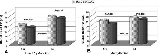
Seven hundred and seventy six thalassemia patients (370 males) were present in the MIOT database having undergone at least one MRI exam. The prevalence of cardiac disease (heart dysfunction and/or arrhythmias requiring medications) was significantly higher in males than in females (males 28% vs females 17%; P<0.0001). The analysis of different chelation treatments did not demonstrate a significant difference between patients with and without cardiac disease (P=0.59), nor between sexes (P=0.46). In addition, there was no difference in the reported compliance to chelation therapy between males and females (P=0.52). Global heart T2* values were significantly lower in both males and females with heart dysfunction (males: 20 ± 15 ms; females: 18 ± 12 ms), compared to those without dysfunction (males: 29 ± 11 ms; females: 27 ± 13 ms) (P<0.0001), but no difference was observed according to sex (Figure 1A). Global heart T2* values were not significantly lower in patients with arrhythmias compared to those without arrhythmias, nor was there a significant difference between sexes (Figure 1B).
The confirmed higher prevalence of cardiac disease in males with thalassemia major was not correlated to a worse compliance to chelation therapy or to an higher cardiac iron burden. Increased survival of female thalassemia major patients seems to not be attributed to lower cardiac iron overload. It can be hypothesized that females tolerate iron toxicity better, possibly as an effect of reduced sensitivity to chronic oxidative stress.
No relevant conflicts of interest to declare.
Author notes
Asterisk with author names denotes non-ASH members.


