Abstract
Abstract 3406
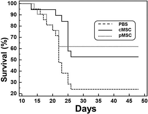
The precise mechanism of action of MSCs after systemic infusion in various tissue injury models is currently undefined. Although trans-differentiation to host tissues has been proposed as one potential mechanism, recent reports have suggested that there is little evidence of persistence of infused MSC after the initial few days in the target tissues making trans-differentiation unlikely. Hence we determined the in vivo distribution kinetics of infused MSC by three techniques: whole-body bioluminescent imaging. (BLI), immune histochemistry (IHC) and quantitative real time polymerase chain reaction (q RT PCR). MSC were labeled with firefly luciferase (ffLuc) using a lentiviral vector and infused to adult female recipients after they were administered 700 cGy radiation (1×10⁶ cells each, n=7). In all mice, strong bioluminescent signals were detected from the chest region at 4 hours after infusion, suggesting an accumulation of infused MSCs in the lungs. The signals rapidly decreased during the first 24 hours, and no bioluminescent signal was detected at 72 hours after infusion. No signals were detectable from other organs (liver, spleen or long bones) at any time point. Ffluc could not be reliably detected in tissues by IHC. Detection of luciferase transcripts in different tissues (lungs, hearts, spleens, livers, guts, muscles, and bone marrow) was performed by quantitative RT PCR at days 1, 4 and 7 (n = 6 each) following infusion. Ffluc transcripts were reliably detected in all animals only in the lungs at 24 hours after MSC infusion, confirming the BLI results. At subsequent time points, ffluc transcripts were detectable at very low levels in a variable proportion of animals from various tissues. In the absence of reliable BLI signals from these extra-pulmonary tissues, we interpret the presence of low levels of ffluc transcripts in these tissues as to have arisen from unviable cells or circulating RNA. Together, these results show that both immortalized MSCs and primary MSCs improve hematopoietic recovery after lethal ionizing radiation, but the infused cells are mostly filtered out by the lungs and their beneficial effect is likely mediated by indirect mechanisms (secondary effector cells in the lungs or secreted cytokines).
No relevant conflicts of interest to declare.
Author notes
Asterisk with author names denotes non-ASH members.


