Abstract
Abstract 563 FN2
FN2
The role of TRIM (tripartite motif) proteins in human cancer is unknown except for TRIM19 (PML, promyelocytic leukemia gene), which acquires oncogenic activity when fused to RARα (retinoic acid receptor alpha) and causes acute promyelocytic leukemia. We have confirmed a putative role of TRIM62 as a tumor suppressor by genetically deleting the TRIM62 locus in a mouse model. TRIM62 maps to chromosome 1p35.1, a genomic region frequently associated with loss of heterozygosity in human cancer, including leukemia.
We assessed TRIM62 expression by high-throughput reverse phase protein array (RPPA) technology in 511 pts. Eleven CD34+ bone marrow (BM) and 10 normal peripheral blood (PB) lymphocyte samples were used as controls. Samples were printed as 5 serial 1:2 dilutions in duplicate using an Aushon 2470 Arrayer. Slides were probed with 195 antibodies against apoptosis, cell cycle, signaling, integrins, and phosphatases among other functional protein groups. Patients were segregated according to TRIM62 levels into TRIM62LOW (lower two tertiles) and TRIM62HIGH (highest tertile).
The level of TRIM62 protein in AML samples was significantly below (p=0.0048) that of CD34+ cells obtained from healthy volunteers., The level of expression was similar in the paired PB and BM derived AML samples (p=0.2433). Cases of AML-M4 and –M5 exhibited lower expression of TRIM62 than other FAB subtypes.,Stratification according to specific cytogenetic abnormalities (p=0.9286), standard cytogenetic risk criteria (favorable, intermediate, poor; p=0.6824), or mutational status (FLT3, NPM1, RAS, p53, or IDH1/2) failed to show statistically significant differences in TRIM62 expression.
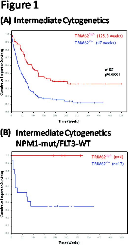
Overall survival (OS) was significantly different in TRIM62LOW and TRIM62HIGH for all patients (40.4 vs. 75.6 weeks, p=0.00002), and this difference was greater among patients with cytogenetically normal AML (CN-AML; 47 vs. 125.3 weeks, p=0.00001; Figure 1`A). TRIM62HIGH patients had significantly longer OS compared with TRIM62LOW patients, regardless of whether they had received standard cytarabine-based chemotherapy (p=0.00003) or epigenetic therapy (i.e. hypomethylating agents with or without histone deacetylase inhibitors; p=0.02). TRIM62LOW was associated with a trend toward shorter remission duration compared with TRIM62HIGH for all cases (38 vs. 63 weeks, p=0.06) and in CN-AML patients (47.7 vs. 58.1 weeks, p=0.09). By multivariate analysis, TRIM62HIGH was identified as an independent favorable risk factor for survival both in the whole cohort (p=0.0085) and in patients with CN-AML (p=0.0014). Notably, TRIM62LOW discriminated a subset of patients with a distinct poor prognosis among those with FLT3MUT (36 vs. 128.5 weeks, p=0.006), FLT3WT (66 vs. 129 weeks, p=0.001), NPM1MUT (34.6 vs. 253.1 weeks, p=0.0026), NPM1WT (51.7 vs. 102.6 weeks, p=0.017), and RASWT (46.3 vs. 110, p=0.0002). Among patients with the most favorable prognosis (FLT3WTNPM1MUT CN-AML), TRIM62HIGH identified a subset of patients for whom stem cell transplant is not recommended: OS for FLT3WTNPM1MUTTRIM62HIGH vs. FLT3WTNPM1MUTTRIM62LOW patients was 100% vs. 35%, after 6 years of follow-up (Figure 1B). When patients with FLT3WTNPM1mutant were excluded from the analysis, TRIM62 still stratified two distinct groups of patients with AML regarding OS, both for the whole cohort (p=0.03) and for those with CN-AML (p=0.0006). By assessing the level of 195 other proteins on the same RPPA chip, we showed that TRIM62 levels correlated significantly and positively (R>0.2) with proteins involved in leukemia stem cell pathways (β-catenin and Notch), migration/homing (fibronectin, integrin β3), hypoxia (VHL, HIF1α), and the p53 pathway (MDM2, p21).
TRIM62 loss, consistent with a role as a tumor suppressor, represents a powerful independent adverse prognostic factor in AML. TRIM62HIGH identifies a population of CN-AML patients with superior outcomes compared with TRIM62LOW amongst those with CN-AML even after stratification by NPM1 and FLT3 mutational status.
No relevant conflicts of interest to declare.
Author notes
Asterisk with author names denotes non-ASH members.

This icon denotes a clinically relevant abstract


