Key Points
Transient inhibition of hedgehog signaling augments hematopoiesis in hPSC-derived EBs.
Hedgehog inhibition initiates an advancement in the developmental state of hematopoietic cells derived from hPSCs.
Abstract
Programs that control early lineage fate decisions and transitions from embryonic to adult human cell types during development are poorly understood. Using human pluripotent stem cells (hPSCs), in the present study, we reveal reduction of Hedgehog (Hh) signaling correlates to developmental progression of hematopoiesis throughout human ontogeny. Both chemical- and gene-targeting–mediated inactivation of Hh signaling augmented hematopoietic fate and initiated transitions from embryonic to adult hematopoiesis, as measured by globin regulation in hPSCs. Inhibition of the Hh pathway resulted in truncation of Gli3 to its repressor, Gli3R, and was shown to be necessary and sufficient for initiating this transition. Our results reveal an unprecedented role for Hh signaling in the regulation of adult hematopoietic specification, thereby demonstrating the ability to modulate the default embryonic programs of hPSCs.
Introduction
Given the ethical limitations of working with the human embryo, the study of human pluripotent stem cells (hPSCs) provides a tractable surrogate with which to study human developmental biology. The development of hematopoietic cells from hPSCs first requires differentiation that produces large numbers of blood progenitors that can give rise to adult hematopoietic cell types. Among the procedures used for differentiating hematopoietic tissue from hPSCs, the most effective involve the addition of ligands that activate evolutionarily conserved signaling pathways,1,2 and also have the ability to control hematopoiesis during development.2-7 The Wnt, Notch, and Hedgehog (Hh) signaling pathways have been implicated as strong regulators of hematopoiesis.8-10 Aside from single-gene knockout experimentation in the murine system, which suggests that these pathways may be dispensable,11,12 and like Hox gene overexpression13 versus allele deletion,14 tempered control of these pathways has a powerful ability to govern HSC self-renewal and differentiation.15,16 However, the specific mechanisms and elements of these highly complex pathways capable of governing developmental hematopoietic cell fate in humans remain undefined. An understanding of this process would be instrumental toward developing rational strategies to regulate early hematopoietic processes from hPSCs toward cell replacement therapies and modeling early events of hematologic disorders, such as hereditary anemia,17 that manifest in utero but affect adult blood cells after birth.
Although there have been studies defining the role of Wnt and Notch signaling in HSC biology and aberrant hematopoietic development such as leukemias,18 comparatively less has been elucidated regarding Hh signaling in human HSCs, although the Hh pathway has been associated with hematopoietic specification during invertebrate embryogenesis19 and in adult vertebrates.20 Furthermore, Hh signaling has also been shown to play a role in adult HSC proliferation and homeostasis.9,21,22 To date, no studies have investigated the role of Hh signaling in early human hematopoietic development or cell fate decisions during hPSC differentiation for any lineage. In the present study, we identify a unique role for Hh regulation in hematopoiesis from hPSCs. We demonstrate that the inhibition of Hh augments blood production, overcomes the embryonically biased differentiation program of hPSCs, and targets globin control regions.
Methods
hPSC culture
The human embryonic stem cell (hESC) lines H1, H9, and CA2 and the induced pluripotent cell line hiPS1.2 were cultured on Matrigel (BD Biosciences)–coated 6-well plates with mouse embryonic fibroblast-conditioned medium supplemented with 8 ng/mL of recombinant human basic fibroblast growth factor (Invitrogen). hPSCs were fed daily with fresh conditioned medium and passaged 1:2 every 6-7 days by dissociation with 200 U/mL of collagenase IV (Invitrogen).
EB formation and hematopoietic differentiation
At day 7, hPSCs were treated with 200 U/mL of collagenase IV for 2-5 minutes and transferred to 6-well ultra-low attachment plates (Corning) in embryoid body (EB) medium (KO-DMEM, 20% FBS [Hyclone], 1% nonessential amino acids, 1mM L-Glut, and 0.1mM beta-mercaptoethanol). On day 1, the medium was changed to differentiation medium: 50 ng/mL of G-CSF (Amgen), 300 ng/mL of SCF (Amgen), 10 ng/mL of IL-3 (R&D Systems), 10 ng/mL of IL-6 (R&D Systems), 25 ng/mL of BMP4 (R&D Systems), and 300 ng/mL of Flt-3L (R&D Systems). EBs were cultured for 15 days with fresh medium added every 3 days. Shh recombinant peptide (R&D Systems) was added at 100 ng/mL, purmorphamine (Calbiochem) at 2μM, and cyclopamine (TRC) at 10μM.
Hemangioblast assay
Hemangioblasts were generated as described previously.23 Briefly, human EBs (hEBs) were cultured in hEB differentiation medium (Stemline II [Sigma-Aldrich], 50 ng/mL of BMP4, and VEGF-165 [R&D Systems]) for 48 hours, and then replaced with fresh medium plus 20 ng/mL of basic fibroblast growth factor. At day 4, EBs were dissociated in 0.4 U/mL of collagenase B (Roche) for 45 minutes, centrifuged at 453g, and filtered through a 70-μm mesh. Dissociated hEBs were then plated in hemangioblast medium (5 × 10⁴ cells in 100 μL of Stemline II, 500 μL of Methocult H4436 [StemCell Technologies], 50 ng/mL of BMP4, VEGF-165, 50 ng/mL of thrombopoietin, and 50 ng/mL of Flt3L). “Hemangioblast” colonies were counted on day 6. 2′-4′ platings were performed by collecting cells and plating in 100 μL of Stemline II, 1.5 mL of H4436, 50 ng/mL of BMP4, VEGF-165, 50 ng/mL of thrombopoietin, 50 ng/mL of Flt3L, and 3 U/mL of erythropoietin. Twenty-eight days later (at the end of 4′ plating), cells were collected and plated in 2 mL of Stemline II, 30 U/mL of erythropoietin, and 100 ng/mL of SCF for 4 days with addition of this medium once at day 2. Analysis of human β-globin (HBB) expression was performed on day 4 or day 32 from the hemangioblast counting step.
CFU assay
hEBs were dissociated with 0.4 U/mL of collagenase B (Roche) for 2 hours at 37°C, followed by treatment with cell dissociation buffer (Invitrogen) for 10 minutes at 37°C, and passed through a 40-μm cell strainer (BD Biosciences). A total of 104 cells were seeded in 1 mL of Methocult H4434 (StemCell Technologies). Erythroid colonies were plucked after 14 days of culture using standard morphologic criteria and analyzed by flow cytometry, quantitative PCR (qPCR) and mass spectroscopic analysis.
siRNA transfection
For siRNA transfections, clumps of hESC colonies were obtained during passage and transferred to 6-well ultra-low attachment plates (Corning). Cells were transfected in EB differentiation medium using lipofectamine (Ambion) and 50-100nM siRNA according to the manufacturer's instructions. After 24 hours, the medium was changed to differentiation medium. The siRNAs used were: Smoothened (Smo) siRNA ID: 1885, 1968 and 202229; Gli3 siRNA ID: 3259, 105974 and 105976; Scrambled siRNA number 1; Ambion). Transfection efficiency was assessed using a SilencerCy3-labeled control scrambled siRNA number 1.
Flow cytometry and sorting
EBs were dissociated with 0.4 U/mL of collagenase B (Roche) for 2 hours at 37°C, followed by 10 minutes at 37°C in cell dissociation buffer (Invitrogen), and passed through a 40-μm cell strainer (BD Biosciences). Single-cell suspensions were resuspended at 1-2 × 10⁵ cells/mL with PBS/3% FBS and stained for 1 hour with fluorochrome-conjugated Abs: CD31-PE (BD Biosciences), CD34-FITC (Miltenyi Biotec), and CD45-APC (Miltenyi Biotec). Cells were washed twice, resuspended in PBS/3% FBS, and stained with 7-amino-actinomycin D (Immunotech) for 10 minutes at room temperature. Cells were analyzed using a FACSCalibur or LSR2 II flow cytometer, with cell sorting performed on a FACSAria (BD Biosciences). Postacquisition analysis was performed using FlowJo Version 9.3 software (TreeStar).
PCR
RNA was extracted using the Total RNA Purification Kit (Norgen Biotek). cDNA synthesis was performed (First-Strand cDNA Synthesis Kit; Invitrogen) and gene expression was analyzed using Platinum SYBR Green (Invitrogen) or GoTaq BRYT Green (Promega) qPCR supermixes on a Stratagene Mx3000P or a Bio-Rad CFX96. Comparative quantification of transcripts was assessed relative to 18S rRNA, GAPDH, and GUSB; relative gene quantification was calibrated against the indicated controls.
Western blotting
EBs were lysed in 1% Triton X-100, 150mM NaCl, 10mM Tris-HCl, 5mM EDTA, 10 mg/mL of protease inhibitors, and 0.5mM PMSF. Thirty micrograms of protein was separated by 6% SDS-PAGE and transferred to PVDF membranes. Two hours blocking with 5% milk was followed by overnight incubation with 1° Abs at 4°C. Abs used were anti-Smo sc-13943 (1:1000; Santa Cruz Biotechnology), anti-tubulin sc-58667 (1:1000; Santa Cruz Biotechnology), anti-Gli3 sc-20 688 (1:500; Santa Cruz Biotechnology), or AF3690 (1:500, R&D Systems). The membranes were washed and stained with HRP-conjugated secondary antibodies (1:10 000) and signals were detected by enhanced chemiluminescence (Pierce), exposed to film, and quantified using ImagePro Version 6.2 software.
ChIP
Briefly, sorted CD45+ cells were cross-linked using 1% formaldehyde. Chromatin was digested in 0.1% SDS to obtain fragments of approximately of 1000 bp. Sonicated DNA was immunoprecipitated using anti-H3K4Me3, anti-H3K27Me3, and anti–rabbit IgG Abs (Santa Cruz Biotechnology). Immunoprecipitated DNA was further reverse cross-linked, purified, and analyzed by qPCR using UDG-Platinum SYBR Green mix (Invitrogen). Relative enrichment was calculated by subtracting signals detected from the specific Ab from signals from the control Ab. Differences were divided by signals observed from 1/15 of the ChIP input.
Immunohistochemistry and immunofluorescence
hEBs were washed 3 times in PBS-T (PBS 0.1% Tween-20), fixed with 4% paraformaldehyde for 2 hours, and snap-frozen in liquid N2. Eight-micrometer cryosections were blocked with 10% rabbit serum and 1% BSA at room temperature for 2 hours. Primary Abs were: rabbit anti-Gli3 ab6050 (1:200; Abcam) and rabbit anti-Patched sc-9016 (1:200; Santa Cruz Biotechnology). Sections were incubated with Abs overnight and stained with secondary antibodies for 1 hour (Alexa Fluor-488–conjugated goat anti–rabbit IgG; Invitrogen) at 2 μg/mL. Slides were mounted and counterstained using Vectashield HardSet Mounting Medium with DAPI (Vector Laboratories). For cellular immunofluorescence studies, samples were fixed in methanol at −20°C for 20 minutes, blocked in 1% FCS/PBS, and stained with goat anti-Gli3 at 1:500 (R&D Systems). Visualization was performed using the secondary antibody Alexa Fluor-647–conjugated donkey anti–goat IgG 1:1000 (Invitrogen). Imaging was performed on an Olympus IX18 microscope and analyzed using ImageJ Version 1.46 software.
Whole-mount immunohistochemistry
Briefly, EBs were fixed using fresh methanol: DMSO (4:1) and stored at 4°C overnight. After fixation, EBs were rehydrated, washed in PBS-T, and incubated twice with blocking solution (PBS-T and 2% milk) for 1 hour. EBs were incubated at 4°C overnight with primary Ab (rabbit anti-Smo, 1:500; Abcam). After washing in PBS-T for 5 hours, EBs were incubated with HRP-conjugated goat anti–rabbit antibody, 1:500 (Bio-Rad) for 5 hours and washed twice in PBS. EBs were stained using the VIP Peroxidase Substrate Kit for (Vector Laboratories) and imaged under a light microscope (SMZ1000; Nikon).
Affymetrix gene-expression profiling
Array data were normalized using the Robust Multichip Averaging (RMA) algorithm with baseline transformation to the median of all samples using GeneSpring Version 11.5.1 software (Agilent Technologies). Hh pathway gene lists were curated from KEGG and applied to clustering analysis, principal component analysis (PCA), and gene set enrichment analysis (GSEA Version 2.0.8). The Pearson correlation coefficient was used for clustering and dendrogram generation. For GSEA, samples were grouped as either embryonic (hPSCs and hEBs) or others and assessed for enrichment using GSEA software (Broad Institute).
Mass spectrometry
Bulk hemangioblast cultures were washed twice in PBS and lysed in 100mM (NH4)HCO3 containing 8M urea and 2mM DTT. Samples were reduced in an equal volume of 100mM (NH4)HCO3 plus 20mM iodoacetamide in the dark for 1 hour (all chemicals from Sigma-Aldrich). Protein tryptic digestion was performed with 400 ng of sequencing-grade trypsin (Promega) dissolved in an equal volume of 100mM (NH4)HCO3 plus 2mM CaCl2 overnight. Samples were then cleared by centrifugation for 20 minutes at 20 000g, snap-frozen in liquid N2, and lyophilized. Liquid chromatography-tandem mass spectrometry analysis of tryptic digests was performed using a combined setup consisting of an HTS-PAL autosampler (CTC Analytics), a Paradigm MS4 HPLC (Michrom BioResources), and an LTQ (Thermo Finnigan) linear ion trap mass spectrometer. Tandem mass spectrometry data were analyzed using the MASCOT search engine against a nonredundant National Center for Biotechnology Information (NCBI) human protein database and a custom-made database consisting of sequenced human globins (HBE, HBG, HBD, HBB, HBZ, and HBA2) verified via cloning and sequencing of corresponding transcripts from the hPSC lines used. MASCOT results were further analyzed with Scaffold3 (Proteome Software) with peptide and protein probability filters at 90%. Unique spectra of different α- and β-globins are shown in relation to the number of unique spectra obtained for housekeeping control β-actin.
Statistical analysis
Results are presented as the means ± SEM of at least 3 independent experiments unless stated otherwise. Significance levels were determined using 2-tailed Student t tests (unless stated otherwise).
Results
Expression signature of the Hh pathway is inversely correlated with human blood development
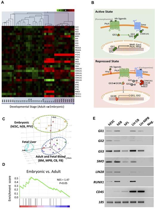
To examine broadly the potential roles of conserved pathways previously associated with early vertebrate development during hematopoiesis, in the present study, we analyzed the expression patterns of components involved in Wnt, Notch, and Hh signaling. Using transcriptome profiles from human cell populations, we evaluated various developmental time points, including: undifferentiated pluripotent hESCs, differentiated hematopoietic hPSC derivatives (differentiated EBs, hemogenic precursors [PFVs], and hPSC-derived hematopoietic progenitors [hPCs]),24 and multiple sources of highly purified putative HSCs (CD34+CD38−Lin−), including human fetal blood, human fetal liver, neonatal cord blood, and adult BM and mobilized peripheral blood, demonstrating in vivo hematopoietic reconstitution capacity (Figure 1A). Hh pathway expression revealed significant patterns relating to human ontogeny with predominant signatures that clustered hESC-derived hematopoietic cells, fetal HSCs, and adult HSC fractions (Figure 1). Progression from early to later developmental stages of human hematopoietic cells was associated with the gradual down-regulation of Hh pathway member expression (Figure 1A) and was further illustrated by PCA analysis (Figure 1C). Mediators of Hh signaling were the highest in hESC-derived hematopoietic cells compared with fractions enriched for adult HSCs (Figure 1D), suggesting that Hh pathway activation is strictly regulated during human development and ontogeny. Key components of the Hh pathway were further validated by PCR using hESCs, hematopoietic hEBs, and somatic human sources (Figure 1E and supplemental Figure 1, available on the Blood Web site; see the Supplemental Materials link at the top of the online article), which verified down-regulation of Hh pathway members during human hematopoietic ontogeny. These data indicate a strong correlation of Hh signaling inhibition during the maturation of the early human hematopoietic lineage that had not been revealed previously.
Analysis of Hh pathway member expression in different hematopoietic developmental stages. (A) Affymetrix U133A arrays were normalized across samples and hierarchical clustering of Hh pathway genes using the Pearson correlation was performed, demonstrating an increased expression in earlier developmental stages. (B) Schematic of Hh signaling indicating genes identified by expression array analysis and small molecules used to agonize (puromorphamine) or antagonize (cyclopamine) the pathway. Green coloring is used for stimulatory and red is used for repressive proteins and processes. Gli3 is shown as a composite because it functions in both stimulating and repressing the pathway. (C) PCA was used to generate unbiased groupings of arrays, which demonstrated clustering based on developmental stage. (D) GSEA shows statistical significance (P < .05) for augmented expression of Hh pathway members in embryonic versus fetal, neonatal, and adult samples. (E) PCR (30-40 cycles) confirming higher expression of the Hh pathway members Gli1/2/3 and SMO at early developmental stages. LIN28 serves as a positive control for early ontogeny, RUNX1 for hematopoietic progenitors, CD45 for later hematopoietic differentiation, and 18S rRNA as a loading control. hFL indicates human fetal liver; hFB, fetal blood; MPB, human mobilized peripheral blood, and CB, human cord blood. All somatic sources except FL were lineage depleted (Lin−).
Analysis of Hh pathway member expression in different hematopoietic developmental stages. (A) Affymetrix U133A arrays were normalized across samples and hierarchical clustering of Hh pathway genes using the Pearson correlation was performed, demonstrating an increased expression in earlier developmental stages. (B) Schematic of Hh signaling indicating genes identified by expression array analysis and small molecules used to agonize (puromorphamine) or antagonize (cyclopamine) the pathway. Green coloring is used for stimulatory and red is used for repressive proteins and processes. Gli3 is shown as a composite because it functions in both stimulating and repressing the pathway. (C) PCA was used to generate unbiased groupings of arrays, which demonstrated clustering based on developmental stage. (D) GSEA shows statistical significance (P < .05) for augmented expression of Hh pathway members in embryonic versus fetal, neonatal, and adult samples. (E) PCR (30-40 cycles) confirming higher expression of the Hh pathway members Gli1/2/3 and SMO at early developmental stages. LIN28 serves as a positive control for early ontogeny, RUNX1 for hematopoietic progenitors, CD45 for later hematopoietic differentiation, and 18S rRNA as a loading control. hFL indicates human fetal liver; hFB, fetal blood; MPB, human mobilized peripheral blood, and CB, human cord blood. All somatic sources except FL were lineage depleted (Lin−).
Hh signaling functionally regulates human blood development
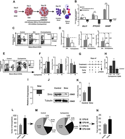
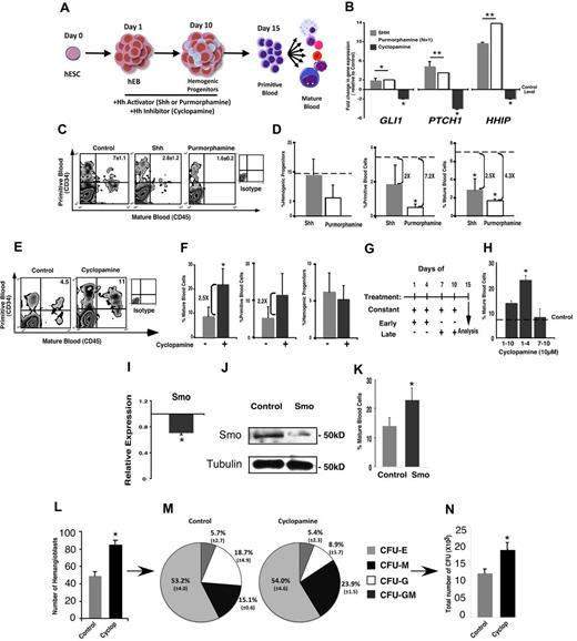
To examine functionally the role of Hh signaling, we used the hEB differentiation model (Figure 2A).24,25 The Hh pathway was activated by optimized addition of Shh ligand (100 ng/mL) or the chemical agonist purmorphamine (2μM) to hEBs (Figure 2B and supplemental Figure 2A-B). Conversely, Hh inhibition was achieved by the addition of the chemical inhibitor cyclopamine (10μM), which binds specifically to and inactivates Smo, causing down-regulation of Hh signaling26-28 (Figure 2B and supplemental Figure 2C). During hPSC-derived hematopoietic differentiation, addition of either Shh or purmorphamine resulted in augmented expression of Gli1, Ptch, and Hhip, indicative of Hh pathway activation (Figure 2B). In contrast, cyclopamine treatment decreased the expression of these genes (Figure 2B), which is consistent with Hh signal inhibition. This inhibitory effect was further validated at the protein level, where Ptch1 and Smo were reduced in the first 5 days of cyclopamine treatment compared with control-treated hEBs (supplemental Figure 3A-B) and were undetectable by day 10 (not shown).
Cyclopamine increases blood differentiation in hEBs and favors adult hematopoiesis. (A) Schematic representation of the hEB differentiation system and treatment with cyclopamine, Shh, or purmorphamine. (B) Activation of the Hh pathway by Shh or purmorphamine results in a strong induction of Gli1, Ptch, and Hhip expression, in contrast to the down-regulation shown after the inhibition of the pathway mediated by cyclopamine. (C) Representative flow cytometry data of the hematopoietic differentiation at day 15 of hEB differentiation after Shh or purmorphamine treatment showing inhibition of hEB-derived hematopoiesis. (D) Reduction in the percentage of mature blood cells (CD45+) and primitive blood cells (CD34+/45+), but not in hemogenic progenitors (CD45−PFV) after Shh or purmorphamine treatment. (E) Representative flow cytometry data of hematopoietic differentiation at day 15 of hEB differentiation after DMSO (control) or cyclopamine treatment showing inhibition of hEB-derived hematopoiesis. (F) Reduction in the percentage of mature blood cells (CD45+) and primitive blood cells (CD34+/45+), but not in hemogenic progenitors (CD45−PFV) after control or cyclopamine treatment. (G-H) Treatment schema (G) and percentage of mature blood cells (H) after constant, early, and late cyclopamine treatment relative to control-treated hEBs (dashed line). Early inhibition of the Hh pathway significantly increases hematopoietic differentiation compared with all other combinations. (I) Inhibition of the Hh pathway by siRNA directed against Smo with a 30% reduction of Smo expression in hEBs on day 2 after the Smo-siRNA transfection. (J) Western-blot showing a reduction in Smo protein in hEBs at day 4 after Smo-siRNA transfection. (K) Increase in the percentage of mature blood cells at day 15 after Smo-siRNA transfection. (L) Number of hemangioblast colonies is increased on cyclopamine treatment compared with control. (M) Pie charts representing the number of individual CFU types generated from control and cyclopamine-treated cells. (N) An increase in total number of CFUs is observed from equal starting numbers of control or cyclopamine-treated hemangioblasts. *P < .05; **P < .01.
Cyclopamine increases blood differentiation in hEBs and favors adult hematopoiesis. (A) Schematic representation of the hEB differentiation system and treatment with cyclopamine, Shh, or purmorphamine. (B) Activation of the Hh pathway by Shh or purmorphamine results in a strong induction of Gli1, Ptch, and Hhip expression, in contrast to the down-regulation shown after the inhibition of the pathway mediated by cyclopamine. (C) Representative flow cytometry data of the hematopoietic differentiation at day 15 of hEB differentiation after Shh or purmorphamine treatment showing inhibition of hEB-derived hematopoiesis. (D) Reduction in the percentage of mature blood cells (CD45+) and primitive blood cells (CD34+/45+), but not in hemogenic progenitors (CD45−PFV) after Shh or purmorphamine treatment. (E) Representative flow cytometry data of hematopoietic differentiation at day 15 of hEB differentiation after DMSO (control) or cyclopamine treatment showing inhibition of hEB-derived hematopoiesis. (F) Reduction in the percentage of mature blood cells (CD45+) and primitive blood cells (CD34+/45+), but not in hemogenic progenitors (CD45−PFV) after control or cyclopamine treatment. (G-H) Treatment schema (G) and percentage of mature blood cells (H) after constant, early, and late cyclopamine treatment relative to control-treated hEBs (dashed line). Early inhibition of the Hh pathway significantly increases hematopoietic differentiation compared with all other combinations. (I) Inhibition of the Hh pathway by siRNA directed against Smo with a 30% reduction of Smo expression in hEBs on day 2 after the Smo-siRNA transfection. (J) Western-blot showing a reduction in Smo protein in hEBs at day 4 after Smo-siRNA transfection. (K) Increase in the percentage of mature blood cells at day 15 after Smo-siRNA transfection. (L) Number of hemangioblast colonies is increased on cyclopamine treatment compared with control. (M) Pie charts representing the number of individual CFU types generated from control and cyclopamine-treated cells. (N) An increase in total number of CFUs is observed from equal starting numbers of control or cyclopamine-treated hemangioblasts. *P < .05; **P < .01.
To define the functional role of Hh signaling during early human hematopoiesis, we analyzed the cellular effects of activating or blocking Hh signaling during hPSC-hematopoiesis. Pathway activation with Shh and purmorphamine caused a significant reduction in mature hematopoietic development (CD45+ blood cells; Figure 2C-D). Similarly, activation with Shh reduced the frequency of CD45+CD34+ primitive blood cells, but not hemogenic precursors (CD45−PFV; Figure 2D). In contrast, antagonism of the Hh pathway with cyclopamine increased the frequency of primitive (CD34+CD45+) and mature (CD45+) hematopoietic cells (Figure 2E-F). Similar to Hh activation, antagonism of the pathway by cyclopamine did not affect the frequency of hemogenic CD45−PFV precursors (Figure 2F). To investigate temporal effects of Hh signaling in hematopoietic development, we treated hEBs throughout hematopoietic program induction (days 1-10) compared with segmented early (days 1-4) and late (days 7-10; Figure 2G) treatments. Frequency of hematopoietic output of mature blood cells (CD45+) was highest on early exposure of hEBs to cyclopamine, whereas late treatment had no effect (Figure 2H). To validate molecularly the ability to increase hESC-derived hematopoiesis via Hh signal inhibition, we performed siRNA-mediated knockdown of Smo (supplemental Figure 4A-C). A reduction in Smo transcription was observed 2 days after Smo-siRNA transfection of hEBs (Figure 2I) and subsequently led to a substantial decrease in Smo protein levels by day 4 (Figure 2J). Similar to the functional effects on hematopoiesis induced by cyclopamine treatment, Smo-siRNA consistently resulted in increased frequency of mature blood cells (Figure 2K) independently of targeted region using various siRNAs (supplemental Figure 4D). These temporal effects indicate that inhibition of Hh signaling is required to induce subsequent increases in hematopoietic differentiation, suggesting that programs regulated by Hh that effect hematopoiesis of hPSCs arise in the earliest phases of lineage commitment.
Consistent with these results, Hh inhibition also induced increased total numbers of hematoendothelial progenitors or hemangioblasts starting from an equivalent input population (Figure 2L) using an established technique for hemangioblast formation.23 However, hematopoietic lineage progenitor potential of individual CFU progeny arising from hemangioblasts was not altered by cyclopamine treatment (Figure 2M), nor was the capacity of CFUs altered on a per-cell basis (frequency of CFU from untreated vs treated hemangioblasts was 81 ± 8.3 and 89 ± 9.4, respectively); but total hematopoietic CFU output was increased on cyclopamine addition (Figure 2N). These results indicate that inhibition of the Hh pathway by cyclopamine treatment or Smo knock-down potently regulates hPSC-derived hematopoietic programming and is restricted to early stages of hematopoietic cell fate decisions of hPSCs.
Inhibition of Hh signaling augments adult versus embryonic globin expression
In all mammals, primitive hematopoietic (erythroid) cells of initial embryonic origin predominantly express ϵ-globin and low levels of γ-globin.29 The transition between primitive (embryonic and fetal) and definitive hematopoiesis in humans is signified by changes in ϵ-globin and γ-globin expression to β-globin.29 The human globin locus contains 5 genes, ϵ (HBE), γ-A (HBG1), γ-G (HBG2), δ (HBD), and β (HBB), that are expressed and strictly regulated in a tissue- and developmentally specific manner.30 Evidence from the mouse, however, has shown the appearance of erythroid cells in the fetal liver that express not only fetal γ-globins, but also adult type globins.31 Presently, it is not known if this occurs in the human fetal liver. To elucidate the effects of Hh inhibition on embryonic versus definitive hematopoietic programs in the human, we used hPSCs, in which human globin gene cluster regulation was used as a genomic and molecular target for measurement.
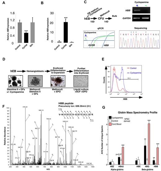
Fractions of blood cells (CD45+) from day 15 hEBs (supplemental Figure 5) were purified for qPCR analysis. A 50% reduction in embryonic (HBE) globin was accompanied by a 20-fold increase in adult globin (HBB) expression on cyclopamine treatment (Figure 3A-B). Alteration in globin expression pattern was selectively associated with Hh inhibition, because activation of Hh by Shh or purmorphamine had no effect on adult globin transcription (Figure 3A-B). In addition to up-regulation at the hEB stage, bulk CFU cultures grown in methylcellulose-based hematopoietic medium were isolated and analyzed for expression of HBB. HBB was amplified from hEBs that had been treated with cyclopamine (Figure 3C). Because of high sequence homology between the different human globins, DNA sequencing of PCR amplicons verified HBB specificity (Figure 3C).
Cyclopamine treatment induces expression of definitive hematopoietic markers. (A-B) qPCR analysis of CD45+ cells sorted from control, cyclopamine-treated, and Shh-treated hEBs demonstrated a significant increase in HBB and lowered HBE on Hh inhibition. (C) hEBs grown in the presence or absence of cyclopamine at days 1-4 were plated into CFU assays and analyzed for HBB expression at day 15. HBB was augmented in bulk CFUs from cyclopamine-treated hEBs only. β-glucuronidase (GUSB) and GAPDH served as control genes. Bottom right: sequencing was used as a means to verify product specificity. Asterisks (*) present in a representative stretch of 16 bases indicate unique nucleotides present in HBB only; lack of any double sequence indicates amplicon purity. (D) Schematic representation of the protocol followed to derive erythroid cells from hEBs (“hemangioblast assay”). (E) Presence of anti-HBB immunoreactivity in control and cyclopamine treatment by flow cytometry. (F) Representative spectrum for HBB identified in cyclopamine-treated samples only. (G) Summary of globin hits relative to control β-actin in cyclopamine-treated versus control samples. Cord blood samples, run in parallel as a comparative control for developmental stage, are shown. A significant increase in HBG- and HBB-specific peptides was observed in cyclopamine-treated samples relative to paired hPSC-derived controls. **P < .01; ***P < .001.
Cyclopamine treatment induces expression of definitive hematopoietic markers. (A-B) qPCR analysis of CD45+ cells sorted from control, cyclopamine-treated, and Shh-treated hEBs demonstrated a significant increase in HBB and lowered HBE on Hh inhibition. (C) hEBs grown in the presence or absence of cyclopamine at days 1-4 were plated into CFU assays and analyzed for HBB expression at day 15. HBB was augmented in bulk CFUs from cyclopamine-treated hEBs only. β-glucuronidase (GUSB) and GAPDH served as control genes. Bottom right: sequencing was used as a means to verify product specificity. Asterisks (*) present in a representative stretch of 16 bases indicate unique nucleotides present in HBB only; lack of any double sequence indicates amplicon purity. (D) Schematic representation of the protocol followed to derive erythroid cells from hEBs (“hemangioblast assay”). (E) Presence of anti-HBB immunoreactivity in control and cyclopamine treatment by flow cytometry. (F) Representative spectrum for HBB identified in cyclopamine-treated samples only. (G) Summary of globin hits relative to control β-actin in cyclopamine-treated versus control samples. Cord blood samples, run in parallel as a comparative control for developmental stage, are shown. A significant increase in HBG- and HBB-specific peptides was observed in cyclopamine-treated samples relative to paired hPSC-derived controls. **P < .01; ***P < .001.
These observed effects on globin regulation by Hh inhibition prompted us to investigate the effects using a more robust erythroid differentiation model23 (Figure 3D). Flow cytometric analysis revealed that 42% of cyclopamine-treated cells stained positively for HBB, indicative of initiation of definitive hematopoietic programs, compared with control (vehicle alone) treatment (Figure 3E).
Because specific antibodies to the various human globins are often criticized for their inability to distinguish between globin subtypes and not to rely on transcript alone, we performed mass spectrometry as an independent assay to ascertain globin profiles and abundance using shotgun proteomic analysis of erythroid cells differentiated as illustrated in Figure 3D compared with control cord blood samples. Globin profiles obtained from protein isolates of these cellular pools showed a decrease in embryonic HBE in cyclopamine-treated samples, a significant increase in fetal HBG, and, most importantly, the presence of unambiguous HBB peptides in cyclopamine-treated samples only from hPSC-derived sources (Figure 3F-G) and bore more similarity to cord blood profiles. These findings not only demonstrate that Hh inhibition via cyclopamine treatment increases hematopoietic differentiation of hEBs, but also conclusively demonstrate an increased capacity for adult hematopoietic specification. We believe this to be the first comprehensive demonstration of adult versus embryonic/fetal globin protein expression profiles induced from hematopoietic cells generated from hPSCs.
Hh inhibition results in cleavage of Gli3 to Gli3R capable of phenocopying the effects of cyclopamine
During in utero development, Hh signals are mediated through complex signaling mechanisms that are cell and lineage specific.32 On binding to Ptch1, Shh relieves repression of Smo, which in turn regulates the Gli family of transcription factors (Gli1, Gli2, and Gli3) that are thought to activate or repress transcription of target genes in a cell context–specific manner.32,33 Regulation of the Gli transcription factors is a key element to understanding the impact of Hh signaling.
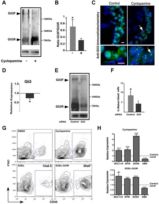
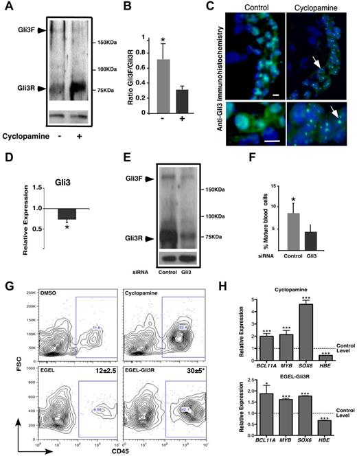
In the absence of Shh, full-length Gli3 is proteolytically processed from its larger activating form to a truncated repressor to inhibit the Hh pathway.34 Because of the effects of Hh inhibition on hPSC hematopoiesis, we examined the levels of the repressor form of Gli3 (Gli3R). Protein analysis of both the full-length and repressor forms of Gli3 (Gli3F and Gli3R, respectively) demonstrated that Gli3 is processed into its repressor form after cyclopamine treatment (Figure 4A-B). Consistent with studies showing strong nuclear localization of truncated Gli repressor forms versus cytoplasmic localization of full-length Gli3,35 immunostaining revealed that Hh inhibition induced nuclear localization of Gli3 in hEBs (Figure 4C). To examine the specific role of Gli3 in hESC-derived hematopoiesis, we targeted Gli3 directly by RNAi. Knock-down of Gli3 reduced both the transcript (Figure 4D) and protein levels after transfection (Figure 4E). Functionally, Gli3 knock-down resulted in a reduction in blood development mediated by Hh inhibition (Figure 4F). These results demonstrate that inhibition of Hh signaling during hESC hematopoiesis is mediated by Gli3.
Gli3 repressor mediates the effect of cyclopamine in hEB-derived hematopoiesis. (A) Western blot for Gli3 showing the full-length (Gli3F) and the Gli3 repressor (Gli3R) forms. In cyclopamine-treated hEBs, the majority of the protein was processed to the shorter Gli3R form. (B) Gli3F: Gli3R ratio demonstrating the processing of protein to the Gli3R form after cyclopamine treatment. (C) Immunostaining for Gli3 in frozen sections from day 5 hEBs showing that cyclopamine treatment induces focal nuclear localization of Gli3. Scale bars indicate 10 μm. (D) Down-regulation of the Gli3 transcript 2 days after Gli3 siRNA transfection. (E) Western blot for Gli3 showing a reduction in the Gli3F and the Gli3R forms at day 4 after transfection. (F) Reduction of mature blood cells (%CD45+) 15 days after transfection. (G) EBs treated with cyclopamine (left) or transfected at ES and EB stages with Gli3R expression vectors exhibit similar increases in CD45+ blood cells. (H) Cyclopamine-treated and Gli3R-overexpressing cells also have augmented expression of the transcription factors Sox6, Bcl11A, and Myb, which are associated with definitive erythropoiesis. Suppression of embryonic HBE globin transcription is also apparent. Bottom panels for A and E are anti-tubulin loading controls. *P < .05; ***P < .001.
Gli3 repressor mediates the effect of cyclopamine in hEB-derived hematopoiesis. (A) Western blot for Gli3 showing the full-length (Gli3F) and the Gli3 repressor (Gli3R) forms. In cyclopamine-treated hEBs, the majority of the protein was processed to the shorter Gli3R form. (B) Gli3F: Gli3R ratio demonstrating the processing of protein to the Gli3R form after cyclopamine treatment. (C) Immunostaining for Gli3 in frozen sections from day 5 hEBs showing that cyclopamine treatment induces focal nuclear localization of Gli3. Scale bars indicate 10 μm. (D) Down-regulation of the Gli3 transcript 2 days after Gli3 siRNA transfection. (E) Western blot for Gli3 showing a reduction in the Gli3F and the Gli3R forms at day 4 after transfection. (F) Reduction of mature blood cells (%CD45+) 15 days after transfection. (G) EBs treated with cyclopamine (left) or transfected at ES and EB stages with Gli3R expression vectors exhibit similar increases in CD45+ blood cells. (H) Cyclopamine-treated and Gli3R-overexpressing cells also have augmented expression of the transcription factors Sox6, Bcl11A, and Myb, which are associated with definitive erythropoiesis. Suppression of embryonic HBE globin transcription is also apparent. Bottom panels for A and E are anti-tubulin loading controls. *P < .05; ***P < .001.
To determine whether the effects of temporal Hh inhibition by short-term cyclopamine treatment operated via Gli3R production, we performed transient overexpression experiments of a truncated form of Gli3 alone (supplemental Figure 6). In agreement with observations after chemical inhibition by cyclopamine (Figure 4G), transient overexpression of Gli3R led to equally significant increases in hematopoietic output (CD45+ mature blood cells) during hEB differentiation (Figure 4G). To determine whether this increase in hematopoietic differentiation via Gli3R also affected the adult hematopoietic program, the expression of transcription factors associated with definitive hematopoiesis were evaluated.31 We observed an increase in Myb, Sox6, and Bcl11a using both chemical inhibition of Hh signaling and Gli3R overexpression in hEBs (Figure 4H). Furthermore, embryonic HBE expression was decreased in hEBs overexpressing Gli3R, similar to that of of Hh using cyclopamine (Figure 4H). These results indicate that Gli3R can phenocopy the effects of cyclopamine.
Hh inhibition targets the globin locus genomically
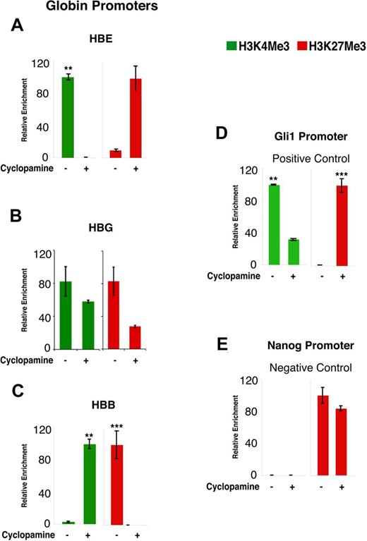
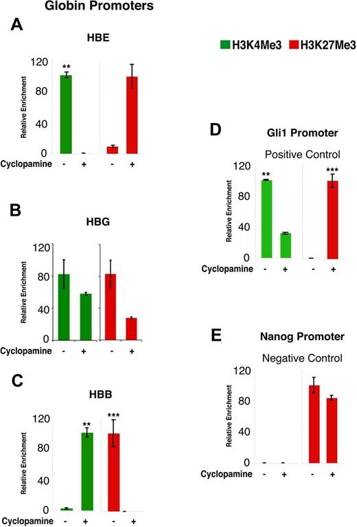
The mammalian globin locus is subjected to strict control by multiple posttranslational histone modifications, including active histone 3 (H3) and lysine 4 (K4) methylation and repressive H3K9 and H3K27 methylation.36 Because we had observed a shift from embryonic to adult globin expression upon inhibition of the Hh pathway, we next aimed to characterize this locus at the chromatin level. ChIP assays were performed using H3K4Me3 and H3K27Me3 Abs from day 15 control and cyclopamine-treated purified CD45+ cells. ChIP results revealed that the fetal HBE promoter was enriched with H3K4Me3 marks in control conditions, but demonstrated repressive (H3K27Me3) marks upon cyclopamine treatment (Figure 5A). In contrast, embryonic HBG had marks suggestive of bivalency, with activating H3K4Me3 marks being similar between treatments, whereas repressive H3K27Me3 marks decreased after Hh inhibition by cyclopamine (Figure 5B). The HBB promoter was repressed in control conditions yet extensively enriched for active H3K4Me3 marks upon Hh inhibition (Figure 5C). The positive control Gli1 promoter was enriched for active H3K4Me3 in control conditions compared with cyclopamine-treated hEBs, which had enhanced repressive H3K27Me3 marks (Figure 5D), demonstrating specificity of the ChIP reaction. IP signals from the promoter of the pluripotency-associated gene Nanog consistently exhibited H3K27Me3 marks after both control and cyclopamine treatment (Figure 5E). These results indicate that inhibition of Hh signaling controls globin preference by depositing active marks on the HBB promoter while repressing the embryonic HBE globin promoter.
Histone-modification studies using H3K4Me3 (active) and H3K27Me3 (repressive) antibodies in CD45+ hEBs. (A) Active HBE promoter marks are present in controls with a strong shift to repressive marks after Hh inhibition. (B) The HBG promoter shows active marks in both control and cyclopamine conditions, but has elevated repressive marks in control conditions. (C) The HBB promoter shows active marks after cyclopamine treatment only, with repressive marks observed in the control condition. (D) Positive control Gli1 promoter was repressed after cyclopamine treatment and uniquely displayed active marks in control conditions. (E) Negative control Nanog promoter exhibited repressive marks irrespective of treatment used for hEBs. Significance was measured with 1-tailed t tests. **P < .01; ***P < .001.
Histone-modification studies using H3K4Me3 (active) and H3K27Me3 (repressive) antibodies in CD45+ hEBs. (A) Active HBE promoter marks are present in controls with a strong shift to repressive marks after Hh inhibition. (B) The HBG promoter shows active marks in both control and cyclopamine conditions, but has elevated repressive marks in control conditions. (C) The HBB promoter shows active marks after cyclopamine treatment only, with repressive marks observed in the control condition. (D) Positive control Gli1 promoter was repressed after cyclopamine treatment and uniquely displayed active marks in control conditions. (E) Negative control Nanog promoter exhibited repressive marks irrespective of treatment used for hEBs. Significance was measured with 1-tailed t tests. **P < .01; ***P < .001.
Discussion
The results of the present study reveal that transient inhibition of the Hh signaling pathway during the initial stages of differentiation allows hPSCs to overcome intrinsic embryonic hematopoietic differentiation programs and initiate an adult hematopoietic program. Hh inhibition also increased hematopoietic differentiation output in a reproducible and robust manner. This was determined using 3 independent measures, transcript, protein, and ChIP, and 3 independent methods, chemical inhibition by cyclopamine, Smo siRNA, and transient overexpression of Gli3R. By overcoming the default embryonic differentiation program intrinsic to hematopoietic cells derived from hPSCs and increasing hematopoietic differentiation from hPSCs, we provide a model with which to study globin switching in the human that sets a foundation for a better understanding of hematopoietic development and disease.
Although these results have a practical impact for the use of hPSCs, we have used this system to further dissect the mechanism of adult versus embryonic developmental hematopoiesis that has not been achieved to date partly because of the lack of human models. The molecular framework of globin regulation has been well studied over the past decades,29 with numerous studies of embryonic versus definitive hematopoietic development in mammals.37,38 However, the identification of upstream or downstream signaling pathways that regulate either of these processes has remained elusive. Based on our present results, we propose a mechanism whereby Hh represents a regulatory pathway capable of initiating an alteration in embryonic versus adult globin expression programs in hPSCs. At this time, it remains unclear whether Hh signaling occurs in a cell-autonomous or a non–cell-autonomous fashion in the hematopoietic lineage and this remains an interesting subject for future studies.
Our present results implicate a role for known elements of Hh signal machinery in hPSCs that regulate early events in adult hematopoietic programming of blood cell fate. We demonstrate that Hh inhibition via cyclopamine induces Gli3 cleavage and results in the alteration of globin regulation by removing repressive H3K27Me3 marks on adult globin loci. In addition, we reveal that Gli3R overexpression is capable of mimicking the effects of chemically induced Hh inhibition. We believe this to be the first study to implicate a signaling pathway of any kind in embryonic versus adult hematopoietic control and to broadly associate a role for Hh during early human hematopoiesis.
Based on previous work in the mouse system,39,40 overcoming the embryonic character of blood development remains a major issue that has yet to be resolved in hPSCs. Although there have been several attempts toward improving protocols for the stepwise differentiation of hPSCs into several lineages, few have attempted to tackle the problem of overcoming an embryonic program that is established during robust self-renewal of PSCs in vitro. These concerns of the adult versus embryonic nature of cell types generated from hPSCs are not limited to the hematopoietic system, as hPSC-derived hepatocytes41,42 or cardiomyocytes43 adopt fetal programs using current protocols, making the study of phase 2 metabolism found only in adult liver cells or toxicity indices specific to adult cardiomyocytes difficult. The ability to generate adult versus embryonic blood cells from hPSCs suggests that strategies to generate adult cell types of other lineages are feasible and would broaden the spectrum of applications for mature cell types derived from hPSCs in the future. Somatic/adult programs for the generation of appropriate cell types that could be applicable for transplantation and drug screening will be possible, in contrast to differentiated cell types that are embryonic in nature. For mature cells of the hematopoietic lineage, the results of the present study establish an approach whereby differentiation of hPSCs to a more adult-like blood fate can be achieved. This will ultimately facilitate the generation of adult blood cells, likely more useful for cell replacement therapies in anemia and correctable thalassemias,44,45 and for in vitro screening and blood toxicity applications in which cells exhibiting adult characteristics are required.46
The online version of this article contains a data supplement.
The publication costs of this article were defrayed in part by page charge payment. Therefore, and solely to indicate this fact, this article is hereby marked “advertisement” in accordance with 18 USC section 1734.
Acknowledgments
The authors thank Dr Marilyne Levadoux-Martin for FACS expertise; Monica Graham for hESC culture; Zoya Shapovalova for expression array analysis; Dr Eva Szabo for critical comments during manuscript preparation; Vanja Avdic, Angelique Schnerch, and Dr Kausalia Vijayaragavan for technical assistance; and Dr Shigeo Hayashi and Kaori Shinmyozu for use of the RIKEN CDB Mass Spectrometry Facility.
This research was supported by funding from the Canadian Cancer Society Research Institute and the Canadian Research Chair Program (to M.B.).
Authorship
Contribution: B.A.S.M., V.R.-M., and S.R. assisted in the design of the study and performed experiments; R.M., J.-H.L., C.A., and G.S. performed experiments; B.A.S.M., S.R., and M.B. contributed to drafts of the manuscript; and M.B. designed the study, supervised the experiments, provided intellectual support for interpretation of the results, and wrote the final version of the manuscript.
Conflict-of-interest disclosure: The authors declare no competing financial interests.
Correspondence: Dr Mickie Bhatia, Stem Cell and Cancer Research Institute, Michael G. DeGroote School of Medicine, McMaster University, 1280 Main St W, MDCL 5029, Hamilton, ON, Canada L8S 4K1; e-mail: mbhatia@mcmaster.ca.
References
Author notes
B.A.S.M., V.R.-M., and S.R. contributed equally to this work.


