Key Points
Plasma concentrations of CXCL9 are elevated at the onset of cGVHD diagnosis, but not in patients with cGVHD for more than 3 months.
Plasma concentrations of CXCL9 are impacted by immunosuppressive therapy.
Abstract
There are no validated biomarkers for chronic GVHD (cGVHD). We used a protein microarray and subsequent sequential enzyme-linked immunosorbent assay to compare 17 patients with treatment-refractory de novo–onset cGVHD and 18 time-matched control patients without acute or chronic GVHD to identify 5 candidate proteins that distinguished cGVHD from no cGVHD: CXCL9, IL2Rα, elafin, CD13, and BAFF. We then assessed the discriminatory value of each protein individually and in composite panels in a validation cohort (n = 109). CXCL9 was found to have the highest discriminatory value with an area under the receiver operating characteristic curve of 0.83 (95% confidence interval, 0.74-0.91). CXCL9 plasma concentrations above the median were associated with a higher frequency of cGVHD even after adjustment for other factors related to developing cGVHD including age, diagnosis, donor source, and degree of HLA matching (71% vs 20%; P < .001). A separate validation cohort from a different transplant center (n = 211) confirmed that CXCL9 plasma concentrations above the median were associated with more frequent newly diagnosed cGVHD after adjusting for the aforementioned factors (84% vs 60%; P = .001). Our results confirm that CXCL9 is elevated in patients with newly diagnosed cGVHD.
Introduction
Improvements in survival following allogeneic hematopoietic cell transplantation (HCT) have been achieved by decreasing early post-HCT toxicities through better HLA matching, improved supportive care, and less toxic conditioning regimens. Despite multiple clinical trials investigating innovative treatments for chronic graft-versus-host disease (cGVHD), standard treatment has not changed in the past 30 years and cGVHD remains the leading cause of morbidity and mortality for long-term transplant survivors.1 The reasons for this lack of improvement are multifactorial and include an incomplete understanding of the pathophysiology as well as inconsistent definitions for diagnostic and response criteria. In 2005, the National Institutes of Health Consensus Development Project on Criteria for Clinical Trials in cGVHD published a series of articles to help standardize the clinical approach to these patients and promoted new interest in this important posttransplant complication.2,3
Acute GVHD (aGVHD) biomarkers have been identified that predict disease occurrence, distinguish new-onset GVHD from non-GVHD, have organ specificity, and can predict treatment response.4-8 There is increasing interest in identifying cGVHD biomarkers that could also provide clinically meaningful information. Several publications have reported discovery of cGVHD biomarkers, but validation studies of biomarkers in independent populations are currently lacking.9-12 Furthermore, newly diagnosed and established cGVHD cases are often studied together, although the pathologic processes culminating in a new diagnosis may be different than those present in established disease. Therefore, we focused on identifying biomarkers for newly diagnosed cGVHD. We interrogated patient samples with a microarray approach to identify candidate proteins elevated in the plasma of patients with newly diagnosed cGVHD. The leading 5 protein candidates were tested in 2 independent populations to validate the findings using high-throughput assays.
Of the 5 proteins, chemokine (C-X-C motif) ligand 9 (CXCL9) had the most significant association with cGVHD. CXCL9 is an interferon-γ–inducible ligand for chemokine (C-X-C motif) receptor 3 (CXCR3), which is expressed on effector CD4+ Th1 cells and CD8+ cytotoxic T lymphocytes. CXCL9 has been shown to influence the interactions and migration patterns of effector T cells to inflamed tissue.13 We found that CXCL9 was elevated in the plasma of all 3 cohorts studied and emerged as the best potential cGVHD biomarker.
Methods
Patients
This study was approved by the institutional review boards (IRBs) of both the University of Michigan (UM) and the Fred Hutchinson Cancer Research Center (FHCRC). Informed consent was obtained from all patients or their legal guardians in accordance with the Declaration of Helsinki. Patient characteristics are summarized in Table 1. The UM discovery cohort consisted of 17 patients with treatment refractory de novo–onset cGVHD (defined as rapidly progressive in severity or refractory to initial therapy) and 18 patients without a history of either aGVHD or cGVHD in order to identify 2 groups most likely to show differences in protein concentrations and to remove biomarkers only associated with aGVHD. The UM validation set was made up of a separate group of 109 patients. There were 45 patients with de novo–onset cGVHD who had prospectively collected plasma samples obtained within 50 days of the onset of cGVHD. There were an additional 64 patients who had plasma samples collected at matched time points to the 45 cGVHD patients but had not developed cGVHD at the time of sample acquisition and any aGVHD had resolved (22%). Both the UM discovery and validation patients provided plasma samples for an IRB-approved biorepository from 2002 to 2008. cGVHD-specific data were retrospectively reviewed by 2 clinicians (C.L.K. and D.R.C.) with expertise in cGVHD who confirmed that patients met the National Institutes of Health (NIH) consensus criteria for diagnosis of the disease and assigned individual organ involvement and global score according to the 2005 NIH Consensus Criteria.2 Details of cGVHD characteristics are provided in supplemental Table 1, available on the Blood Web site.
A second independent validation set was composed of 211 patients treated at FHCRC from 2008 to 2011. The FHCRC validation cohort included samples obtained at the time of enrollment on an IRB-approved long-term follow-up study. Patients entered this study from 3 months to 66 months posttransplant; thus, there was greater heterogeneity in timing of sample acquisition relative to cGVHD onset. Therefore, we divided the FHCRC cohort into 3 groups: controls without cGVHD, newly diagnosed cGVHD (sample obtained within 90 days of diagnosis), and those with established cGVHD (sample obtained 3-36 months post-cGVHD diagnosis). Time to sample acquisition relative to HCT and diagnosis of cGVHD for both cohorts are provided in Table 2. In contrast to the UM patients, the FHCRC cGVHD cohort included all types of cGVHD presentation (de novo, quiescent, and progressive). In both the UM and FHCRC cohorts, the onset of cGVHD was defined as the first time the NIH consensus criteria for diagnosis of cGVHD occurred,2 which was not necessarily when a patient first received systemic therapy.
Antibody array and ELISA
Plasma samples in the discovery set were analyzed using a customized quantitative microarray dotted with 130 antibodies that targeted a diverse group of proteins detailed in supplemental Table 2 (RayBiotech, Norcross, GA). Briefly, we used an array of matched-pair antibodies for detection of each target protein. Samples (50 μL) were incubated with the arrays, nonspecific proteins were washed off, and detection was carried out using a cocktail of biotinylated antibodies, followed by a streptavidin-conjugated fluor. Signals were visualized using a fluorescence laser scanner and quantified by comparison with array-specific protein standard curves. Proteins that could distinguish between the cGVHD-positive and cGVHD-negative groups with a P value ≤ .1 met the threshold for validation with enzyme-linked immunosorbent assay (ELISA).
Validation of the proteins of interest from the microarray was performed with a sequential ELISA protocol to maximize the number of measured analytes per sample by reusing the same aliquot consecutively in individual ELISA plates. Commercial antibody pairs were available for CXCL9 (RayBiotech), elafin, interleukin 2 receptor α (IL2Rα), and soluble B-cell–activating factor (BAFF) (R&D Systems, Minneapolis, MN). The specificity of the capture and detection antibodies for CXCL9 from RayBiotech is as follows. For capture antibody: host, mouse; isotype, mouse immunoglobulin G1; κ, immunogen, baculovirus-expressed full-length recombinant human CXCL9 protein; clonality, monoclonal. For detection antibody: host, mouse; isotype, mouse immunoglobulin G1; κ, immunogen, baculovirus-expressed full-length recombinant human CXCL9 protein; Clonality: Monoclonal. These antibodies have shown <0.1% cross-reactivity with many human CXC chemokines (CXCL1, CXCL2, CXCL3, CXCL4/PF4, CXCL7, and CXCL10) as well as a variety of other immunologic proteins. Samples and standards were analyzed in duplicate according to a previously described protocol.14
In addition, because CD13 has been reported to be elevated in patients at onset of cGVHD,11 we developed a novel sandwich ELISA using 2 mouse anti–human CD13 monoclonal antibodies directed at distinct epitopes of CD13 to analyze CD13 plasma concentrations in the discovery set. Briefly, plates were coated with anti-CD13 antibody WM1515 in carbonate buffer and then blocked with a blocking solution devoid of animal protein (Vector Laboratories, Burlingame, CA). Test samples were applied and CD13 was detected using a biotinylated anti-CD13 antibody termed 591.1D7.34 that was generated in the Fox laboratory, followed by streptavidin/horseradish peroxidase and TMB substrate. We used the same technique for measuring CD13 concentrations in the validation cohort as CD13 met our a priori criteria for a candidate biomarker. Plasma samples were run by a technician blinded to clinical factors or case/control status.
Statistical methods
Differences in the groups with and without cGVHD were compared with Student t tests for continuous variables and with Fisher’s exact tests for categorical variables. Differences in patient characteristics between training and validation sets were assessed with a Breslow-Day test for homogeneity of the odds ratios. Median protein concentrations were compared using the Wilcoxon-Mann-Whitney test. The χ2 test was used for unadjusted comparison of proportions. Logistic regression with adjustment for clinical factors known to be related to cGVHD in the 2 cohorts was used to compare proportions of patients with cGVHD in the high vs low CXCL9 groups, classified by division at the median. A probability level of <.05 was considered to be statistically significant. P values were not corrected for multiple comparisons in a priori analyses. Receiver operating characteristic (ROC) area under the curves (AUC) were estimated nonparametrically.
Results
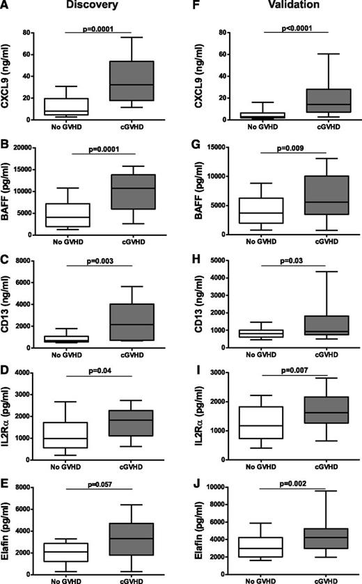
We hypothesized that samples at onset of de novo cGVHD from patients who ultimately developed treatment-refractory disease would be most likely to contain cGVHD-specific biomarkers. Using the protein microarray (supplemental Table 2) and subsequent ELISA workflow outlined, we identified 5 proteins (out of the 131 tested; 130 from the microarray + CD13, which was measured separately) that distinguished refractory cGVHD patients at disease onset from patients who never had aGVHD or cGVHD: CXCL9, IL2Rα, elafin, CD13, and BAFF (Figure 1A-E).
Biomarkers at onset of cGVHD. (A-E) ELISA results of median plasma concentrations of CXCL9 (A), BAFF (B), CD13 (C), IL2Rα (D), and elafin (E) in the no cGVHD patients (n = 18) and refractory de novo cGVHD patients (n = 17) from the discovery cohort. (F-J) ELISA results of median plasma concentrations of CXCL9 (F), BAFF (G), CD13 (H), IL2Rα (I), and elafin (J) in the non-cGVHD patients (n = 64) and de novo cGVHD patients (n = 45) from the validation cohort. Data are illustrated as box and whisker plots with the whiskers indicating the 90th and 10th percentiles.
Biomarkers at onset of cGVHD. (A-E) ELISA results of median plasma concentrations of CXCL9 (A), BAFF (B), CD13 (C), IL2Rα (D), and elafin (E) in the no cGVHD patients (n = 18) and refractory de novo cGVHD patients (n = 17) from the discovery cohort. (F-J) ELISA results of median plasma concentrations of CXCL9 (F), BAFF (G), CD13 (H), IL2Rα (I), and elafin (J) in the non-cGVHD patients (n = 64) and de novo cGVHD patients (n = 45) from the validation cohort. Data are illustrated as box and whisker plots with the whiskers indicating the 90th and 10th percentiles.
We then measured concentrations of these 5 proteins in samples from the validation cohort of UM patients. Of note, patients in the cGVHD group were older and more likely to have received a transplant for a malignant condition than the no-cGVHD controls. Otherwise, there were no statistically significant differences between the patients with cGVHD and without cGVHD based on donor type, graft source, HLA match, or conditioning intensity. Likewise, samples were collected at similar times for both the cGVHD cases and controls. Samples were obtained at a median of 154 days after HCT in the cGVHD group compared with 135 days after HCT in the no-cGVHD group (P = .25). As in the discovery set, all 5 candidate proteins were significantly elevated in patients with newly diagnosed de novo–onset cGVHD compared with those without cGVHD (Figure 1F-J), validating our initial findings. As others have also reported, we found an association of higher CD13 concentrations in patients whose cGVHD included liver involvement compared with cGVHD patients without liver involvement (median 1382 vs 725 ng/mL; P < .0001).9
To better define the potential clinical utility of these proteins elevated at the onset of cGVHD, we performed area under the ROC curve analyses for each protein comparing no cGVHD to de novo–onset cGVHD. The AUCs were similar for IL2Rα, elafin, CD13, and BAFF and ranged from 0.62–0.67 while the AUC for CXCL9 was 0.83 (supplemental Figure 1A). Given the similar AUCs for 4 of the proteins, we combined them into a composite panel (without CXCL9), which provided an improved AUC of 0.74. When CXCL9 was added to the composite panel, the AUC improved further to 0.83 but was not better than CXCL9 alone (supplemental Figure 1B). Because there was no additional diagnostic value to using the composite panels, we determined that CXCL9 had the best correlation with de novo–onset cGVHD and further analyses were confined to CXCL9.
Next, we determined that the median CXCL9 plasma concentration provide an 87% sensitivity and a 77% specificity for identifying de novo cGVHD (supplemental Table 3). We then assessed the correlation of CXCL9 plasma concentrations and diagnosis of cGVHD by χ2 analysis. CXCL9 plasma concentrations above the median (6.5 pg/mL) were strongly associated with the presence of newly diagnosed cGVHD (71% vs 20%; P < .001), a finding that remained statistically significant (P < .001) after adjusting for potential confounding factors associated with the development of cGVHD (patient age, graft source [bone marrow/cord blood vs peripheral blood HCT], HLA match [matched sibling vs other] and diagnosis [malignant vs nonmalignant]) (Table 3).
Finally, we assessed if CXCL9 concentrations were associated with other factors. Since changes in CXCL9 concentrations may reflect differences in immune recovery, we first analyzed for an association of CXCL9 concentrations and absolute lymphocyte count, and found none. We then examined whether CXCL9 concentrations were higher as time post-HCT increased, an alternative way to look for an association with CXCL9 and immune recovery. In the cGVHD patients, we did not detect an association of CXCL9 concentration and time post-HCT. Therefore, we concluded that CXCL9 elevated concentrations at the time of de novo cGVHD were due to the presence of the disease. We then sought to further validate CXCL9 as a marker of cGVHD activity in a second, more heterogeneous, independent cohort.
We obtained 211 samples from the FHCRC for validation. Unlike the UM cohort, the FHCRC validation cohort included patients with any type of cGVHD presentation (de novo, quiescent, or progressive). In order to create more homogenous subsets within the FHCRC cohort, we divided the cGVHD patients into a newly diagnosed group (within 90 days of diagnosis; n = 86) and an established cGVHD group (diagnosed 3-36 months prior to sample acquisition; n = 92). Control patients (n = 33) did not have cGVHD, but prior treated aGVHD was allowed (73%). The median plasma concentration of CXCL9 was significantly higher in the FHCRC cohort than in the UM cohort (26 vs 6.5 pg/mL; P < .0001), presumably reflecting the differences in the 2 populations described above. For our initial analysis of this independent cohort, we limited comparisons to no cGVHD controls vs newly diagnosed patients because they were most similar to the UM cohort. Despite differences in absolute values of CXCL9, as in the UM results, CXCL9 plasma concentrations were significantly higher in patients with newly diagnosed cGVHD compared with the no-cGVHD patients (P = .003; Figure 2). Area under the ROC curve analyses for CXCL9 comparing controls with no cGVHD with patients with newly diagnosed cGVHD revealed an AUC of 0.68 with a sensitivity and specificity at the median of 59% and 70%, respectively (supplemental Table 3). Given the similarity of these results to those seen in the UM validation set, we performed an identical adjusted χ2 analysis for the FHCRC newly diagnosed cGVHD patients. As in the UM analysis, CXCL9 plasma concentrations above the median were strongly associated with the presence of cGVHD (84% vs 60%; P = .001; Table 3).
CXCL9 is elevated in newly diagnosed cGVHD from an independent cohort. ELISA results of median plasma concentrations of CXCL9 from no cGVHD patients (n = 33) and newly diagnosed cGVHD patients (n = 86) in a second validation cohort from the FHCRC. Data are illustrated as box and whisker plots with the whiskers indicating the 90th and 10th percentiles.
CXCL9 is elevated in newly diagnosed cGVHD from an independent cohort. ELISA results of median plasma concentrations of CXCL9 from no cGVHD patients (n = 33) and newly diagnosed cGVHD patients (n = 86) in a second validation cohort from the FHCRC. Data are illustrated as box and whisker plots with the whiskers indicating the 90th and 10th percentiles.
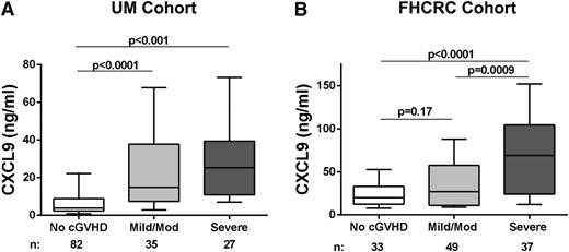
Given the strong correlation between CXCL9 plasma concentrations above the median and the presence of newly diagnosed cGVHD, we evaluated whether CXCL9 plasma concentrations were also associated with cGVHD severity at diagnosis. Very few patients in either the UM cohort (n = 4) or the newly diagnosed FHCRC cohort (n = 3) had mild cGVHD, so those patients were combined with patients who presented with moderate cGVHD. In both the UM cohort and FHCRC cohorts, CXCL9 plasma concentrations were significantly higher in patients who presented with severe cGVHD compared with no cGVHD group (P < .0001 and P = .0009 respectively; Figure 3A-B). Although UM patients who presented with mild/moderate cGVHD had significantly higher CXCL9 plasma concentrations compared with no cGVHD controls (P < .001; Figure 3A), we were unable to reproduce this finding in the FHCRC patients with mild/moderate cGVHD (P = .17; Figure 3B).
Increased CXCL9 levels are associated with increased cGVHD severity. (A) ELISA results of median plasma concentration of CXCL9 from no GVHD (n = 82), mild/moderate cGVHD (n = 35), and severe cGVHD (n = 27) from the entire UM cohort. (B) ELISA results of median plasma levels of CXCL9 from no GVHD (n = 33), mild/moderate (mild/mod) cGVHD (n = 49), and severe cGVHD (n = 37) from the newly diagnosed FHCRC cohort. Data are illustrated as box and whisker plots with the whiskers indicating the 90th and 10th percentiles.
Increased CXCL9 levels are associated with increased cGVHD severity. (A) ELISA results of median plasma concentration of CXCL9 from no GVHD (n = 82), mild/moderate cGVHD (n = 35), and severe cGVHD (n = 27) from the entire UM cohort. (B) ELISA results of median plasma levels of CXCL9 from no GVHD (n = 33), mild/moderate (mild/mod) cGVHD (n = 49), and severe cGVHD (n = 37) from the newly diagnosed FHCRC cohort. Data are illustrated as box and whisker plots with the whiskers indicating the 90th and 10th percentiles.
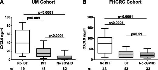
Finally, because previously reported biomarkers for both acute and chronic GVHD have been shown to decrease following initiation of immunosuppressive therapy (IST),10,16 we analyzed the effect of treatment with IST on CXCL9 concentrations. In the UM cohort, where samples were obtained closer to the time of onset and possible initiation of therapy, we found that median CXCL9 concentrations were higher in patients not on IST (n = 19) compared with patients on IST (n = 43; 39 vs 15 ng/mL; P = .009); furthermore, both groups had higher concentrations than the no cGVHD controls (n = 82, 4 ng/mL; P < .001 for both comparisons; Figure 4A). We performed the same analysis in the newly diagnosed FHCRC cohort. As in the UM cohort, patients not on IST at the time of sample acquisition (n = 43) had higher CXCL9 concentrations than patients on IST (n = 43; 77 vs 23 ng/mL; P < .0001; Figure 4B) and the no cGVHD controls (n = 33; 20 ng/mL; P < .0001). Unlike the UM cohort however, concentrations of CXCL9 in patients on IST was not higher than the no cGVHD controls (P = .51). This result might be explained by differences in the intensity and duration of IST between the cohorts. UM patients on IST were generally not on systemic steroids at the time of sample acquisition (84%), whereas only 2% of FHCRC patients were not treated with steroids when samples were acquired. Taken together, this finding suggests that intensity and duration of cGVHD treatment lowers CXCL9 concentrations. Lastly, because both cohorts consisted entirely of patients with multiorgan involvement, we could not validate CXCL9 as a biomarker with target organ specificity (data not shown).
CXCL9 is influenced by immunosuppression therapy. (A) ELISA results from the UM cohort of median plasma concentrations of CXCL9 from cGVHD patients on no immunosuppressive therapy at the time of sample acquisition (n = 19), cGVHD on any immunosuppressive therapy at the time of sample acquisition (n = 43), and patients with no cGVHD (n = 82). (B) ELISA results from the FHCRC of median plasma concentrations of CXCL9 from cGVHD patients on no immunosuppressive therapy at the time of sample acquisition (n = 43), cGVHD on any immunosuppressive therapy at the time of sample acquisition (n = 43), and patients with no cGVHD (n = 33). Data are illustrated as box and whisker plots with the whiskers indicating the 90th and 10th percentiles.
CXCL9 is influenced by immunosuppression therapy. (A) ELISA results from the UM cohort of median plasma concentrations of CXCL9 from cGVHD patients on no immunosuppressive therapy at the time of sample acquisition (n = 19), cGVHD on any immunosuppressive therapy at the time of sample acquisition (n = 43), and patients with no cGVHD (n = 82). (B) ELISA results from the FHCRC of median plasma concentrations of CXCL9 from cGVHD patients on no immunosuppressive therapy at the time of sample acquisition (n = 43), cGVHD on any immunosuppressive therapy at the time of sample acquisition (n = 43), and patients with no cGVHD (n = 33). Data are illustrated as box and whisker plots with the whiskers indicating the 90th and 10th percentiles.
We also were able to study CXCL9 concentrations in the FHCRC patients with established cGVHD (n = 92; sample obtained 3-36 months after cGVHD diagnosis). CXCL9 plasma concentrations in this group of patients with long-standing and treated cGVHD were not statistically different compared with the no-cGVHD controls (P = .18). Likewise, there was no correlation between CXCL9 plasma concentrations above the median and the presence of cGVHD (Table 3) or by disease severity (data not shown).
Discussion
Discovery of valid and reproducible biomarkers for cGVHD remains a significant challenge. Compared with aGVHD, cGVHD is clinically more heterogeneous and can involve many more target organs, often simultaneously. Additionally, the timing of sample acquisition for biomarker assessment is also critical. Once immunosuppression has been initiated, the biomarker pattern may change, as has been previously been observed with BAFF plasma concentrations after patients are treated with corticosteroids10 and was observed in our study as well. Therefore, one of the strengths of our study design was the inclusion of only de novo cGVHD in the first validation cohort, when the length of prior therapy was minimized. Another strength of our study was that we were then able to reproduce the strong correlation of CXCL9 with cGVHD in a second more heterogeneous cohort. Taken together, these findings provide convincing evidence that elevated CXCL9 concentrations are a marker for newly diagnosed cGVHD.
CXCL9 is an interferon-γ–inducible chemokine that binds to CXCR3, its only known receptor. CXCR3 expression can be rapidly induced in both CD4+ type 1 helper cells as well as CD8+ cytotoxic lymphocytes following dendritic cell activation of naïve lymphocytes.13 In both human and mouse autoimmune disease studies, the binding of CXCL9 to CXCR3 promotes lymphocyte migration to inflamed tissues.17,18 CXCR3 has also been shown to be critical for the recruitment of alloreactive T cells in aGVHD,19,20 whereas CXCL9 has been shown to be elevated in tissue samples from patients with oral,21 ocular,22 and cutaneous23 cGVHD. We found that the plasma of patients with newly diagnosed cGVHD, but not established cGVHD, contains higher concentrations of CXCL9 than patients without cGVHD. These results suggest that this T-cell chemoattractant is involved in the initiating steps of the cGVHD disease process, particularly around the time that clinical manifestations are first noted. The role of CXCL9 in the pathophysiology of cGVHD after the disease is well established and systemic therapy has been given is not as clear. Given the well-described relationship of CXCL9-CXCR3 in Th1-mediated disease states, it is intriguing to speculate that the Th1 pathways may be important during the early stages of cGVHD.
One other group reported that in a study of 28 patients with cGVHD, CXCL9 serum concentrations were associated with cGVHD involving the skin, but not other phenotypes.23 Our cohort did not include patients with isolated skin involvement, which precluded us from performing the same analysis. However, as noted, we did not find that CXCL9 correlated with any particular organ involvement (data not shown). Given our large sample size and reproducibility of our results in independent validation cohorts, we believe that CXCL9 may be useful as a marker of cGVHD that presents with a variety of clinical phenotypes. Of note, the same group also reported an association of CXCL10 and CXCL11 and cGVHD.23 Though CXCL10 was in the discovery array, we did not find a difference in CXCL10 levels in our discovery experiments and CXCL11 was not included in our discovery array and, therefore, neither marker was pursued further.
Several limitations should be noted. First, although we included a large number of candidate biomarkers in our discovery array, our approach was not unbiased in that we preselected the candidates for study. Thus, proteins not included in our array but associated with cGVHD were missed. In addition, a previous study demonstrated a strong correlation of BAFF/B-cell ratios with active cGVHD.11 Our study did not include analyses such as B-cell enumeration, so we cannot confirm the BAFF/B-cell ratio correlation. Using plasma protein concentrations alone, we found that CXCL9 had the highest AUC and best sensitivity and specificity for diagnosis of cGVHD of the 5 proteins tested. A direct comparison of the diagnostic utility of CXCL9 compared with BAFF/B-cell ratios could be useful. Next, this study does not address whether CXCL9 can predict the development of cGVHD as our focus was on samples obtained at the time of diagnosis. Furthermore, although 2 independent validation cohorts were included, the similarities in HCT practices, such as the paucity of haploidentical donors and bone marrow and cord blood stem cell sources, may limit the generalizability of the findings to patients who develop cGVHD subsequent to transplants where these factors are present, if they impact cGVHD. Finally, as noted in “Results,” the median concentrations of CXCL9 around the time of diagnosis of cGVHD were significantly different in the 2 validation cohorts. We suspect these differences can be explained by differences in the timing of sample acquisition relative to the onset of cGVHD (there was a wider window for the FHCRC patients), the inclusion in the FHCRC cohort of patients with cGVHD presentation other than de novo, the impact of immunosuppressive therapy (including intensity or duration treatment), or center effects due to other differences between the cohorts that we have not yet been able to define. We believe that the difference in median values highlights the importance of internally controlling each experiment and testing samples blinded to clinical characteristics, which maximizes the interpretability of validation studies.
An additional challenge for future cGVHD biomarker discovery will be to determine whether specific biomarkers correlate with clinically relevant outcomes such as clinical phenotypes or treatment response. Our study was designed to identify a marker of overall cGVHD activity as all of our patients had at least 2 target organ involvement at disease onset. Therefore, this study was unable to determine if a particular organ manifestation may be driving CXCL9 concentrations at disease onset. Furthermore, the current NIH consensus criteria for treatment response have not been validated, and publications from the Chronic GVHD Consortium have raised the concern that these measures may in fact not be valid.24,25 The association of CXCL9 concentrations with treatment response would be best analyzed in a controlled context, such as a correlative study in a clinical treatment trial.
The clinical utility of CXCL9 as a biomarker for cGVHD needs to be further defined in future studies. These studies should include serial measurement at important times, such as prior to the typical onset of cGVHD, to determine if elevations precede overt clinical manifestations. If that were the case, CXCL9 levels could be used to guide clinical trials testing pre-emptive strategies. For example, if the CXCL9 level exceeded a certain threshold, planned immunosuppression taper could be delayed. Second, CXCL9 may correlate with clinical outcomes, a possibility suggested by the correlation of high urinary CXCL9 levels and renal allograft rejection.26 In addition, confirmation of CXCL9 as a cGVHD biomarker opens up the possibility of new therapeutic avenues, if further evidence can be developed implicating the CXCL9-CXCR3 pathway in cGVHD pathogenesis. For example, bortezomib, which has shown promise for aGVHD prevention in two clinical trials,27,28 was recently shown to inhibit T-cell chemotactic movements, decrease expression of CXCR3, and decrease the secretion of CXCL9 by activated T cells in a mouse study.29 Other CXCL9 and CXCR3 inhibitors are under development, raising the possibility of translating these findings into a future clinical trial.
In conclusion, CXCL9 was the best biomarker for newly diagnosed cGVHD out of 131 candidates we tested. Future directions should include prospective and serial evaluations of CXCL9 in order to further define its clinical utility, potentially as a predictive biomarker prior to the onset of cGVHD. Our findings also implicate the CXCL9-CXCR3 pathway in the pathogenesis of cGVHD, which we believe warrants further exploration. Finally, it may be fruitful to expand the search for cGVHD biomarkers using unbiased large-scale proteomic discovery approaches that could detect additional proteins and potentially reveal important pathways in the development of cGVHD that are currently unknown.
The online version of this article contains a data supplement.
The publication costs of this article were defrayed in part by page charge payment. Therefore, and solely to indicate this fact, this article is hereby marked “advertisement” in accordance with 18 USC section 1734.
Acknowledgments
The authors would like to thank the clinicians of the University of Michigan Blood and Marrow Transplant program and at the Fred Hutchinson Cancer Research Center, the University of Michigan clinical research support staff, the University of Michigan BMT division data managers, and the members of the Paczesny laboratory.
We would like to acknowledge our funding sources, including RayBiotech (Biomarker Discovery Pilot grant) (C.L.K. and S.P.), Briskin/Schlafer Pediatric Oncology Investigators Fund (C.L.K.), University of Michigan Pediatric Hematology/Oncology Junior Faculty Grant (C.L.K.), and the National Marrow Donor Program (Amy Strelzer Manasevit Scholars grant 200513 and grant R01CA174667) (S.P.) (grants R01CA118953 and U54CA163438) (S.J.L.). The Chronic GVHD Consortium (U54 CA163438) is a part of the NIH Rare Diseases Clinical Research Network, supported through collaboration between the NIH Office of Rare Diseases Research at the National Center for Advancing Translational Science and the National Cancer Institute. The content is solely the responsibility of the authors and does not necessarily represent the official views of the NIH.
Authorship
Contribution: C.L.K. conceived and planned the study design, performed clinical data collection and quality assurance, interpreted the data, and wrote the manuscript; D.A.F. and R.M. developed and performed the CD13 ELISAs; T.M.B, B.E.S., and X.C. were the study statisticians and wrote the manuscript; L.C. assisted with the statistical analysis and wrote the manuscript; M.C., B.J.F., P.P., and K.L. performed ELISA experiments, participated in research discussions, and wrote the manuscript; D.R.C. performed clinical data quality assurance and wrote the manuscript; J.E.L., J.L.M.F., P.J.M., M.E.F., and J.A.H. contributed to patient accrual, clinical data collection and quality assurance, and research discussion and wrote the manuscript; S.J.L. planned the study design, interpreted the data, and wrote the manuscript; and S.P. conceived and planned the study design, supervised the experiments, interpreted the data, and wrote the manuscript.
Conflict-of-interest disclosure: The authors declare no competing financial interests.
Correspondence: Carrie L. Kitko, Blood and Marrow Transplant Program, University of Michigan Comprehensive Cancer Center, Room 5303, 1500 E. Medical Center Dr, Ann Arbor, MI 48109; e-mail: ckitko@med.umich.edu; or Sophie Paczesny, Bone Marrow and Stem Cell Transplantation Program, Indiana University Melvin and Bren Simon Cancer Center and Wells Center for Pediatric Research, 1044 W Walnut St, Room 425, Indianapolis, IN 46202; e-mail: sophpacz@iu.edu.


