Abstract
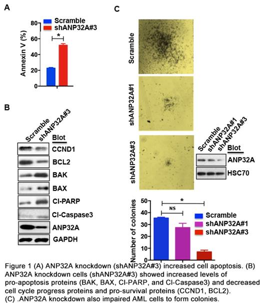
Genetic alterations as the initiator inducing leukemia also cause disorder of gene expression. Cooperation of these factors is known to be essential for leukemia development and remains to be further elucidated. In this study, we discovered that ANP32A dysregulation might contribute to myeloid leukemia. ANP32A expression was unanimously elevated in multiple online leukemia datasets and its upregulation was confirmed in human primary myeloid leukemia cells. Although ectopic expression of ANP32A did not promote leukemic cell proliferation, it did increase cell resistance to drug treatment such as TPA, Ara-C, VP16, and BCL2 inhibitor. Interestingly, ANP32A knockdown reduced cell proliferation and impaired colony formation in soft agar in various leukemic cells. ANP32A knockdown did so by inducing apoptosis and cell cycle arrest at G1 phase evidenced by upregulation of pro-apoptosis genes (BAK, BAD, cleaved caspase 3 and PARP) and downregulation of pro-survival or cell cycle progress genes (BCL2, CDK4, CCND1). The function of ANP32A knockdown to induce apoptosis and reduce colony formation was verified in human primary acute myeloid leukemia (AML) cells (Figure 1). However, re-introduction of BCL2, CDK4, or CCND1 failed to restore the impaired ability of colony formation by ANP32A knockdown, suggesting that apoptosis and cell cycle arrest may not be the direct effect of ANP32A knockdown.
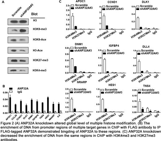
To probe how ANP32A would affect cell proliferation, we performed microarray analysis to identify potential ANP32A target genes including APOC1 and CCL26. Indeed, reintroduction of APOC1 or CCL26 significantly rescued colony formation ability while knockdown of APOC1 or CCL26 alone was sufficient to reduce colony formation. Further gene set enrichment analysis (GSEA) also revealed Notch signaling and histone modification signatures in ANP32A knockdown cells. To support this, ANP32A knockdown reduced intracellular Notch and NOTCH1 ovexpression significantly recovered the ability of colony formation. Furthermore, ANP32A knockdown also led to a global alteration of histone modifications including decrease of H3K9-acetylation (H3K9ac), H3K27-trimethylation (H3K27me3), and H3K4-trimethylation (H3K4me3) and increase of H3K9-trimethylation (H3K9me3). In fact, ChIP-PCR demonstrated that ANP32A bound to the promoter region of multiple target genes identified in microarray analysis and ANP32A downregulation caused decreased H3K27me3 and H3K4me3 at the same sites (Figure 2). These findings suggest that ANP32A may bind to the chromosome and involve histone modifications.
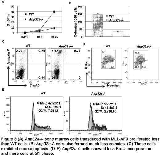
To further test the potential function of ANP32A in leukemogenesis, we took advantage of MLL-AF9 fusion gene to immortalize bone marrow cells in vitro and compared wild-type (WT) cells to Anp32a knockout (Anp32a-/-) cells. We found that MLL-AF9-transduced Anp32a-/- bone marrow cells reproduced all phenotypes of leukemic cells with ANP32A knockdown: Anp32a-/- cells proliferated less, formed less colonies, exhibited more apoptosis, and showed cell cycle arrest at G1 phase compared to WT cells (Figure 3). These observations suggest a potential role of ANP32A in MLL-AF9-induced acute myeloid leukemia.
Taken together, our studies have revealed ANP32A as a potential novel player to cooperate with other genetic alterations to induce leukemia. ANP32A upregulation may somehow involve histone modifications at genomic level that ultimately alter the expression of multiple genes and facilitate leukemic cell proliferation and survival. Thus ANP32A may serve as a potential target for developing novel therapy or biomarker for diagnosis and prognosis.
No relevant conflicts of interest to declare.
Author notes
Asterisk with author names denotes non-ASH members.


