Abstract
Background. Chromosomal abnormalities (CA) are the most prognostic factor in acute myeloid leukemia (AML). However, it is still not clear whether the cytogenetic risk groups established for patients (pts), treated by standard chemotherapy, are so optimal for pts undergoing allogeneic hematopoietic stem cell transplantation (alloHSCT). Recent studies have confirmed negative effects of both monosomal and complex karyotypes (MK, CK) on outcomes after alloHSCT [1,2]. Meantime, some investigators consider that immune effects of alloHSCT can reduce negative impact of these CA [3].
Aim. To evaluate impact of the CA in AML pts with adverse cytogenetic risk group on outcome after alloHSCT.
Material and Methods. In this study, outcomes of alloHSCT, which were performed in a single institution between 2009 and 2014 years for 97 AML pts have been analyzed. Patients and transplantation characteristics are presented in Table I. The probabilities of overall survival (OS), leukemia free survival (LFS), cumulative incidence of relapse (CIR) were evaluated for different cytogenetic groups.
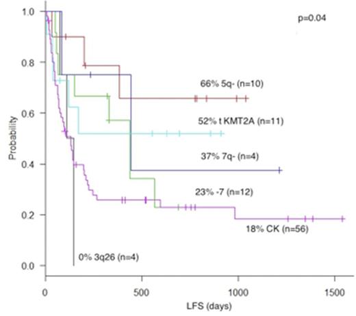
Results. According to univariateanalysis, the probabilities of 4-year OS in pts with 5q-, KMT2A translocations and monosomy 7 were 66%, 59% and 56%, respectively. At the same time, OS in pts with CK, 7q- and 3q26 rearrangements were lower i.e. 33%, 25% and 25%, respectively (p=0.01). Multivariate analysis showed, that clinical stage at HSCT, age and HSC source are independent predictors of OS in AML pts (Table 2). Four-year LFS were various in pts with different CA. The higher LFS was noted in pts with 5q- and KMT2A translocations (66% and 52%, respectively), whereas lower LFS were in pts with 3q26 rearrangements, CK, monosomy 7 and 7q- (0, 18%, 23% and 37%, respectively) (Fig. 1). Besides, LFS distinguished between groups with CK+ and CK- (18% vs 41%, p=0.008), as well as with MK+ and MK- (17% vs 30%, p=0.04). Multivariate analysis evidenced clinical stage at HSCT, cytogenetic groups, MK and number of transplanted CD34+ cells to be independent predictor of LFS in AML pts (Table 2).Cumulative incidence of relapses in pts transplanted in remission (n=42) was higher in those with CK+ (55% vs 14%, p=0.03) and MK+ (75% vs 31%, p=0.013).
Discussion. The study showed that 4-year OS in AML pts with 5q-, KMT2A translocations and monosomy 7 significantly distinguished from those with CK, 7q- and 3q26 rearrangements. Furthermore, OS depended on clinical stage at HSCT, patient's age and HSC source. On the other hand, EFS differed from all above-mentioned cytogenetic groups, including MK. Finally EFS depended on clinical stage at HSCT and number of transplanted CD34+ cells.
Conclusion. On the basis of this data, a conclusion may be drawn that alloHSCT in AML pts with adverse CA should be performed at complete remission, with bone marrow as HSC source and enough number of transplanted CD34+ cells.
Reference.
1. Hemmatti et al. Eur J Haematol. 2013; 92:102-10.
2. Fang et al. Blood 2011; 118:1490-4.
3. Guo et al. Biol Blood Marrow Transplant 2014; 20: 690-5.
Patients and Transplant characteristics
| Patients, n (%) . | 97 (100) . |
|---|---|
| Gender, n (%) Male Female | 53 (54.6) 44 (45.4) |
| Age at HSCT, median, (range) years | 25 (1.5-60) |
| Cytogenetics, n (%) 3q26 rearrangements Deletion 5q (sole) Monosomy 7 (sole) Deletion 7q (sole) KMT2A translocation Complex karyotype ³3 CA Monosomal karyotype | 4 (4) 10 (10.3) 12 (12.4) 4 (4) 11 (11.3) 56 (58) 18 (18.5) |
| Time from diagnosis to HSCT, median (range) days | 477 (47 - 3482) |
| Clinical stage at HSCT, n (%) CR1 ³CR2 Active disease | 29 (30) 13 (13) 55 (57) |
| HSC source, n (%) Bone marrow Peripheral blood BM+PB | 54 (56) 34 (35) 9 (9) |
| Conditioning regimen, n (%) MA Non-MA | 35 (36) 62 (64) |
| Donor type, n (%) HLA-id sibling Matched unrelated Haploidentical | 17 (17.5) 53 (54.6) 27 (27.8) |
| Number of transplanted CD34+ cells median (range), x106/kg | 6.1 (1.5-17.9) |
Multivariate analyses
| . | HR . | 95% CI . | P . |
|---|---|---|---|
| Overall survival | |||
| Clinical stage at HSCT | 2.65 | 1.72-4.09 | 0.00001 |
| Median age <18 years) | 2.05 | 1.05-3.99 | 0.034 |
| HSC source | 1.66 | 1.12-2.45 | 0.011 |
| Cytogenetic groups | 1.72 | 0.81-3.66 | 0.15 |
| Median number of transplanted CD34+ cells >6x10⁶/kg | 1.77 | 0.91-3.42 | 0.08 |
| Leukemia-free survival | |||
| Clinical stage at HSCT | 2.59 | 1.8-3.7 | 0.0001 |
| Cytogenetic groups | 1.31 | 1.03-1.69 | 0.031 |
| Monosomal karyotype | 1.88 | 0.97-3.66 | 0.044 |
| Median number of transplanted CD34+ cells >6x10⁶/kg | 2.78 | 1.51-5.11 | 0.0003 |
Leukemia-free survival for AML patients with different cytogenetic groups after alloHSCT
Leukemia-free survival for AML patients with different cytogenetic groups after alloHSCT
No relevant conflicts of interest to declare.
Author notes
Asterisk with author names denotes non-ASH members.


