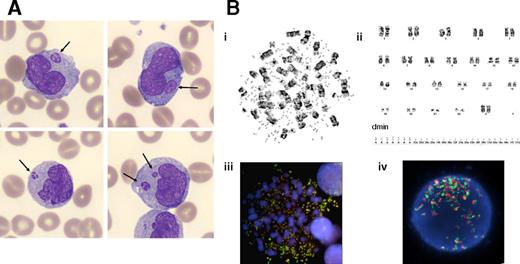
A 61-year-old woman with a history of hepatic hemangioma and cholecystectomy developed epigastric pain for several weeks. Her blood tests showed hemoglobin 74 g/L, platelets 127 × 109/L, and white blood cells 104.6 × 10⁹/L with 51% monoblasts. The bone marrow was hypercellular with 76% monoblasts and promonocytes. Peripheral blood and bone marrow smears showed that some monoblasts had micronuclei, which are small intracellular nucleus-like structures expelled from the nucleus (panel A; Giemsa stain, original magnification ×100). Immunophenotyping revealed that these cells were positive for HLA-DR, CD13, CD33 (very bright), CD14, CD11b, CD36, and CD64, confirming the diagnosis of acute myeloid leukemia M5 subtype (AML-M5). G-banding analysis of bone marrow cells showed 14 metaphases with double-minute chromosomes (dmin) (panel Bi-ii): 46,XX,del(13)(q14q21),5∼25dmin[3]/46,XX,5∼70dmin[11]/46,XX[6]. Fluorescence in situ hybridization revealed MYC amplification (panel Biii-iv).
A dmin is a small, paired, usually spherical chromatin particle that lacks a centromere, and it represents a form of extrachromosomal gene amplification (most commonly MYC). In general, dmin associated with micronuclei are rare in hematologic malignancies, are never described in AML-M5, and are usually associated with an unfavorable prognosis. Our patient achieved hematologic and cytogenetic remission after induction therapy, which consisted of conventional intensive chemotherapy for elderly patients with AML. She is currently receiving consolidation treatment before an allogeneic stem cell transplantation.
For additional images, visit the ASH IMAGE BANK, a reference and teaching tool that is continually updated with new atlas and case study images. For more information visit http://imagebank.hematology.org.


