Anemia of inflammation (AI) is a highly prevalent comorbidity in patients affected by chronic inflammatory disorders, such as chronic kidney disease, inflammatory bowel disease, or cancer, that negatively affect disease outcome and quality of life. The pathophysiology of AI is multifactorial, with inflammatory hypoferremia and iron-restricted erythropoiesis playing a major role in the context of disease-specific factors. Here, we review the recent progress in our understanding of the molecular mechanisms contributing to iron dysregulation in AI, the impact of hypoferremia and anemia on the course of the underlying disease, and (novel) therapeutic strategies applied to treat AI.

Anemia of inflammation (AI) is a highly prevalent comorbidity in patients with chronic inflammatory disorders, autoimmune diseases, infections, and cancer, which causes a severe additional burden on the recovery process from their primary disease. According to the latest report of the World Health Organization (WHO), anemia affects 24.8% of the worldwide population, of which most cases are caused by malnutrition.1 AI ranks in the second place, but its actual prevalence is difficult to assess because it often coexists with iron deficiency anemia (IDA). AI adversely affects disease outcomes and quality of life (QoL) through fatigue, dizziness, and functional cognitive impairment.2 The pathophysiology of AI is multifactorial and includes iron retention in macrophages of the reticuloendothelial system, reduction in circulatory half-life of erythrocytes, inadequate production and activity of erythropoietin (EPO), and impaired proliferation and differentiation of erythroid progenitor cells. The exact pathomechanisms involved often depend on the underlying primary disease. In this review, we focus on the prevalent diseases associated with AI and outline the most important molecular mechanisms that contribute to inflammatory hypoferremia and iron-restricted erythropoiesis.3,4 

Diagnosis of AI

Iron-restricted erythropoiesis is an integral part of the pathophysiology of both AI and IDA, rendering differential diagnosis difficult specifically when both diseases coexist.5,6 Especially in low-income countries, where the burden of infectious diseases is high and true iron deficiency emerges because of poor nutritional iron availability and/or chronic blood loss, for example, owing to helminth infestations, AI and IDA frequently coaffect the same individuals. AI with combined IDA is also found in patients affected by inflammatory diseases and in those with chronic blood loss because of menstruation, repeated venesections, or disease-associated bleeding. As established by the WHO, anemia is defined as a hemoglobin (Hb) level of <120 g/L in women (<110 g/L in pregnant women) and <130 g/L in men. However, the dependency of the Hb value on multiple parameters, such as age, pregnancy, socioeconomic status, ethnicity, or altitude of living,7 complicates the universal definition of normal Hb concentration in healthy individuals.

The underlying causes of erythropoietic iron restriction differ between AI and IDA, and their knowledge is of importance for proper diagnosis and treatment. Iron restriction in AI mainly evolves from iron retention in macrophages and, only to a minor extent, from reduced dietary iron absorption. This state is referred to as functional iron deficiency and is hallmarked by iron storage in macrophages.3 It contrasts with absolute iron deficiency that occurs because of a disequilibrium between iron uptake, iron utilization, and iron loss that is observed in IDA.4 Notably, iron deficiency in response to EPO treatment (eg, when pulsatile erythropoiesis exceeds iron supply) or in adolescence may also cause “functional” iron deficiency, whereby, in this setting, tissue iron stores are depleted, as reflected by reduced circulating ferritin levels.8 

Consistent with hypoferremia, individuals affected by AI show low transferrin saturation (TfSat) but normal or decreased transferrin levels. This differs from true IDA, in which transferrin levels are increased. Furthermore, ferritin levels are decreased in IDA, whereas ferritin levels are normal or increased owing to macrophage iron accumulation in AI.9 AI is further characterized by increased levels of inflammatory markers, such as interleukin 6 (IL-6) and C-reactive protein. Additional biochemical markers and hematologic indices are available for the differential diagnosis of AI and IDA and have been extensively reviewed elsewhere.10,11 

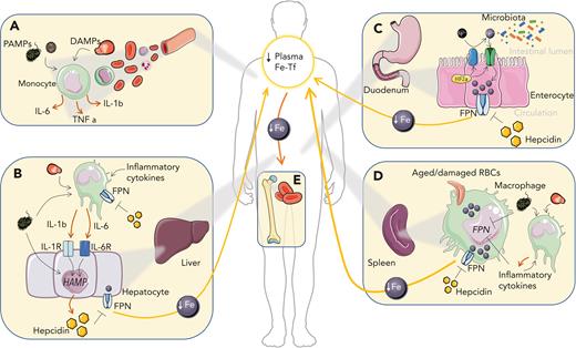
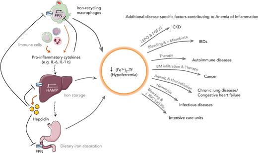
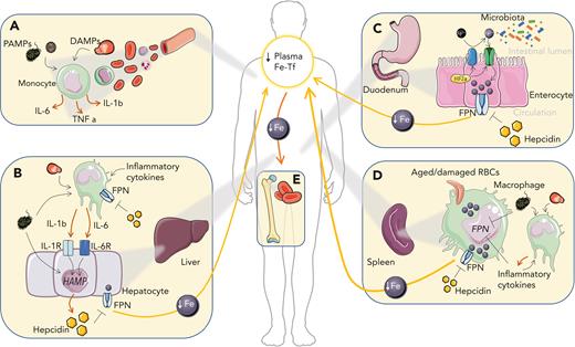
Inflammation induces functional iron deficiency, a state in which total body iron levels remain largely unchanged, and iron is redistributed into macrophages. Macrophages play a critical role in maintaining iron homeostasis by recycling 20 to 25 mg of iron from senescent red blood cells (RBCs) daily. Iron released from Hb during this process is either stored in the cellular iron storage protein ferritin or exported via ferroportin (FPN) into the bloodstream, where it is bound to transferrin. Iron-bound transferrin can be taken up by all tissues, including the erythropoietic bone marrow (BM). For comparison, only 1 to 2 mg of dietary iron is absorbed per day, which merely compensates for blood losses or the desquamation of skin or intestinal cells.12 Because iron export from macrophages and iron absorption from intestinal enterocytes are controlled by FPN, pathways reducing FPN expression affect body iron homeostasis and iron delivery to tissues. Inflammation induces the expression of the iron hormone hepcidin,13 which subsequently reduces FPN expression, and results in macrophage iron retention, reduced dietary iron absorption, and consequently the development of hypoferremia. The resulting restricted iron supply for erythropoiesis causes the gradual development of AI (summarized in Figure 1).

Figure 1.

Iron-related mechanisms contributing to the generation of AI. (A) In circulation, pathogen-associated molecular patterns (PAMPs) and damage-associated molecular patterns (DAMPs) activate myeloid cells leading to the production of proinflammatory cytokines. (B) Hepatocyte stimulation with proinflammatory cytokines, especially IL-6 and IL-1β, and PAMPs stimulates the expression of the liver iron hormone hepcidin, which promotes autocrine and paracrine iron retention due to the degradation of the iron exporter FPN. Kupffer cell stimulation with proinflammatory cytokines, PAMPs and DAMPs further feeds cytokine-mediated hepcidin upregulation in the liver. (C) Increased hepcidin levels in circulation block dietary iron import by degrading FPN. Local intestinal inflammation promotes HIF-2α (hypoxia-inducible factor 2α) stabilization and duodenal cytochrome b (DCYTB)/divalent iron transporter 1 (DMT1)–mediated iron absorption and may alter the composition of the intestinal microbiota. (D) In the spleen, where macrophages turn over large amounts of iron owing to the recycling of RBCs, iron accumulates in response to cytokines, hepcidin, PAMPs, and DAMPs, which decreases FPN-mediated iron export and affects additional pathways that cause iron retention. (E) Macrophage iron retention and decreased iron absorption reduce plasma iron levels and limit iron availability for erythroid progenitors, where iron is required for heme biosynthesis. As a consequence, anemia develops. Fe, iron; Fe-Tf, iron transferrin; TNF, tumor necrosis factor.

Close modal

Inflammation-driven iron retention in macrophages is considered to represent an innate immune mechanism in vertebrates, also known as “nutritional immunity.” It limits iron availability for the proliferation and pathogenicity of extracellular microorganisms.14 However, some bacteria (eg, Salmonella typhimurium) circumvent the host’s defense strategy by invading and proliferating within phagosomal vacuoles of macrophages where they benefit from the increased intracellular iron content.15,16 On the other hand, increased hepcidin protects against siderophilic extracellular pathogens by limiting the appearance of non–transferrin-bound iron in the plasma that promotes their rapid growth.17,18 Accordingly, iron supplementation can promote the pathogenicity of such microbes. This problem is exemplified by iron supplementation studies in geographic areas where the burden of infectious diseases is high.19,20 For example, in the Pemba trial, routine dietary iron fortification in children from areas with high rates of malaria resulted in an increased risk of infection-driven morbidity and death,21 a result replicated in other populations.22-25 Despite this finding, the WHO recommends iron interventions in vulnerable populations, such as children and women of reproductive age,26-28 because benefits of iron interventions are generally considered to outweigh their risks.

Control of iron fluxes by FPN under inflammatory conditions

The only known iron exporter, FPN, is the gatekeeper of systemic iron fluxes. It is predominantly expressed in cell types with an iron export function, such as macrophages, duodenal enterocytes, and hepatocytes. During inflammation, cell surface expression of FPN is reduced by transcriptional and posttranslational mechanisms.

FPN is the target receptor for the liver-expressed iron hormone hepcidin, which upon binding occludes iron efflux29 and promotes internalization and degradation of FPN.30 Hepcidin transcription is activated by inflammatory cytokines that are secreted from innate immunity effector cells. IL-6–induced activation of the signal transducer and activator of transcription 3 (STAT3) pathway is a potent inflammation-driven pathway to induce hepcidin transcription,31,32 as shown both in humans and in animal models of AI.13 However, in complex inflammatory settings, Il6 or Hamp deficiency only partially protect against anemia triggered by injection of heat-killed Brucella abortus, supporting nonoverlapping roles of IL-6 and hepcidin in the etiology of AI.33 Similarly, neither IL-6 nor hepcidin were shown to be required for the development of anemia in aged mice.34 Thus, other cytokines and pathogen-driven immune stimuli contribute to hypoferremia and anemia development.35,36 For example, IL-1β contributes to hepcidin upregulation in cell-based assays and mice,37,38 although the responsible signaling mediators are still under discussion. Notably, IL-10 dose-dependently induced anemia in patients with inflammatory bowel disease (IBD) by promoting macrophage iron uptake and retention.39 Furthermore, PAMPs, such as bacterial cell wall components, have been proposed to directly promote hepcidin transcription upon receptor stimulation (eg, Toll-like receptors [TLRs]), in macrophages and hepatocytes, independent of the downstream-activated cytokines.40,41 Iron excess, in contrast, increases hepcidin levels via the bone morphogenetic protein/small mothers against decapentaplegic (BMP/SMAD) signaling pathway. Inflammatory activation of hepcidin transcription requires BMP/SMAD signaling because hepcidin upregulation does not occur in the absence of the BMP-responsive element in the hepcidin promoter.42-44 Furthermore, mice deficient for proteins mutated in hereditary hemochromatosis, such as hemojuvelin (a BMP receptor coreceptor) or HFE, show reduced BMP/SMAD signaling and attenuated hepcidin and hypoferremia responses in conditions of acute inflammation.45,46 Although most hepcidin is produced in hepatocytes, small amounts are also synthesized in monocytes and macrophages, where it acts in an autocrine manner to repress FPN-mediated iron export.47 Whether elevated hepcidin levels exert bactericidal activity in vivo is poorly understood. Its antimicrobial effect has been reported previously in vitro.48,49 

Hepcidin promotes the degradation of FPN30 and occludes iron efflux by binding in a central cavity between the N and C domains.29 The affinity of hepcidin seems to be increased in the presence of iron, suggesting that iron-loaded FPN molecules are targeted for degradation. This notion is supported by observations that hepcidin-mediated, but not steady-state, FPN degradation is affected by cellular iron deficiency.50 Hepcidin binding induces lysine ubiquitination on an intracellular loop connecting the 2 halves of FPN.51 Specific ubiquitin ligases involved in FPN degradation have been described recently.52,53 

A second prominent mechanism for inhibiting iron export via FPN and inducing hypoferremia is the direct transcriptional repression of Fpn by proinflammatory cytokines and bacterial components in macrophages and hepatocytes.54-57 Inflammation-mediated Fpn transcriptional downregulation alone can be suffice to cause hypoferremia54,57,58 and eventually AI in the absence of hepcidin activation.

Molecules released during tissue damage, named DAMPs or alarmins, arise in response to prototypical inflammatory immune responses and contribute to inflammation.59 These include intracellular components, such as metabolites or chromatin-associated proteins, that play physiological roles, but display “cytokine-like” functions in the extracellular space. DAMPs and alarmins can induce anemia, as exemplified by the finding that the binding of CpG-containing DNA to TLR9 in RBCs promotes erythrophagocytosis by splenic macrophages, resulting in acute anemia60 that may contribute to the reduced circulatory half-life of RBC found in AI. This is in line with the observation that activation of TLR7 and TLR9 signaling promotes the differentiation of specialized phagocytes responsible for increased erythrophagocytosis and the development of anemia.61 

CKD

Chronic kidney disease (CKD) is a progressive, multifactorial disorder that can lead to renal failure over time. Anemia is highly prevalent in patients with CKD, ranging from 21% to 62% in different cohorts,62 and its prevalence increases upon CKD progression. Lower Hb levels correlate with a reduced QoL and the emergence of secondary complications, such as cardiovascular disease and mortality.63-65 In CKD, anemia caused by progressive kidney damage leads to reduced EPO production and hyporesponsiveness.63 In addition, uremic inhibitors of erythropoiesis (eg, indoxyl sulfate66), functional and/or absolute iron deficiency, and vitamin deficiencies (folate, cobalamine, vitamin D) contribute to anemia severity. Hepcidin levels in patients with CKD are elevated by different mechanisms. First, CKD results in reduced renal EPO secretion. EPO induces the hormone erythroferrone (ERFE) in erythroid progenitors, which enhances iron availability for erythropoiesis by suppressing hepcidin expression.67 Accordingly, in patients with CKD who are anemic, serum EPO and ERFE levels positively correlated. However, hepcidin levels vary between different cohorts,68-70 which is in line with the fact that in addition to regulation by ERFE, hepcidin levels are also affected by hypoxia and anemia-inducible factors, such as platelet-derived growth factor BB (PDGF-BB), growth-differentiation factor 15 (GDF15), or sexual hormones.71-74 Because hepcidin is cleared by the kidneys, the progression of renal disease is also associated with increased hepcidin,75,76 which is further aggravated by concomitant inflammation often linked to the underlying disease or dialysis.62,63 Additionally, blood loss during hemodialysis and repeated diagnostic blood sampling along with occult gastrointestinal bleeding contributes to iron losses that aggravate anemia and reduce EPO responsiveness.77 More recently, FGF23 (fibroblast growth factor 23) has been implicated in the establishment of AI in CKD. This bone-secreted hormone maintains phosphate and vitamin D homeostasis by targeting the kidney to modulate phosphate and calcium tubular reabsorption. Kidney damage decreases the expression of the renal FGF23 coreceptor αKlotho, thereby reducing the FGF23 response. To maintain mineral homeostasis, FGF23 levels increase.78,79 Elevated FGF23 levels, which also occur in response to iron deficiency and inflammation, affect hematopoietic stem cell differentiation and EPO regulation.80 In line with the proposed role of FGF23 in CKD, pharmacological inhibition of FGF23 stimulates erythropoiesis and counteracts anemia and iron deficiency in a CKD mouse model.81 

In CKD, anemia is mainly treated with a combination of iron supplementation and erythropoiesis-stimulating agents (ESAs), a therapy that increases Hb levels and QoL.82 However, ∼20% of patients respond poorly, probably because of low-grade inflammation that directly affects hepcidin production and erythropoiesis.13,83 

IBDs

IBDs, which mainly comprise ulcerative colitis and Crohn disease (CD), are conditions of cytokine-driven chronic gastrointestinal inflammation of multifactorial but distinct etiopathogenesis. Genetic susceptibility, environmental cues, and the gut microbiome can contribute to their pathogenesis. In CD, lesions mostly affect the terminal ileum, whereas in ulcerative colitis, they are limited to the colon and rectum.84 About two-thirds of patients with IBD present with anemia at diagnosis, which is considered the most common extraintestinal complication in IBD, significantly impairing patients’ QoL. The correction of anemia improves QoL independent of disease activity.85,86 Several mechanisms likely contribute to anemia in patients with IBD: (1) malnutrition (including iron) due to impaired intestinal absorption, (2) medication-associated toxicity (eg, azathioprine and methotrexate), (3) surgical procedures (eg, extensive bowel resection), (4) chronic intestinal bleeding from disease-associated ulcerations, and (5) inflammation.87 

During the inflammatory process, infiltrating immune cells deplete oxygen in the local epithelial microenvironment, causing localized hypoxia.88 Hypoxia and proinflammatory cytokines promote HIF stabilization in phagocytes.89 Additionally, HIF activation increases iron absorption by controlling the messenger RNA expression of target genes responsible for duodenal iron transport (DMT1, Dcytb,90,91 and FPN92). Because iron export is inhibited by hepcidin-mediated FPN degradation, iron accumulates in enterocytes. Therefore, in IBDs, the sloughing of iron-loaded damaged enterocytes in response to inflammation and/or hypoxia affects the intestinal microbiome. Intestinal bacteria (eg, Lactobacilli) release metabolites that reduce duodenal iron absorption by suppressing intestinal HIF-2α activity to ensure sufficient bacterial iron supply.93 Intestinal iron accumulation in patients with active IBD94 affects gut microbiota diversity, their metabolic profile, and thus, disease activity.95,96 Accordingly, IV vs oral iron supplementation exerts contrasting effects on microbiome composition and disease activity in patients with IBD.95,97 

Autoimmune diseases

Pathologic immune activation causes loss of tolerance and targeting of self-antigens. Manifestations range from asymptomatic disease to severe autoimmune disorders causing systemic and local inflammation that affect multiple organs. Both genetic and environmental factors contribute to autoimmunity. Although the exact disease mechanisms differ between various autoimmune disorders, increased levels of inflammatory mediators (eg, TNF family members and interleukins) or JAK/STAT pathway activation are commonly observed.98,99 

AI has been reported in 30% to 60% of patients with rheumatoid arthritis (RA), whereas IDA only occurs in a minority of patients, mainly because of therapy-induced, gastrointestinal, or urogenital blood losses. Disease severity is associated with reduced Hb levels and a higher prevalence of anemia.100,101 Both autoimmune disease per se or side effects of immunosuppressive therapies can negatively affect BM cellularity, erythropoiesis, and erythrocyte half-life.99,102 IL-6 plays a central role in RA pathogenesis103 because it promotes joint inflammation and the development of anemia via hepcidin induction. Consistently, patients with RA show elevated hepcidin levels that correlate with disease activity, as well as with comorbidities such as coronary atherosclerosis and osteoporosis.104-106 The most effective way to combat AI in autoimmune diseases is to treat the underlying inflammation with (1) systemic immunosuppressive drugs, such as corticosteroids or methotrexate; or (2) cytokine or immune-signaling neutralizing antibodies (eg, TNFα, IL-6, IL-17, IL-23), JAK inhibitors, or combinations thereof.103,107,108 However, in a substantial proportion of patients, these therapeutic strategies fail, and both inflammation and anemia persist. These patients may receive IV iron sometimes in combination with ESAs; however, the efficacy of these therapies is limited in more advanced inflammatory disease states.102,109 

Cancer

Anemia is observed in 40% to 80% of patients with cancer,110,111 varying in severity depending on tumor site, stage, patient age, and treatments.112 It is one of the most frequent secondary problems and is closely related to disease progression. Anemia of cancer is caused by multiple mechanisms, including inflammation, BM invasion by tumor cells, and vitamin deficiencies. It is aggravated by anticancer therapies, which often damage erythroid progenitor cells and/or reduce erythrocyte half-life.113,114 Moreover, tumor cells can produce cytokines that negatively affect erythroid progenitor cell differentiation. Patients with cancer often present with increased hepcidin levels, and accordingly, hepcidin neutralization with monoclonal antibodies promotes iron mobilization.115 

Despite high hepcidin levels116 and functional iron deficiency, several clinical trials demonstrated that IV (but not oral) iron administration increases the hematopoietic response.117 Iron chelation has been tested in other studies to restrict the growth of very iron-avid tumors,118 despite possible worsening of patients’ anemia. Iron homeostasis must be balanced in patients with cancer to ensure iron supply for erythropoiesis without promoting tumor growth. To date, end point data on the effects of iron supplementation or iron chelation are missing to fully estimate treatment effects on the course of the malignant disease and concurrent anemia.

Chronic lung diseases

Chronic lung diseases affect respiratory capacity, cardiovascular functionality, and thus, blood oxygenation with the emergence of tissue hypoxia over time. Patients with chronic lung diseases, such as cystic fibrosis, pulmonary arterial hypertension, or chronic obstructive pulmonary disease (COPD), are often anemic, with or without iron deficiency, and iron deficiency and/or anemia can aggravate the pulmonary disease.119-124 Although the prevalence may vary across different disease entities and populations, the negative impact of anemia on QoL is almost universal. Anemia is best studied in COPD, a disease hallmarked by persistent airflow limitation due to chronic inflammation, oxidative stress, and an imbalance of proteases and antiproteases.125 The prevalence of anemia in patients with COPD varies between 7.5% and 33% and increases over time with worsening functional performance and increased symptoms.126-128 WHO Hb cutoffs may fail to accurately determine the prevalence of anemia in these patients because of the underlying hypoxemia. Hypoxia increases EPO levels and erythropoiesis and represses hepcidin by ERFE-dependent mechanisms to increase iron availability for erythroid precursors.129 Moreover, hypoxia and iron deficiency affect pulmonary remodeling and increase pulmonary pressures with negative effects on cardiopulmonary function.122 Importantly, secondary polycythemia resulting from persisting hypoxia and/or inhalative noxes may further affect pulmonary circulation and oxygenation.130,131 

Systemic inflammation can be causative of COPD, with the pulmonary manifestation being part of multiple organ involvement, or a consequence of the disease due to systemic “spill-over” of pulmonary inflammation and infections, especially during exacerbations.132,133 Increased IL-6 and hepcidin levels in patients with COPD are likely the most relevant iron-related mechanisms that lead to the establishment of anemia that is often combined with absolute iron deficiency.128,134,135 Additionally, age contributes to anemia in patients with COPD,126 and the prevalence of anemia is higher in older adults.136 Because hepcidin levels do not increase with age, this may reflect a preponderant contribution of other mechanisms to the establishment of AI, such as clonal hematopoiesis of indeterminate potential and “inflammaging,” but also related to weaning renal and liver function.137 The intricate relationship between aging, inflammation, and anemia has recently been reviewed by Girelli et al.137,138 

Despite the high prevalence of anemia in patients with COPD, it is frequently disregarded as a treatable problem in clinical practice. In 1 trial, treatment of anemia with a combination of IV iron and ESAs improved dyspnea and overall QoL.119 However, the long-term effects of such interventions on cardiovascular activity or pulmonary circulatory pressures are lacking. Importantly, it is essential to provide a good diagnostic workup of the causes of anemia in COPD because treatment strategies and efficacy may differ according to the presence of absolute or functional iron deficiency or other coexisting pathologies affecting erythropoiesis.139 Accordingly, treatment failure in a subgroup of patients with COPD may be explained by EPO resistance or iron trapping in macrophages as a consequence of inflammation.134,140 

Congestive heart failure (CHF)

CHF is a chronic progressive condition in which the heart cannot pump enough blood to meet the body’s requirements. Anemia and iron deficiency frequently affect patients with CHF (∼30% in stable and ∼50% in hospitalized patients), and its prevalence is positively associated with the progression of the disease.141 The major cause of anemia in CHF is AI, but combined true iron deficiency and hemodilution along with reduced renal function are also of importance. Specifically, patients with CHF and reduced ejection fraction have increased markers of inflammation,142 and accordingly, increased hepcidin levels are observed in patients with more advanced disease.143 Importantly, IV iron supplementation can improve clinical parameters and the prognosis of CHF.144 Regular evaluation of iron status and treatment of eventual iron deficiency is recommended by current guidelines.145 

Infectious diseases

In high-income countries, infectious diseases are less commonly considered as major contributors to the prevalence of AI,146 although AI is frequently found in association with acute or chronic infections.147 With the emergence of COVID-19, the interplay between infection, iron homeostasis, and anemia received increased attention. Meta-analyses reported a 25% prevalence of anemia in patients with COVID-19,148 whereby 70% of patients who were anemic had AI.149 In hospitalized patients, the presence/development of anemia and/or functional iron deficiency was associated with more severe disease and negative outcome of COVID-19.150-152 As in other infectious diseases, severe COVID-19 resulted in a hyperinflammatory state with high serum ferritin levels and reduced TfSat.149,153 In a study performed in hospitalized patients with COVID-19, IL-6 significantly correlated with hepcidin levels. However, it was only poorly correlated with serum iron levels, suggesting that additional hepcidin-independent mechanisms contribute to the establishment of hypoferremia and AI.154 

HIV infections, which cause AIDS, are a global health care challenge. Anemia is highly prevalent (47% in adults155) and one of the most common hematological anomalies in patients with AIDS, affecting clinical progression and QoL.156,157 Anemia is far more frequent in patients not receiving antiretroviral therapy but may also be found in patients undergoing treatment owing to therapy-related toxicity or nutritional deficiencies.156,158 In patients with advanced AIDS, hepcidin levels directly correlate with AI prevalence and inversely with CD4 T-cell counts. The increased hepcidin levels in progressed HIV infections159 result in iron retention in infected cells, which may further enforce viral replication.160 Control of infection, improvement of inflammation, and correction of iron imbalances upon antiretroviral therapy cannot always resolve anemia,161 indicating a multifactorial etiology.

Intensive care units (ICUs)

Anemia is common in critically ill patients admitted to the ICU. Two-thirds of the patients present with only mild anemia upon admission, whereas after 1 week, up to 97% of the patients are anemic.162,163 Although ICU anemia is associated with worse outcomes, it is still controversial whether this reflects a causal relationship or if anemia is just an indicator of disease severity.164,165 The etiopathogenesis of anemia in ICU-admitted patients is multifactorial and involves bleeding-associated and iatrogenic loss of RBCs, along with the typical features of AI and decreased RBC circulatory half-life. The latter may be linked to bacterial DNA sensing by TLR9 on RBCs, subsequently promoting erythrophagocytosis.60 Patients from multiple cohorts show decreased plasma iron, transferrin, and Hb levels, as well as increased levels of ferritin, IL-6, and hepcidin, and these parameters were linked to the outcome from sepsis.163,166,167 Furthermore, a high TfSat in these patients is associated with reduced survival, possibly reflecting either a pathogenic effect of higher iron availability or inappropriate macrophage iron handling after transfusion.167,168 Recent results also suggest that during resolution of sepsis and progression to chronic critical illness, persistent inflammation is triggered by continuous exposure to alarmins and danger-associated molecular patterns that skew hematopoiesis toward myelopoiesis and chronic anemia.169,170 

Guidelines emphasize the need for limited blood losses and recommend conservative treatment of anemia with RBC transfusions with low thresholds for the initiation of therapy, depending also on the underlying comorbidities (Hb, 7-8 g/dL).171,172 This is in line with the observation that restrictive RBC transfusion strategies were associated with lower mortality than liberal blood transfusion strategy. Notably, a randomized controlled trial of IV iron did not show benefit regarding anemia treatment in ICU patients (IRONMAN study),173 which may be linked to inflammation-driven iron trapping in macrophages. This is in agreement with a meta-analysis indicating that iron supplementation in ICU patients did not improve RBC requirements.174 The addition of ESAs has been shown to result in reduced RBC needs in some, but not all, studies.175 However, in 1 study, reduced mortality with ESA treatment was observed, which may be linked to anti-inflammatory activities of EPO in critically ill patients.176,177 Interestingly, in a study involving 399 patients with prolonged ICU admission and the need for anemia treatment, a hepcidin and EPO level–guided diagnostic protocol demonstrated reduced mortality at 3 months in treated patients.178 Thus, iron supplementation and/or ESA may be beneficial in subgroups of critically ill patients and in those with prolonged ICU stay. However, more powerful trials are required to clearly identify the patients who will benefit from the abovementioned strategies.

The most effective treatment for AI is the cure for the underlying disease and the resolution of inflammation. In those cases where this is not possible, particularly when anemia is mild and does not severely affect the patient’s QoL, treatment may be omitted. Unfortunately, data are lacking on how concomitant anemia and/or iron deficiency affect the course or the symptoms of the underlying disease. In principle, 3 treatment options exist for AI: RBC transfusion, iron supplementation, and ESAs (extensively discussed in recent reviews).3,4,139,179 RBC transfusions are restricted to patients with severe anemia (Hb, <8 g/L) and are only a temporary strategy because of the increased mortality in specific conditions.180,181 ESAs, such as erythropoietin, stimulate the BM to produce more RBCs. These drugs counteract the antiproliferative effects of proinflammatory cytokines and stimulate iron uptake and heme biosynthesis in erythroid precursors. However, ESAs were associated with unfavorable clinical outcomes, particularly in patients with an initially poor response.182 Oral and IV iron supplementation is used to combat erythropoietic iron restriction. However, iron absorption and export from macrophages require the surface expression of FPN, which is inhibited by inflammation. Thus, the efficacy of these treatments is often not satisfactory, especially in patients with more advanced inflammation and functional iron deficiency, given the ensuing substantial side effects.109 To overcome the disadvantages of conventional treatments in AI, novel strategies are under development that directly benefit from the growing knowledge of the pathophysiology underlying AI (Table 1).

Despite our dynamically expanding understanding of the molecular mechanisms underlying AI, the impact of anemia and/or iron deficiency on the course of the underlying disease and the effect of therapeutic strategies remain unclear. Suitable diagnostic algorithms for the differentiation of AI with or without concomitant IDA must be developed because these clinical states require different therapeutic approaches. Although RBC transfusions are indicated in emergency situations of life-threatening severe anemia, iron supplementation and ESA therapy remain the mainstay of therapy. However, in the setting of ongoing inflammation, and also due to disease-specific factors, their therapeutic effect can remain unsatisfactory.

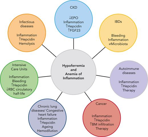
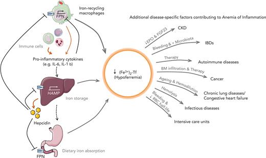
Therefore, a personalized approach based on knowledge of the underlying pathophysiological mechanisms, clinical features of the underlying disease, and diagnostic results is essential to determine the optimal therapeutic approach (summary in Figure 2). Recent scientific discoveries have paved the way for novel emerging therapies, including those targeting the hepcidin/FPN axis, hypoxia-induced pathways, or immune cell signaling cascades. We are urgently awaiting their clinical evaluation. These therapies must be evaluated for their capacity to overcome AI-driven macrophage iron retention and/or immune-mediated repression of erythropoiesis and how such effects will affect the course of the specific disease underlying AI.

Figure 2.

Leading pathophysiological mechanisms contributing to hypoferremia and AI in CKD, IBDs, autoimmune diseases, cancer, chronic lung diseases, CHF, infectious diseases,and ICUs.

Figure 2.

Leading pathophysiological mechanisms contributing to hypoferremia and AI in CKD, IBDs, autoimmune diseases, cancer, chronic lung diseases, CHF, infectious diseases,and ICUs.

Close modal

The authors thank Matthias W. Hentze for the critical reading of the manuscript.

M.U.M. acknowledges funding from the Deutsche Forschungsgemeinschaft (SFB1118; FerrOs-FOR5146) and the Federal Ministry of Education and Research (NephrESA Nr 031L0191C; DZL TLRC-H). O.M. received support from the European Hematology Association (RG66) and by an Olympia Morata-Programme Fellowship given by the Medical Faculty of the University of Heidelberg. G.W. was supported by the Christian Doppler Society.

Contribution: O.M. and M.U.M. were responsible for conception and design; and O.M., G.W., and M.U.M. wrote the manuscript and approved its final version.

Conflict-of-interest disclosure: The authors declare no competing financial interests.

Correspondence: Martina U. Muckenthaler, Im Neuenheimer Feld 350, 69120 Heidelberg, Germany; e-mail: martina.muckenthaler@med.uni-heidelberg.de.

1.
McLean
E
,
Cogswell
M
,
Egli
I
,
Wojdyla
D
,
de Benoist
B
.
Worldwide prevalence of anaemia, WHO Vitamin and Mineral Nutrition Information System, 1993-2005
.
Publ Health Nutr
.
2009
;
12
(
4
):
444
-
454
.
2.
James
SL
,
Abate
D
,
Abate
KH
, et al
.
Global, regional, and national incidence, prevalence, and years lived with disability for 354 diseases and injuries for 195 countries and territories, 1990-2017: a systematic analysis for the Global Burden of Disease Study 2017
.
Lancet
.
2018
;
392
(
10159
):
1789
-
1858
.
3.
Weiss
G
,
Ganz
T
,
Goodnough
LT
.
Anemia of inflammation
.
Blood
.
2019
;
133
(
1
):
40
-
50
.
4.
Camaschella
C
.
Iron deficiency
.
Blood
.
2019
;
133
(
1
):
30
-
39
.
5.
Goodnough
LT
,
Nemeth
E
,
Ganz
T
.
Detection, evaluation, and management of iron-restricted erythropoiesis
.
Blood
.
2010
;
116
(
23
):
4754
-
4761
.
6.
Weiss
G
.
Anemia of chronic disorders: new diagnostic tools and new treatment strategies
.
Semin Hematol
.
2015
;
52
(
4
):
313
-
320
.
7.
Gassmann
M
,
Mairbäurl
H
,
Livshits
L
, et al
.
The increase in hemoglobin concentration with altitude varies among human populations
.
Ann N Y Acad Sci
.
2019
;
1450
(
1
):
204
-
220
.
8.
Skikne
BS
,
Flowers
CH
,
Cook
JD
.
Serum transferrin receptor: a quantitative measure of tissue iron deficiency
.
Blood
.
1990
;
75
(
9
):
1870
-
1876
.
9.
Theurl
I
,
Mattle
V
,
Seifert
M
,
Mariani
M
,
Marth
C
,
Weiss
G
.
Dysregulated monocyte iron homeostasis and erythropoietin formation in patients with anemia of chronic disease
.
Blood
.
2006
;
107
(
10
):
4142
-
4148
.
10.
Nairz
M
,
Theurl
I
,
Wolf
D
,
Weiss
G
.
Iron deficiency or anemia of inflammation? : differential diagnosis and mechanisms of anemia of inflammation
.
Wien Med Wochenschr
.
2016
;
166
(
13-14
):
411
-
423
.
11.
Cappellini
MD
,
Comin-Colet
J
,
de Francisco
A
, et al
.
Iron deficiency across chronic inflammatory conditions: international expert opinion on definition, diagnosis, and management
.
Am J Hematol
.
2017
;
92
(
10
):
1068
-
1078
.
12.
Muckenthaler
MU
,
Rivella
S
,
Hentze
MW
,
Galy
B
.
A red carpet for iron metabolism
.
Cell
.
2017
;
168
(
3
):
344
-
361
.
13.
Nemeth
E
,
Rivera
S
,
Gabayan
V
, et al
.
IL-6 mediates hypoferremia of inflammation by inducing the synthesis of the iron regulatory hormone hepcidin
.
J Clin Invest
.
2004
;
113
(
9
):
1271
-
1276
.
14.
Weinberg
ED
.
Nutritional immunity: host’s attempt to withhold iron from microbial invaders
.
JAMA
.
1975
;
231
(
1
):
39
-
41
.
15.
Lim
D
,
Kim
KS
,
Jeong
J-H
, et al
.
The hepcidin-ferroportin axis controls the iron content of Salmonella-containing vacuoles in macrophages
.
Nat Commun
.
2018
;
9
(
1
):
2091
.
16.
Nairz
M
,
Metzendorf
C
,
Vujic-Spasic
M
, et al
.
Cell-specific expression of Hfe determines the outcome of Salmonella enterica serovar Typhimurium infection in mice
.
Haematologica
.
2021
;
106
(
12
):
3149
-
3161
.
17.
Arezes
J
,
Jung
G
,
Gabayan
V
, et al
.
Hepcidin-induced hypoferremia is a critical host defense mechanism against the siderophilic bacterium Vibrio vulnificus
.
Cell Host Microbe
.
2015
;
17
(
1
):
47
-
57
.
18.
Stefanova
D
,
Raychev
A
,
Arezes
J
, et al
.
Endogenous hepcidin and its agonist mediate resistance to selected infections by clearing non–transferrin-bound iron
.
Blood
.
2017
;
130
(
3
):
245
-
257
.
19.
Hoffmann
A
,
Haschka
D
,
Valente de Souza
L
, et al
.
Baseline iron status and presence of anaemia determine the course of systemic Salmonella infection following oral iron supplementation in mice
.
EBioMedicine
.
2021
;
71
:
103568
.
20.
Mwangi
MN
,
Phiri
KS
,
Abkari
A
, et al
.
Iron for Africa-report of an expert workshop
.
Nutrients
.
2017
;
9
(
6
):
576
.
21.
Sazawal
S
,
Black
RE
,
Ramsan
M
, et al
.
Effects of routine prophylactic supplementation with iron and folic acid on admission to hospital and mortality in preschool children in a high malaria transmission setting: community-based, randomised, placebo-controlled trial
.
Lancet
.
2006
;
367
(
9505
):
133
-
143
.
22.
Malan
L
,
Baumgartner
J
,
Calder
PC
,
Zimmermann
MB
,
Smuts
CM
.
n-3 long-chain PUFAs reduce respiratory morbidity caused by iron supplementation in iron-deficient South African schoolchildren: a randomized, double-blind, placebo-controlled intervention
.
Am J Clin Nutr
.
2015
;
101
(
3
):
668
-
679
.
23.
Soofi
S
,
Cousens
S
,
Iqbal
SP
, et al
.
Effect of provision of daily zinc and iron with several micronutrients on growth and morbidity among young children in Pakistan: a cluster-randomised trial
.
Lancet
.
2013
;
382
(
9886
):
29
-
40
.
24.
Zimmermann
MB
,
Chassard
C
,
Rohner
F
, et al
.
The effects of iron fortification on the gut microbiota in African children: a randomized controlled trial in Côte d'Ivoire
.
AJCN (Am J Clin Nutr)
.
2010
;
92
(
6
):
1406
-
1415
.
25.
Esan
MO
,
van Hensbroek
MB
,
Nkhoma
E
, et al
.
Iron supplementation in HIV-infected Malawian children with anemia: a double-blind, randomized, controlled trial
.
Clin Infect Dis
.
2013
;
57
(
11
):
1626
-
1634
.
26.
WHO Guidelines Approved by the Guidelines Review Committee
. Guideline: Daily Iron Supplementation in Infants and Children.
World Health Organization
;
2016
.
27.
WHO Guidelines Approved by the Guidelines Review Committee
. Guideline: Daily Iron Supplementation in Adult Women and Adolescent Girls.
World Health Organization
;
2016
.
28.
WHO Guidelines Approved by the Guidelines Review Committee
. Guideline: Daily Iron Supplementation in Adult Women and Adolescent Girls.
World Health Organization
;
2016
.
29.
Billesbølle
CB
,
Azumaya
CM
,
Kretsch
RC
, et al
.
Structure of hepcidin-bound ferroportin reveals iron homeostatic mechanisms
.
Nature
.
2020
;
586
(
7831
):
807
-
811
.
30.
Nemeth
E
,
Tuttle
MS
,
Powelson
J
, et al
.
Hepcidin regulates cellular iron efflux by binding to ferroportin and inducing its internalization
.
Science
.
2004
;
306
(
5704
):
2090
-
2093
.
31.
Verga Falzacappa
MV
,
Vujic Spasic
M
,
Kessler
R
,
Stolte
J
,
Hentze
MW
,
Muckenthaler
MU
.
STAT3 mediates hepatic hepcidin expression and its inflammatory stimulation
.
Blood
.
2007
;
109
(
1
):
353
-
358
.
32.
Wrighting
DM
,
Andrews
NC
.
Interleukin-6 induces hepcidin expression through STAT3
.
Blood
.
2006
;
108
(
9
):
3204
-
3209
.
33.
Gardenghi
S
,
Renaud
TM
,
Meloni
A
, et al
.
Distinct roles for hepcidin and interleukin-6 in the recovery from anemia in mice injected with heat-killed Brucella abortus
.
Blood
.
2014
;
123
(
8
):
1137
-
1145
.
34.
Bryan
JM
,
Jacqueline
ML
,
Olivier
DP
, et al
.
Investigation of the role of interleukin-6 and hepcidin antimicrobial peptide in the development of anemia with age
.
Haematologica
.
2013
;
98
(
10
):
1633
-
1640
.
35.
Armitage
AE
,
Eddowes
LA
,
Gileadi
U
, et al
.
Hepcidin regulation by innate immune and infectious stimuli
.
Blood
.
2011
;
118
(
15
):
4129
-
4139
.
36.
Nairz
M
,
Schroll
A
,
Haschka
D
, et al
.
Lipocalin-2 ensures host defense against Salmonella typhimurium by controlling macrophage iron homeostasis and immune response
.
Eur J Immunol
.
2015
;
45
(
11
):
3073
-
3086
.
37.
Lee
P
,
Peng
H
,
Gelbart
T
,
Wang
L
,
Beutler
E
.
Regulation of hepcidin transcription by interleukin-1 and interleukin-6
.
Proc Natl Acad Sci U S A
.
2005
;
102
(
6
):
1906
-
1910
.
38.
Shanmugam
NK
,
Chen
K
,
Cherayil
BJ
.
Commensal bacteria-induced interleukin 1β (IL-1β) secreted by macrophages up-regulates hepcidin expression in hepatocytes by activating the bone morphogenetic protein signaling pathway
.
J Biol Chem
.
2015
;
290
(
51
):
30637
-
30647
.
39.
Tilg
H
,
Ulmer
H
,
Kaser
A
,
Weiss
G
.
Role of IL-10 for induction of anemia during inflammation
.
J Immunol
.
2002
;
169
(
4
):
2204
-
2209
.
40.
Lee
YS
,
Kim
YH
,
Jung
YS
, et al
.
Hepatocyte toll-like receptor 4 mediates lipopolysaccharide-induced hepcidin expression
.
Exp Mol Med
.
2017
;
49
(
12
):
e408
.
41.
Peyssonnaux
C
,
Zinkernagel
AS
,
Datta
V
,
Lauth
X
,
Johnson
RS
,
Nizet
V
.
TLR4-dependent hepcidin expression by myeloid cells in response to bacterial pathogens
.
Blood
.
2006
;
107
(
9
):
3727
-
3732
.
42.
Verga Falzacappa
MV
,
Casanovas
G
,
Hentze
MW
,
Muckenthaler
MU
.
A bone morphogenetic protein (BMP)-responsive element in the hepcidin promoter controls HFE2-mediated hepatic hepcidin expression and its response to IL-6 in cultured cells
.
J Mol Med (Berl)
.
2008
;
86
(
5
):
531
-
540
.
43.
Casanovas
G
,
Mleczko-Sanecka
K
,
Altamura
S
,
Hentze
MW
,
Muckenthaler
MU
.
Bone morphogenetic protein (BMP)-responsive elements located in the proximal and distal hepcidin promoter are critical for its response to HJV/BMP/SMAD
.
J Mol Med (Berl)
.
2009
;
87
(
5
):
471
-
480
.
44.
Matak
P
,
Chaston
TB
,
Chung
B
,
Srai
SK
,
McKie
AT
,
Sharp
PA
.
Activated macrophages induce hepcidin expression in HuH7 hepatoma cells
.
Haematologica
.
2009
;
94
(
6
):
773
-
780
.
45.
Fillebeen
C
,
Wilkinson
N
,
Charlebois
E
,
Katsarou
A
,
Wagner
J
,
Pantopoulos
K
.
Hepcidin-mediated hypoferremic response to acute inflammation requires a threshold of Bmp6/Hjv/Smad signaling
.
Blood
.
2018
;
132
(
17
):
1829
-
1841
.
46.
Roy
CN
,
Custodio
AO
,
de Graaf
J
, et al
.
An Hfe-dependent pathway mediates hyposideremia in response to lipopolysaccharide-induced inflammation in mice
.
Nat Genet
.
2004
;
36
(
5
):
481
-
485
.
47.
Theurl
I
,
Theurl
M
,
Seifert
M
, et al
.
Autocrine formation of hepcidin induces iron retention in human monocytes
.
Blood
.
2008
;
111
(
4
):
2392
-
2399
.
48.
Krause
A
,
Neitz
S
,
Mägert
HJ
, et al
.
LEAP-1, a novel highly disulfide-bonded human peptide, exhibits antimicrobial activity
.
FEBS Lett
.
2000
;
480
(
2-3
):
147
-
150
.
49.
Park
CH
,
Valore
EV
,
Waring
AJ
,
Ganz
T
.
Hepcidin, a urinary antimicrobial peptide synthesized in the liver
.
J Biol Chem
.
2001
;
276
(
11
):
7806
-
7810
.
50.
Link
C
,
Knopf
JD
,
Marques
O
,
Lemberg
MK
,
Muckenthaler
MU
.
The role of cellular iron deficiency in controlling iron export
.
Biochim Biophys Acta Gen Subj
.
2021
;
1865
(
3
):
129829
.
51.
Qiao
B
,
Sugianto
P
,
Fung
E
, et al
.
Hepcidin-induced endocytosis of ferroportin is dependent on ferroportin ubiquitination
.
Cell Metabol
.
2012
;
15
(
6
):
918
-
924
.
52.
Traeger
L
,
Wiegand
SB
,
Sauer
AJ
, et al
.
UBA6 and NDFIP1 regulate the degradation of ferroportin
.
Haematologica
.
2021
;
107
(
2
):
478
-
488
.
53.
Jiang
L
,
Wang
J
,
Wang
K
, et al
.
RNF217 regulates iron homeostasis through its E3 ubiquitin ligase activity by modulating ferroportin degradation
.
Blood
.
2021
;
138
(
8
):
689
-
705
.
54.
Guida
C
,
Altamura
S
,
Klein
FA
, et al
.
A novel inflammatory pathway mediating rapid hepcidin-independent hypoferremia
.
Blood
.
2015
;
125
(
14
):
2265
-
2275
.
55.
Liu
XB
,
Nguyen
NB
,
Marquess
KD
,
Yang
F
,
Haile
DJ
.
Regulation of hepcidin and ferroportin expression by lipopolysaccharide in splenic macrophages
.
Blood Cells Mol Dis
.
2005
;
35
(
1
):
47
-
56
.
56.
Naz
N
,
Malik
IA
,
Sheikh
N
, et al
.
Ferroportin-1 is a ‘nuclear’-negative acute-phase protein in rat liver: a comparison with other iron-transport proteins
.
Lab Invest
.
2012
;
92
(
6
):
842
-
856
.
57.
Laftah Abas
H
,
Sharma
N
,
Brookes Matthew
J
, et al
.
Tumour necrosis factor α causes hypoferraemia and reduced intestinal iron absorption in mice
.
Biochem J
.
2006
;
397
(
1
):
61
-
67
.
58.
Ludwiczek
S
,
Aigner
E
,
Theurl
I
,
Weiss
G
.
Cytokine-mediated regulation of iron transport in human monocytic cells
.
Blood
.
2003
;
101
(
10
):
4148
-
4154
.
59.
Kapurniotu
A
,
Gokce
O
,
Bernhagen
J
.
The multitasking potential of alarmins and atypical chemokines
.
Front Med
.
2019
;
6
:
3
.
60.
Lam
LKM
,
Murphy
S
,
Kokkinaki
D
, et al
.
DNA binding to TLR9 expressed by red blood cells promotes innate immune activation and anemia
.
Sci Transl Med
.
2021
;
13
(
616
):
eabj1008
.
61.
Akilesh
HM
,
Buechler
MB
,
Duggan
JM
, et al
.
Chronic TLR7 and TLR9 signaling drives anemia via differentiation of specialized hemophagocytes
.
Science
.
2019
;
363
(
6423
):
eaao5213
.
62.
Babitt
JL
,
Eisenga
MF
,
Haase
VH
, et al
.
Controversies in optimal anemia management: conclusions from a Kidney Disease: Improving Global Outcomes (KDIGO) Conference
.
Kidney Int
.
2021
;
99
(
6
):
1280
-
1295
.
63.
Begum
S
,
Latunde-Dada
GO
.
Anemia of inflammation with an emphasis on chronic kidney disease
.
Nutrients
.
2019
;
11
(
10
):
2424
.
64.
Ueda
N
,
Takasawa
K
.
Impact of inflammation on ferritin, hepcidin and the management of iron deficiency anemia in chronic kidney disease
.
Nutrients
.
2018
;
10
(
9
):
1173
.
65.
Inker
LA
,
Grams
ME
,
Levey
AS
, et al
.
Relationship of estimated GFR and albuminuria to concurrent laboratory abnormalities: an individual participant data meta-analysis in a global consortium
.
Am J Kidney Dis
.
2019
;
73
(
2
):
206
-
217
.
66.
Hamza
E
,
Metzinger
L
,
Metzinger-Le Meuth
V
.
Uremic toxins affect erythropoiesis during the course of chronic kidney disease: a review
.
Cells
.
2020
;
9
(
9
):
2039
.
67.
Kautz
L
,
Jung
G
,
Valore
EV
,
Rivella
S
,
Nemeth
E
,
Ganz
T
.
Identification of erythroferrone as an erythroid regulator of iron metabolism
.
Nat Genet
.
2014
;
46
(
7
):
678
-
684
.
68.
Honda
H
,
Kobayashi
Y
,
Onuma
S
, et al
.
Associations among erythroferrone and biomarkers of erythropoiesis and iron metabolism, and treatment with long-term erythropoiesis-stimulating agents in patients on hemodialysis
.
PLoS One
.
2016
;
11
(
3
):
e0151601
.
69.
Mark
RH
,
Maxime
R
,
Kristine
C
, et al
.
Levels of the erythropoietin-responsive hormone erythroferrone in mice and humans with chronic kidney disease
.
Haematologica
.
2018
;
103
(
4
):
e141
-
e142
.
70.
Spoto
B
,
Kakkar
R
,
Lo
L
, et al
.
Serum erythroferrone levels associate with mortality and cardiovascular events in hemodialysis and in CKD patients: a two cohorts study
.
J Clin Med
.
2019
;
8
(
4
):
523
.
71.
Liu
Q
,
Davidoff
O
,
Niss
K
,
Haase
VH
.
Hypoxia-inducible factor regulates hepcidin via erythropoietin-induced erythropoiesis
.
J Clin Invest
.
2012
;
122
(
12
):
4635
-
4644
.
72.
Piperno
A
,
Galimberti
S
,
Mariani
R
, et al
.
Modulation of hepcidin production during hypoxia-induced erythropoiesis in humans in vivo: data from the HIGHCARE project
.
Blood
.
2011
;
117
(
10
):
2953
-
2959
.
73.
Sonnweber
T
,
Nachbaur
D
,
Schroll
A
, et al
.
Hypoxia induced downregulation of hepcidin is mediated by platelet derived growth factor BB
.
Gut
.
2014
;
63
(
12
):
1951
-
1959
.
74.
Roth
MP
,
Meynard
D
,
Coppin
H
.
Regulators of hepcidin expression
.
Vitam Horm
.
2019
;
110
:
101
-
129
.
75.
Weiss
G
,
Theurl
I
,
Eder
S
, et al
.
Serum hepcidin concentration in chronic haemodialysis patients: associations and effects of dialysis, iron and erythropoietin therapy
.
Eur J Clin Invest
.
2009
;
39
(
10
):
883
-
890
.
76.
Valenti
L
,
Messa
P
,
Pelusi
S
,
Campostrini
N
,
Girelli
D
.
Hepcidin levels in chronic hemodialysis patients: a critical evaluation
.
Clin Chem Lab Med
.
2014
;
52
(
5
):
613
-
619
.
77.
Wish
JB
,
Aronoff
GR
,
Bacon
BR
, et al
.
Positive iron balance in chronic kidney disease: how much is too much and how to tell?
.
Am J Nephrol
.
2018
;
47
(
2
):
72
-
83
.
78.
Babitt
JL
,
Sitara
D
.
Crosstalk between fibroblast growth factor 23, iron, erythropoietin, and inflammation in kidney disease
.
Curr Opin Nephrol Hypertens
.
2019
;
28
(
4
):
304
-
310
.
79.
Czaya
B
,
Faul
C
.
The role of fibroblast growth factor 23 in inflammation and anemia
.
Int J Mol Sci
.
2019
;
20
(
17
):
4195
.
80.
Coe
LM
,
Madathil
SV
,
Casu
C
,
Lanske
B
,
Rivella
S
,
Sitara
D
.
FGF-23 is a negative regulator of prenatal and postnatal erythropoiesis
.
J Biol Chem
.
2014
;
289
(
14
):
9795
-
9810
.
81.
Agoro
R
,
Montagna
A
,
Goetz
R
, et al
.
Inhibition of fibroblast growth factor 23 (FGF23) signaling rescues renal anemia
.
Faseb J
.
2018
;
32
(
7
):
3752
-
3764
.
82.
Weir
MR
.
Managing anemia across the stages of kidney disease in those hyporesponsive to erythropoiesis-stimulating agents
.
Am J Nephrol
.
2021
;
52
(
6
):
450
-
466
.
83.
Yilmaz
MI
,
Solak
Y
,
Covic
A
,
Goldsmith
D
,
Kanbay
M
.
Renal anemia of inflammation: the name is self-explanatory
.
Blood Purif
.
2011
;
32
(
3
):
220
-
225
.
84.
Sturm
A
,
Maaser
C
,
Mendall
M
, et al
.
European Crohn’s and Colitis Organisation topical review on IBD in the elderly
.
J Crohns Colitis
.
2016
;
11
(
3
):
263
-
273
.
85.
Dignass
AU
,
Gasche
C
,
Bettenworth
D
, et al
.
European consensus on the diagnosis and management of iron deficiency and anaemia in inflammatory bowel diseases
.
J Crohns Colitis
.
2015
;
9
(
3
):
211
-
222
.
86.
Dibb
M
,
Subramanian
S
.
Anaemia in inflammatory bowel disease
.
Frontline Gastroenterol
.
2014
;
5
(
3
):
190
-
196
.
87.
Gasche
C
,
Lomer
MC
,
Cavill
I
,
Weiss
G
.
Iron, anaemia, and inflammatory bowel diseases
.
Gut
.
2004
;
53
(
8
):
1190
-
1197
.
88.
Campbell
EL
,
Bruyninckx
WJ
,
Kelly
CJ
, et al
.
Transmigrating neutrophils shape the mucosal microenvironment through localized oxygen depletion to influence resolution of inflammation
.
Immunity
.
2014
;
40
(
1
):
66
-
77
.
89.
Peyssonnaux
C
,
Datta
V
,
Cramer
T
, et al
.
HIF-1alpha expression regulates the bactericidal capacity of phagocytes
.
J Clin Invest
.
2005
;
115
(
7
):
1806
-
1815
.
90.
Mastrogiannaki
M
,
Matak
P
,
Keith
B
,
Simon
MC
,
Vaulont
S
,
Peyssonnaux
C
.
HIF-2alpha, but not HIF-1alpha, promotes iron absorption in mice
.
J Clin Invest
.
2009
;
119
(
5
):
1159
-
1166
.
91.
Shah
YM
,
Matsubara
T
,
Ito
S
,
Yim
S-H
,
Gonzalez
FJ
.
Intestinal hypoxia-inducible transcription factors are essential for iron absorption following iron deficiency
.
Cell Metabol
.
2009
;
9
(
2
):
152
-
164
.
92.
Taylor
M
,
Qu
A
,
Anderson
ER
, et al
.
Hypoxia-inducible factor-2α mediates the adaptive increase of intestinal ferroportin during iron deficiency in mice
.
Gastroenterology
.
2011
;
140
(
7
):
2044
-
2055
.
93.
Das
NK
,
Schwartz
AJ
,
Barthel
G
, et al
.
Microbial metabolite signaling is required for systemic iron homeostasis
.
Cell Metabol
.
2020
;
31
(
1
):
115
-
130.e116
.
94.
Bonovas
S
,
Fiorino
G
,
Allocca
M
, et al
.
Intravenous versus oral iron for the treatment of anemia in inflammatory bowel disease: a systematic review and meta-analysis of randomized controlled trials
.
Medicine (Baltim)
.
2016
;
95
(
2
):
e2308
.
95.
Lee
T
,
Clavel
T
,
Smirnov
K
, et al
.
Oral versus intravenous iron replacement therapy distinctly alters the gut microbiota and metabolome in patients with IBD
.
Gut
.
2017
;
66
(
5
):
863
-
871
.
96.
Mahalhal
A
,
Williams
JM
,
Johnson
S
, et al
.
Oral iron exacerbates colitis and influences the intestinal microbiome
.
PLoS One
.
2018
;
13
(
10
):
e0202460
.
97.
Richards
T
,
Breymann
C
,
Brookes
MJ
, et al
.
Questions and answers on iron deficiency treatment selection and the use of intravenous iron in routine clinical practice
.
Ann Med
.
2021
;
53
(
1
):
274
-
285
.
98.
Lee
KH
,
Ahn
BS
,
Cha
D
, et al
.
Understanding the immunopathogenesis of autoimmune diseases by animal studies using gene modulation: a comprehensive review
.
Autoimmun Rev
.
2020
;
19
(
3
):
102469
.
99.
Levine
AB
,
Erkan
D
.
Clinical assessment and management of cytopenias in lupus patients
.
Curr Rheumatol Rep
.
2011
;
13
(
4
):
291
-
299
.
100.
Wilson
A
,
Yu
HT
,
Goodnough
LT
,
Nissenson
AR
.
Prevalence and outcomes of anemia in rheumatoid arthritis: a systematic review of the literature
.
Am J Med
.
2004
;
116
(
suppl 7A
):
50s
-
57s
.
101.
Scholz
GA
,
Leichtle
AB
,
Scherer
A
, et al
.
The links of hepcidin and erythropoietin in the interplay of inflammation and iron deficiency in a large observational study of rheumatoid arthritis
.
Br J Haematol
.
2019
;
186
(
1
):
101
-
112
.
102.
Weiss
G
,
Schett
G
.
Anaemia in inflammatory rheumatic diseases
.
Nat Rev Rheumatol
.
2013
;
9
(
4
):
205
-
215
.
103.
Schett
G
,
McInnes
IB
,
Neurath
MF
.
Reframing immune-mediated inflammatory diseases through signature cytokine hubs
.
N Engl J Med
.
2021
;
385
(
7
):
628
-
639
.
104.
Sato
H
,
Takai
C
,
Kazama
JJ
, et al
.
Serum hepcidin level, iron metabolism and osteoporosis in patients with rheumatoid arthritis
.
Sci Rep
.
2020
;
10
(
1
):
9882
.
105.
Khalaf
W
,
Al-Rubaie
HA
,
Shihab
S
.
Studying anemia of chronic disease and iron deficiency in patients with rheumatoid arthritis by iron status and circulating hepcidin
.
Hematol Rep
.
2019
;
11
(
1
):
7708
.
106.
Abdel-Khalek
MA
,
El-Barbary
AM
,
Essa
SA
,
Ghobashi
AS
.
Serum hepcidin: a direct link between anemia of inflammation and coronary artery atherosclerosis in patients with rheumatoid arthritis
.
J Rheumatol
.
2011
;
38
(
10
):
2153
-
2159
.
107.
Wolfe
RM
,
Ang
DC
.
Biologic therapies for autoimmune and connective tissue diseases
.
Immunol Allergy Clin
.
2017
;
37
(
2
):
283
-
299
.
108.
Her
M
,
Kavanaugh
A
.
Alterations in immune function with biologic therapies for autoimmune disease
.
J Allergy Clin Immunol
.
2016
;
137
(
1
):
19
-
27
.
109.
De Souza
LV
,
Hoffmann
A
,
Fischer
C
, et al
.
Comparative analysis of oral and intravenous iron therapy in rat models of inflammatory anemia and iron deficiency
.
Haematologica
.
Published online 7 July 2022
.
110.
Gaspar
BL
,
Sharma
P
,
Das
R
.
Anemia in malignancies: pathogenetic and diagnostic considerations
.
Hematology
.
2015
;
20
(
1
):
18
-
25
.
111.
Bohlius
J
,
Bohlke
K
,
Castelli
R
, et al
.
Management of cancer-associated anemia with erythropoiesis-stimulating agents: ASCO/ASH clinical practice guideline update
.
Blood Adv
.
2019
;
3
(
8
):
1197
-
1210
.
112.
Macciò
A
,
Madeddu
C
,
Gramignano
G
, et al
.
The role of inflammation, iron, and nutritional status in cancer-related anemia: results of a large, prospective, observational study
.
Haematologica
.
2015
;
100
(
1
):
124
-
132
.
113.
Steinmetz
T
,
Totzke
U
,
Schweigert
M
, et al
.
A prospective observational study of anaemia management in cancer patients - results from the German Cancer Anaemia Registry
.
Eur J Cancer Care
.
2011
;
20
(
4
):
493
-
502
.
114.
Waters
JS
,
O’Brien
MER
,
Ashley
S
, et al
.
Management of anemia in patients receiving chemotherapy
.
J Clin Oncol
.
2002
;
20
(
2
):
601
-
603
.
115.
Vadhan-Raj
S
,
Abonour
R
,
Goldman
JW
, et al
.
A first-in-human phase 1 study of a hepcidin monoclonal antibody, LY2787106, in cancer-associated anemia
.
J Hematol Oncol
.
2017
;
10
(
1
):
73
.
116.
Cheng
Z
,
Yan
M
,
Lu
Y
,
Pan
XT
.
Expression of serum BMP6 and hepcidin in cancer-related anemia
.
Hematology
.
2020
;
25
(
1
):
134
-
138
.
117.
Busti
F
,
Marchi
G
,
Ugolini
S
,
Castagna
A
,
Girelli
D
.
Anemia and iron deficiency in cancer patients: role of iron replacement therapy
.
Pharmaceuticals
.
2018
;
11
(
4
):
94
.
118.
Brown
RAM
,
Richardson
KL
,
Kabir
TD
,
Trinder
D
,
Ganss
R
,
Leedman
PJ
.
Altered iron metabolism and impact in cancer biology, metastasis, and immunology
.
Front Oncol
.
2020
;
10
:
476
.
119.
Silverberg
DS
,
Mor
R
,
Weu
MT
,
Schwartz
D
,
Schwartz
IF
,
Chernin
G
.
Anemia and iron deficiency in COPD patients: prevalence and the effects of correction of the anemia with erythropoiesis stimulating agents and intravenous iron
.
BMC Pulm Med
.
2014
;
14
(
1
):
24
.
120.
von Drygalski
A
,
Biller
J
.
Anemia in cystic fibrosis (CF): prevalence, mechanisms and correlation with pulmonary function
.
Blood
.
2006
;
108
(
11
):
1286
.
121.
Rhodes
CJ
,
Wharton
J
,
Howard
L
,
Gibbs
JS
,
Vonk-Noordegraaf
A
,
Wilkins
MR
.
Iron deficiency in pulmonary arterial hypertension: a potential therapeutic target
.
Eur Respir J
.
2011
;
38
(
6
):
1453
-
1460
.
122.
Sonnweber
T
,
Pizzini
A
,
Tancevski
I
,
Löffler-Ragg
J
,
Weiss
G
.
Anaemia, iron homeostasis and pulmonary hypertension: a review
.
Internal and Emergency Medicine
.
2020
;
15
(
4
):
573
-
585
.
123.
Xanthouli
P
,
Theobald
V
,
Benjamin
N
, et al
.
Prognostic impact of hypochromic erythrocytes in patients with pulmonary arterial hypertension
.
Respir Res
.
2021
;
22
(
1
):
288
.
124.
Tilea
I
,
Petra
DN
,
Serban
RC
, et al
.
Short term impact of iron deficiency in different subsets of patients with precapillary pulmonary hypertension from an Eastern European Pulmonary Hypertension Referral Center
.
Int J Gen Med
.
2021
;
14
:
3355
-
3366
.
125.
Fischer
BM
,
Pavlisko
E
,
Voynow
JA
.
Pathogenic triad in COPD: oxidative stress, protease-antiprotease imbalance, and inflammation
.
Int J Chronic Obstr Pulm Dis
.
2011
;
6
:
413
-
421
.
126.
Sarkar
M
,
Rajta
PN
,
Khatana
J
.
Anemia in chronic obstructive pulmonary disease: prevalence, pathogenesis, and potential impact
.
Lung India
.
2015
;
32
(
2
):
142
-
151
.
127.
Balasubramanian
A
,
Henderson
RJ
,
Putcha
N
, et al
.
Haemoglobin as a biomarker for clinical outcomes in chronic obstructive pulmonary disease
.
ERJ Open Res
.
2021
;
7
(
3
):
00068-2021
-
02021
.
128.
Pizzini
A
,
Aichner
M
,
Sonnweber
T
,
Tancevski
I
,
Weiss
G
,
Löffler-Ragg
J
.
The significance of iron deficiency and anemia in a real-life COPD cohort
.
Int J Med Sci
.
2020
;
17
(
14
):
2232
-
2239
.
129.
Shah
YM
,
Xie
L
.
Hypoxia-inducible factors link iron homeostasis and erythropoiesis
.
Gastroenterology
.
2014
;
146
(
3
):
630
-
642
.
130.
Vlahakos
V
,
Marathias
K
,
Lionaki
S
,
Loukides
S
,
Zakynthinos
S
,
Vlahakos
D
.
The paradigm shift from polycythemia to anemia in COPD: the critical role of the renin-angiotensin system inhibitors
.
Expert Rev Respir Med
.
2022
;
16
(
4
):
391
-
398
.
131.
Kent
BD
,
Mitchell
PD
,
McNicholas
WT
.
Hypoxemia in patients with COPD: cause, effects, and disease progression
.
Int J Chronic Obstr Pulm Dis
.
2011
;
6
:
199
-
208
.
132.
Fabbri
LM
,
Rabe
KF
.
From COPD to chronic systemic inflammatory syndrome?
.
Lancet
.
2007
;
370
(
9589
):
797
-
799
.
133.
Markoulaki
D
,
Kostikas
K
,
Papatheodorou
G
, et al
.
Hemoglobin, erythropoietin and systemic inflammation in exacerbations of chronic obstructive pulmonary disease
.
Eur J Intern Med
.
2011
;
22
(
1
):
103
-
107
.
134.
John
M
,
Hoernig
S
,
Doehner
W
,
Okonko
DD
,
Witt
C
,
Anker
SD
.
Anemia and inflammation in COPD
.
Chest
.
2005
;
127
(
3
):
825
-
829
.
135.
Tandara
L
,
Grubisic
TZ
,
Ivan
G
, et al
.
Systemic inflammation up-regulates serum hepcidin in exacerbations and stabile chronic obstructive pulmonary disease
.
Clin Biochem
.
2015
;
48
(
18
):
1252
-
1257
.
136.
Hanneke
JCMW
,
Melanie
MvdK
,
Theo de
W
, et al
.
Association of anemia with health-related quality of life and survival: a large population-based cohort study
.
Haematologica
.
2019
;
104
(
3
):
468
-
476
.
137.
Girelli
D
,
Marchi
G
,
Camaschella
C
.
Anemia in the elderly
.
Hemasphere
.
2018
;
2
(
3
):
e40
.
138.
Girelli
D
,
Busti
F
.
Anemia and adverse outcomes in the elderly: a detrimental inflammatory loop?
.
Haematologica
.
2019
;
104
(
3
):
417
-
419
.
139.
Lanser
L
,
Fuchs
D
,
Kurz
K
,
Weiss
G
.
Physiology and inflammation driven pathophysiology of iron homeostasis-mechanistic insights into anemia of inflammation and its treatment
.
Nutrients
.
2021
;
13
(
11
):
3732
.
140.
Boutou
AK
,
Pitsiou
GG
,
Stanopoulos
I
,
Kontakiotis
T
,
Kyriazis
G
,
Argyropoulou
P
.
Levels of inflammatory mediators in chronic obstructive pulmonary disease patients with anemia of chronic disease: a case-control study
.
QJM
.
2012
;
105
(
7
):
657
-
663
.
141.
Anand
IS
,
Gupta
P
.
Anemia and iron deficiency in heart failure: current concepts and emerging therapies
.
Circulation
.
2018
;
138
(
1
):
80
-
98
.
142.
Niethammer
M
,
Sieber
M
,
von Haehling
S
, et al
.
Inflammatory pathways in patients with heart failure and preserved ejection fraction
.
Int J Cardiol
.
2008
;
129
(
1
):
111
-
117
.
143.
Jankowska
EA
,
Malyszko
J
,
Ardehali
H
, et al
.
Iron status in patients with chronic heart failure
.
Eur Heart J
.
2012
;
34
(
11
):
827
-
834
.
144.
von Haehling
S
,
Ebner
N
,
Evertz
R
,
Ponikowski
P
,
Anker
SD
.
Iron deficiency in heart failure: an overview
.
JACC Heart Fail
.
2019
;
7
(
1
):
36
-
46
.
145.
Ponikowski
P
,
Voors
AA
,
Anker
SD
, et al
.
2016 ESC Guidelines for the diagnosis and treatment of acute and chronic heart failure: the Task Force for the diagnosis and treatment of acute and chronic heart failure of the European Society of Cardiology (ESC) developed with the special contribution of the Heart Failure Association (HFA) of the ESC
.
Eur Heart J
.
2016
;
37
(
27
):
2129
-
2200
.
146.
Safiri
S
,
Kolahi
A-A
,
Noori
M
, et al
.
Burden of anemia and its underlying causes in 204 countries and territories, 1990-2019: results from the Global Burden of Disease Study 2019
.
J Hematol Oncol
.
2021
;
14
(
1
):
185
.
147.
Valente de Souza
L
,
Hoffmann
A
,
Weiss
G
.
Impact of bacterial infections on erythropoiesis
.
Expert Rev Anti Infect Ther
.
2021
;
19
(
5
):
619
-
633
.
148.
Zuin
M
,
Rigatelli
G
,
Quadretti
L
,
Fogato
L
,
Zuliani
G
,
Roncon
L
.
Prognostic role of anemia in COVID-19 patients: a meta-analysis
.
Infect Dis Rep
.
2021
;
13
(
4
):
930
-
937
.
149.
Bellmann-Weiler
R
,
Lanser
L
,
Barket
R
, et al
.
Prevalence and predictive value of anemia and dysregulated iron homeostasis in patients with COVID-19 infection
.
J Clin Med
.
2020
;
9
(
8
):
2429
.
150.
Wang
C
,
Deng
R
,
Gou
L
, et al
.
Preliminary study to identify severe from moderate cases of COVID-19 using combined hematology parameters
.
Ann Transl Med
.
2020
;
8
(
9
):
593
.
151.
Taneri
PE
,
Gómez-Ochoa
SA
,
Llanaj
E
, et al
.
Anemia and iron metabolism in COVID-19: a systematic review and meta-analysis
.
Eur J Epidemiol
.
2020
;
35
(
8
):
763
-
773
.
152.
Lanser
L
,
Burkert
FR
,
Bellmann-Weiler
R
, et al
.
Dynamics in anemia development and dysregulation of iron homeostasis in hospitalized patients with COVID-19
.
Metabolites
.
2021
;
11
(
10
):
653
.
153.
Bergamaschi
G
,
Borrelli de Andreis
F
,
Aronico
N
, et al
.
Anemia in patients with Covid-19: pathogenesis and clinical significance
.
Clin Exp Med
.
2021
;
21
(
2
):
239
-
246
.
154.
Hippchen
T
,
Altamura
S
,
Muckenthaler
MU
,
Merle
U
.
Hypoferremia is associated with increased hospitalization and oxygen demand in COVID-19 patients
.
HemaSphere
.
2020
;
4
(
6
):
e492
.
155.
Cao
G
,
Wang
Y
,
Wu
Y
,
Jing
W
,
Liu
J
,
Liu
M
.
Prevalence of anemia among people living with HIV: a systematic review and meta-analysis
.
EClinicalMedicine
.
2022
;
44
:
101283
.
156.
Belperio
PS
,
Rhew
DC
.
Prevalence and outcomes of anemia in individuals with human immunodeficiency virus: a systematic review of the literature
.
Am J Med
.
2004
;
116
(
suppl 7A
):
27s
-
43s
.
157.
Wisaksana
R
,
Sumantri
R
,
Indrati
AR
, et al
.
Anemia and iron homeostasis in a cohort of HIV-infected patients in Indonesia
.
BMC Infect Dis
.
2011
;
11
(
1
):
213
.
158.
Moore
RD
,
Keruly
J
,
Richman
DD
,
Creagh-Kirk
T
,
Chaisson
RE
.
Natural history of advanced HIV disease in patients treated with zidovudine
.
The Zidovudine Epidemiology Study Group. Aids
.
1992
;
6
(
7
):
671
-
677
.
159.
Armitage
AE
,
Stacey
AR
,
Giannoulatou
E
, et al
.
Distinct patterns of hepcidin and iron regulation during HIV-1, HBV, and HCV infections
.
Proc Natl Acad Sci U S A
.
2014
;
111
(
33
):
12187
-
12192
.
160.
Xu
M
,
Kashanchi
F
,
Foster
A
, et al
.
Hepcidin induces HIV-1 transcription inhibited by ferroportin
.
Retrovirology
.
2010
;
7
(
1
):
104
.
161.
Quiros-Roldan
E
,
Castelli
F
,
Lanza
P
, et al
.
The impact of antiretroviral therapy on iron homeostasis and inflammation markers in HIV-infected patients with mild anemia
.
J Transl Med
.
2017
;
15
(
1
):
256
.
162.
Rawal
G
,
Kumar
R
,
Yadav
S
,
Singh
A
.
Anemia in intensive care: a review of current concepts
.
J Crit Care Med (Targu Mures)
.
2016
;
2
(
3
):
109
-
114
.
163.
Jiang
Y
,
Jiang
F-Q
,
Kong
F
, et al
.
Inflammatory anemia-associated parameters are related to 28-day mortality in patients with sepsis admitted to the ICU: a preliminary observational study
.
Ann Intensive Care
.
2019
;
9
(
1
):
67
.
164.
Hayden
SJ
,
Albert
TJ
,
Watkins
TR
,
Swenson
ER
.
Anemia in critical illness: insights into etiology, consequences, and management
.
Am J Respir Crit Care Med
.
2012
;
185
(
10
):
1049
-
1057
.
165.
McEvoy
MT
,
Shander
A
.
Anemia, bleeding, and blood transfusion in the intensive care unit: causes, risks, costs, and new strategies
.
Am J Crit Care
.
2013
;
22
(
suppl 6
):
eS1
-
eS13
. quiz eS14.
166.
Boshuizen
M
,
Binnekade
JM
,
Nota
B
, et al
.
Iron metabolism in critically ill patients developing anemia of inflammation: a case control study
.
Ann Intensive Care
.
2018
;
8
(
1
):
56
.
167.
Brandtner
A
,
Tymoszuk
P
,
Nairz
M
, et al
.
Linkage of alterations in systemic iron homeostasis to patients’ outcome in sepsis: a prospective study
.
J Intensive Care
.
2020
;
8
(
1
):
76
.
168.
Tacke
F
,
Nuraldeen
R
,
Koch
A
, et al
.
Iron parameters determine the prognosis of critically ill patients
.
Crit Care Med
.
2016
;
44
(
6
):
1049
-
1058
.
169.
Hawkins
RB
,
Raymond
SL
,
Stortz
JA
, et al
.
Chronic critical illness and the persistent inflammation, immunosuppression, and catabolism syndrome
.
Front Immunol
.
2018
;
9
:
1511
.
170.
Hayden
SJ
,
Albert
TJ
,
Watkins
TR
,
Swenson
ER
.
Anemia in critical illness: insights into etiology, consequences, and management
.
Am J Respir Crit Care Med
.
2012
;
185
(
10
):
1049
-
1057
.
171.
Lasocki
S
,
Pène
F
,
Ait-Oufella
H
, et al
.
Management and prevention of anemia (acute bleeding excluded) in adult critical care patients
.
Ann Intensive Care
.
2020
;
10
(
1
):
97
.
172.
Carson
JL
,
Stanworth
SJ
,
Dennis
JA
, et al
.
Transfusion thresholds for guiding red blood cell transfusion
.
Cochrane Database Syst Rev
.
2021
;
12
(
12
):
Cd002042
.
173.
Litton
E
,
Baker
S
,
Erber
WN
, et al
.
Intravenous iron or placebo for anaemia in intensive care: the IRONMAN multicentre randomized blinded trial: a randomized trial of IV iron in critical illness
.
Intensive Care Med
.
2016
;
42
(
11
):
1715
-
1722
.
174.
Shah
A
,
Roy
NB
,
McKechnie
S
,
Doree
C
,
Fisher
SA
,
Stanworth
SJ
.
Iron supplementation to treat anaemia in adult critical care patients: a systematic review and meta-analysis
.
Crit Care
.
2016
;
20
(
1
):
306
.
175.
Prakash
D
.
Anemia in the ICU: anemia of chronic disease versus anemia of acute illness
.
Crit Care Clin
.
2012
;
28
(
3
):
333
-
343
. v.
176.
Corwin
HL
,
Gettinger
A
,
Fabian
TC
, et al
.
Efficacy and safety of epoetin alfa in critically ill patients
.
N Engl J Med
.
2007
;
357
(
10
):
965
-
976
.
177.
Nairz
M
,
Schroll
A
,
Moschen
AR
, et al
.
Erythropoietin contrastingly affects bacterial infection and experimental colitis by inhibiting nuclear factor-κB-inducible immune pathways
.
Immunity
.
2011
;
34
(
1
):
61
-
74
.
178.
Lasocki
S
,
Asfar
P
,
Jaber
S
, et al
.
Impact of treating iron deficiency, diagnosed according to hepcidin quantification, on outcomes after a prolonged ICU stay compared to standard care: a multicenter, randomized, single-blinded trial
.
Crit Care
.
2021
;
25
(
1
):
62
.
179.
Pasricha
SR
,
Tye-Din
J
,
Muckenthaler
MU
,
Swinkels
DW
.
Iron deficiency
.
Lancet
.
2021
;
397
(
10270
):
233
-
248
.
180.
Webster
NR
.
Stranger danger-mortality after transfusions
.
Br J Anaesth
.
2017
;
118
(
3
):
280
-
282
.
181.
Villanueva
C
,
Colomo
A
,
Bosch
A
, et al
.
Transfusion strategies for acute upper gastrointestinal bleeding
.
N Engl J Med
.
2013
;
368
(
1
):
11
-
21
.
182.
Solomon
SD
,
Uno
H
,
Lewis
EF
, et al
.
Erythropoietic response and outcomes in kidney disease and type 2 diabetes
.
N Engl J Med
.
2010
;
363
(
12
):
1146
-
1155
.
183.
Bandach
I
,
Segev
Y
,
Landau
D
.
Experimental modulation of interleukin 1 shows its key role in chronic kidney disease progression and anemia
.
Sci Rep
.
2021
;
11
(
1
):
6288
.
184.
Petzer
V
,
Tymoszuk
P
,
Asshoff
M
, et al
.
A fully human anti-BMP6 antibody reduces the need for erythropoietin in rodent models of the anemia of chronic disease
.
Blood
.
2020
;
136
(
9
):
1080
-
1090
.
185.
Pergola
PE
,
Devalaraja
M
,
Fishbane
S
, et al
.
Ziltivekimab for treatment of anemia of inflammation in patients on hemodialysis: results from a phase 1/2 multicenter, randomized, double-blind, placebo-controlled trial
.
J Am Soc Nephrol
.
2021
;
32
(
1
):
211
-
222
.
186.
Renders
L
,
Budde
K
,
Rosenberger
C
, et al
.
First-in-human phase I studies of PRS-080#22, a hepcidin antagonist, in healthy volunteers and patients with chronic kidney disease undergoing hemodialysis
.
PLoS One
.
2019
;
14
(
3
):
e0212023
.
187.
Sheetz
M
,
Barrington
P
,
Callies
S
, et al
.
Targeting the hepcidin-ferroportin pathway in anaemia of chronic kidney disease
.
Br J Clin Pharmacol
.
2019
;
85
(
5
):
935
-
948
.
188.
Eckardt
KU
,
Agarwal
R
,
Aswad
A
, et al
.
Safety and efficacy of vadadustat for anemia in patients undergoing dialysis
.
N Engl J Med
.
2021
;
384
(
17
):
1601
-
1612
.
189.
Chen
N
,
Hao
C
,
Liu
BC
, et al
.
Roxadustat treatment for anemia in patients undergoing long-term dialysis
.
N Engl J Med
.
2019
;
381
(
11
):
1011
-
1022
.
190.
Singh
AK
,
Carroll
K
,
Perkovic
V
, et al
.
Daprodustat for the treatment of anemia in patients undergoing dialysis
.
N Engl J Med
.
2021
;
385
(
25
):
2325
-
2335
.
191.
Shu
W
,
Pang
Z
,
Xu
C
, et al
.
Anti-TNF-α monoclonal antibody therapy improves anemia through downregulating hepatocyte hepcidin expression in inflammatory bowel disease
.
Mediat Inflamm
.
2019
;
2019
:
4038619
.
192.
Cavallaro
F
,
Duca
L
,
Pisani
LF
, et al
.
Anti-TNF-mediated modulation of prohepcidin improves iron availability in inflammatory bowel disease, in an IL-6-mediated fashion
.
Chin J Gastroenterol Hepatol
.
2017
;
2017
:
6843976
.
193.
Bergamaschi
G
,
Di Sabatino
A
,
Albertini
R
, et al
.
Prevalence and pathogenesis of anemia in inflammatory bowel disease. Influence of anti-tumor necrosis factor-alpha treatment
.
Haematologica
.
2010
;
95
(
2
):
199
-
205
.
194.
Park
K-J
,
Jin
H-M
,
Cho
Y-N
, et al
.
Clinical and hematological effects of tocilizumab on serum hepcidin, anemia response and disease activity in patients with active rheumatoid arthritis
.
J Rheum Dis
.
2016
;
23
(
1
):
37
-
46
.
195.
Isaacs
JD
,
Harari
O
,
Kobold
U
,
Lee
JS
,
Bernasconi
C
.
Effect of tocilizumab on haematological markers implicates interleukin-6 signalling in the anaemia of rheumatoid arthritis
.
Arthritis Res Ther
.
2013
;
15
(
6
):
R204
.
196.
Macciò
A
,
Madeddu
C
,
Gramignano
G
,
Mulas
C
,
Sanna
E
,
Mantovani
G
.
Efficacy and safety of oral lactoferrin supplementation in combination with rHuEPO-beta for the treatment of anemia in advanced cancer patients undergoing chemotherapy: open-label, randomized controlled study
.
Oncol
.
2010
;
15
(
8
):
894
-
902
.
197.
Schuster
M
,
Rigas
JR
,
Orlov
SV
, et al
.
ALD518, a humanized anti-IL-6 antibody, treats anemia in patients with advanced non-small cell lung cancer (NSCLC): results of a phase II, randomized, double-blind, placebo-controlled trial
.
J Clin Oncol
.
2010
;
28
(
suppl 15
):
7631
.
198.
van Eijk
LT
,
John
AS
,
Schwoebel
F
, et al
.
Effect of the antihepcidin Spiegelmer lexaptepid on inflammation-induced decrease in serum iron in humans
.
Blood
.
2014
;
124
(
17
):
2643
-
2646
.
199.
Wish
JB
.
Debate: are hydroxylase inhibitors stabilizers a viable alternative to erythropoiesis-stimulating agents in the management of anemia in CKD? CON
.
Am J Nephrol
.
2022
;
53
(
5
):
366
-
371
.
200.
Singh
AK
.
Debate: are HIF stabilizers a viable alternative to ESAs in the management of anemia in CKD? PRO
.
Am J Nephrol
.
2022
;
53
(
5
):
361
-
365
.
Sign in via your Institution