Abstract
Juvenile myelomonocytic leukemia (JMML) is a deadly pediatric cancer characterized by splenomegaly, excessive accumulation of monocytes/macrophages and myelodysplastic features. Mutations in the RAS pathway are the major drivers, with NF1 loss-of-function (NF1LOF) mutations found in up to 20% of patients who experience particularly dismal outcomes. Neurofibromin1 (NF1) gene negatively regulates RAS/MAPK/PI3K pathways by converting active Ras-GTP to inactive Ras-GDP to control proliferation/differentiation of immature myeloid cells (Side et al., 1998). In JMML patients, NF1mutations results in either termination of protein translation or translation of truncated, non-functional protein that upregulates the RAS pathway. Hematopoietic stem cell transplantation is the only curative option, but relapse occurs in ~50% of patients. Therefore, there is an urgent need for novel therapeutic strategies. However, to date, clarifying the mechanism of disease development has been challenging due to low patients’ sample availability and lack of reliable humanized animal models which are the gold standard for studying rare diseases.
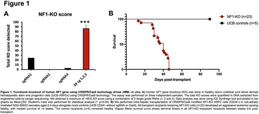
While there has been some success in developing transgenic (Nf1LOF) and xenograft (Ptpn11, Nras, and Kras mutated) murine models of JMML (Liu et al., 2016, Krombholz et al., 2016, Yoshimi et al., 2017), there is still a need for a reliable preclinical humanized model. We used CRISPR/Cas9 to create NF1LOF in healthy juvenile hematopoietic stem and progenitor cells (HSPCs). We knocked out (KO) the human NF1 gene in HSPCs from healthy donor umbilical cord blood (UCBs) by designing single guide RNAs targeting NF1 Exon 2 to mimic the loss of protein function found in cells of JMML patients. We consistently obtained KO scores of ~85% in modified UCB HSPCs (Fig.1A). We confirmed the loss of full-length NF1 protein and presence of a truncated protein in the modified cells by western blot. Additionally, we observed significant hypersensitivity of NF1-KO cells to GM-CSF in colony-forming unit assays in vitro, a unique characteristic feature of JMML patient cells (Emanuel et al., 1991).
Thereafter, we assessed the ability of NF1-KO HSPCs to initiate leukemia in humanized immunodeficient NSG-SGM3 neonates. We performed intrahepatic transplantation of the NF1LOF UCBs at post-natal days 2-4. We observed signs of leukemia (including weight loss, weakness, monocytosis in blood, anemia, enlarged spleen and liver) in all mice transplanted with modified NF1-KO cells, with a median survival of 5 weeks (Fig.1B). Significant splenomegaly was observed in all leukemic mice, along with the abrogation of lymphocyte production and an accumulation of myeloid cells (CD33+, CD13+, CD33+CD13+, CD14+, CD16+ and CD14+CD16+) and NK cells (CD56+/BrightCD16+) in the bone marrow and spleen of all leukemic mice, as observed by flow cytometry. This phenotype is consistent with that observed in patient-derived xenograft models of JMML (Krombholz et al., 2016). Extensive leukemia infiltration was also observed in liver and lungs, similar to what is observed in JMML patients. We are currently assessing the long-term leukemia initiation potential of CRISPR-modified NF1-KO cells through secondary transplants.
In conclusion, we have successfully developed a novel humanized mouse model of JMML carrying NF1LOF mutation using CRISPR/Cas9 genome editing technology, with features (splenomegaly, monocytosis, and GM-CSF hypersensitivity) that have been reported as unique characteristics of this disease. This provides a new tool to investigate mechanisms of disease development, epigenetic dysregulation pathways, and to ultimately identify potential targets and test therapeutic interventions.
Disclosures
No relevant conflicts of interest to declare.
Author notes
Asterisk with author names denotes non-ASH members.


