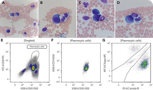
A 48-year-old man, originally from Kenya, presented with fever and fatigue and was found to have marked anemia (hemoglobin 7.1 g/dL), moderate thrombocytopenia (83 × 109/L), hepatosplenomegaly, and diffuse lymphadenopathy. Upon lymph node biopsy, human herpesvirus 8 (HHV8)-associated multicentric Castleman disease (MCD) and multifocal Kaposi sarcoma (KS) were diagnosed. Notably, immunostains on the lymph node highlighted numerous HHV8+ λ-restricted plasmablastic cells within mantle zones. Furthermore, markedly high HHV8 viral load was detected in plasma, HIV serology was negative, and Epstein-Barr virus DNA was undetectable. Numerous cytokines were elevated including interleukin-6 (IL-6) and IL-10. Serum protein electrophoresis and immunofixation showed polyclonal gammopathy including monoclonal IgM λ. Peripheral blood morphologic evaluation showed atypical large cells with round or ovoid nuclei with nucleoli and abundant deep-blue cytoplasm with occasional vacuoles (panels A-D: Wright-Giemsa stain; panels A and B-D: 20× and 100× objectives, respectively). Concurrent flow cytometry showed 1% λ-monotypic cells expressing CD19 (dim), CD38, CD45 (dim), CD138 (partial, predominantly negative) while lacking CD20 and CD56 (panels E-G; FITC, fluorescein isothiocyanate; IC, intracytoplasmic; PE, phycoerythrin). The morphologic and immunophenotypic findings were consistent with circulating plasmablastic cells/HHV-8-infected viroblasts.
This case illustrates a very rare presentation of HHV8-associated MCD and KS in an HIV-negative individual with circulating plasmablastic cells/viroblasts during active disease. These cells might mimic plasmablastic leukemia or lymphoma, underscoring the importance of a thorough and detailed correlation with clinicopathologic and laboratory findings.
For additional images, visit the ASH Image Bank, a reference and teaching tool that is continually updated with new atlas and case study images. For more information, visit https://imagebank.hematology.org.


