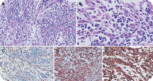
A 6-year-old boy with 22q11.2 distal microdeletion syndrome (characterized by telomeric microdeletions in chromosome 22 encompassing INI1/SMARCB1 gene and associated with a high incidence of malignant rhabdoid tumors) presented with back pain and vertebral lesions. Vertebral biopsy (panel A; original magnification ×400; hematoxylin and eosin [H&E] stain) showed small to medium cells with compact chromatin with characteristic rhabdoid morphology (abundant eosinophilic cytoplasm and eccentric nuclei) in a subset (panel B; original magnification ×600; H&E stain) and loss of INI1 expression (panel C; original magnification ×400; INI1 immunohistochemistry [IHC] stain). The neoplastic cells were positive for CD5 (panel D; original magnification ×400; CD5 IHC stain), CD3 (panel E; original magnification ×400; CD3 IHC stain), CD2, CD4 (partial weak), CD45, and CD43, and they were negative for CD7, CD8, CD30, anaplastic lymphoma kinase, CD56, CD57, Epstein-Barr encoding region in situ hybridization, and keratin. Ki-67 showed a high proliferation rate. T-cell receptor-γ polymerase chain reaction showed a clonal rearrangement pattern. At 13-month follow-up, the patient was in complete remission after cyclophosphamide, doxorubicin, vincristine, and prednisone therapy, followed by allogeneic hematopoietic stem cell transplant.
This novel peripheral T-cell lymphoma, not otherwise specified, exhibited rhabdoid histomorphology with loss of INI1 expression. Malignant rhabdoid tumors (characterized by rhabdoid cells, loss of INI1 expression, and biallelic inactivation of the INI1 gene) are described in the central nervous system, kidney, and soft tissue. Limited data exist regarding hematological neoplasms associated with biallelic loss of the INI1 gene. This case raises awareness of this rare entity and the utility of INI1 immunohistochemistry to facilitate its diagnosis.
For additional images, visit the ASH Image Bank, a reference and teaching tool that is continually updated with new atlas and case study images. For more information, visit https://imagebank.hematology.org.


