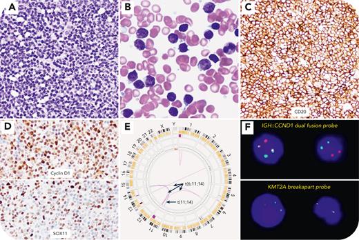
A 75-year-old man presented with widespread lymphadenopathy above and below the diaphragm. A left axillary lymph node biopsy revealed classic mantle cell lymphoma (MCL). The bone marrow (BM) was nearly completely replaced by small lymphoid cells (panel A, hematoxylin and eosin stain, original magnification ×500), which comprised 92% of the differential (panel B, Giemsa stain, original magnification ×1000). Flow cytometry showed 86% λ-restricted B cells that expressed CD5, CD19, CD20, CD38, CD43, and ROR1 and that were negative for CD10, CD11c, CD23, and CD200. No increase in blasts or monocytic cells were seen. Immunohistochemistry confirmed CD20 (panel C, original magnification ×500), cyclin D1, and SOX11 expression (panel D, original magnification ×500), consistent with MCL. Karyotyping showed 45,X,-Y[4]/45,idem,del(11)(q22q24)[15]/46,XY[1]. Optical genome mapping identified t(11;14)(q13;q32)/CCND1::IGH, t(6;11;14)(q21;q23.2;q31.3) that involved the KMT2A gene (potential fusion partners: SLC22A16 and FOXN3 on chromosomes 6 and 14, respectively) and gain of 3q13.33q29 (panel E). Fluorescence in situ hybridization confirmed CCND1::IGH and KMT2A rearrangements in 67.5% and 63% of the BM cells, respectively (panel F). A next-generation sequencing study identified variants of unknown significance, namely ATM p.R2227G, ATM p.L2033P, BCOR p.P1223fs∗37, MEF2B p.K23R, and SPEN p.A3340T. Immunostains for CD34, CD117, myeloperoxidase, lysozyme, and TdT were negative. The patient was treated with acalabrutinib and rituximab and achieved complete remission 4 months later.
KMT2A rearrangements are typically seen in myeloid and immature lymphoid neoplasms but have been reported in rare cases of high-grade B-cell lymphoma and diffuse large B-cell lymphoma. This is the first report of KMT2A rearrangement in MCL.
For additional images, visit the ASH Image Bank, a reference and teaching tool that is continually updated with new atlas and case study images. For more information, visit https://imagebank.hematology.org.


