Abstract
Although erythrocytes readily metabolize fructose, it has not been known how this sugar gains entry to the red blood cell. We present evidence indicating that human erythrocytes express the fructose transporter GLUT5, which is the major means for transporting fructose into the cell. Immunoblotting and immunolocalization experiments identified the presence of GLUT1 and GLUT5 as the main facilitative hexose transporters expressed in human erythrocytes, with GLUT2 present in lower amounts. Functional studies allowed the identification of two transporters with different kinetic properties involved in the transport of fructose in human erythrocytes. The predominant transporter (GLUT5) showed an apparent Km for fructose of approximately 10 mmol/L. Transport of low concentrations of fructose was not affected by 2-deoxy–D-glucose, a glucose analog that is transported by GLUT1 and GLUT2. Similarly, cytochalasin B, a potent inhibitor of the functional activity of GLUT1 and GLUT2, did not affect the transport of fructose in human erythrocytes. The functional properties of the fructose transporter present in human erythrocytes are consistent with a central role for GLUT5 as the physiological transporter of fructose in these cells.
CELLULAR GLUCOSE uptake is mediated by specific transporters that transfer it through the lipid barrier. Two glucose transporter families have been identified in mammalian cells that differ both in structure and function; the sodium-glucose cotransporters and the facilitative glucose transporters.1-4 The sodium-glucose cotransporters correspond to a transport system that is restricted in expression to the small intestine and kidney and consist of a family of proteins with the capacity to transport glucose against a concentration gradient.4 The transport of glucose is coupled to the simultaneous transport of sodium ions down a concentration gradient. The facilitative glucose transporters (GLUTs) allow the transport of glucose down a concentration gradient (facilitated transport) and are present in all cells and tissues.1-3 Six facilitative glucose transporter isoforms have been characterized in mammalian cells, of which five, GLUT1 through GLUT5, are mainly expressed on the cell membrane, and one, GLUT7, is restricted in expression to the internal membranes of the endoplasmic reticulum.5 Each glucose transporter has a characteristic tissue distribution and mammalian cells generally express more than one transporter isoform concomitantly.
In human erythrocytes, the transport of glucose is mediated by the glucose transporter isoform GLUT1, of which there are about 300,000 per cell.6 As a consequence of this high level of expression, the initial studies that led to the functional characterization of the facilitative glucose transporters were performed using human erythrocytes and later using highly purified preparations of GLUT1 reconstituted in lipid vesicles. Although current evidence suggests that GLUT1 is universally expressed in mammalian cells, this isoform is still known as the “erythrocyte” glucose transporter.
The high glucose transport capacity of the human erythrocyte is consistent with its dependence on glucose for generating energy. The erythrocyte is also capable of metabolizing fructose by at least two routes.7 Fructose is phosphorylated to fructose-6-phosphate by hexokinase,8,9 an enzyme that has an important role in the metabolism of glucose. Fructose-3–phosphate, a phosphorylated sugar that is not a known intermediary of glucose metabolism, has also been identified in human erythrocytes.10 Although the physiological source of the intracellular fructose was not identified in these studies, generation of fructose-3–phosphate in vitro in erythrocytes incubated in the presence of extracellular fructose is compatible with the existence of fructose transporters operating in the erythrocyte plasma membrane. Two facilitative hexose transporter isoforms have the capacity to transport fructose, GLUT2, and GLUT5. GLUT2 is a low affinity transporter of glucose that also transports fructose with lower affinity. The hexose transporter GLUT5 is unable to transport glucose and is instead a high affinity transporter of fructose with a Km approximately 10 times lower than GLUT2.11
In the course of studies of the expression of facilitative hexose transporters in human breast tumors, we12 and others13 have noted that erythrocytes in blood vessels show strong immunohistochemical reactivity for GLUT5. We have studied in detail the expression of facilitative hexose transporters on human erythrocytes and kinetically and functionally characterized the transport and uptake of fructose. The immunolocalization studies indicated the presence of GLUT2 and GLUT5 in human erythrocytes. The kinetic characteristics of the transport of fructose by human erythrocytes show that they express a high affinity transporter of fructose, and because the transport of fructose is minimally affected by compounds known to block the function of GLUT1 and GLUT2, our results indicate that GLUT5 is the primary physiological transporter of fructose in these cells.
MATERIALS AND METHODS
Erythrocytes.Outdated human blood was obtained from the Blood Bank at Hospital Regional, Valdivia. Blood samples were washed three times in phosphate-buffered saline (PBS) pH 7.4 by centrifugation at 2,000g for 5 minutes. The final pellet was resuspended in PBS and stored at 4°C until use. Erythrocytes were used within 2 weeks of preparation.
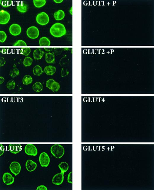
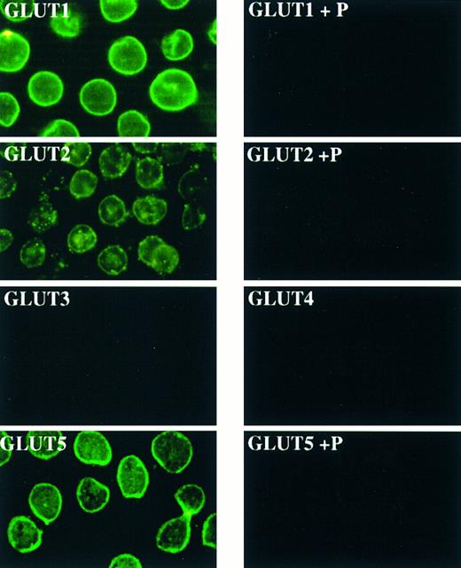
The fructose transporter GLUT5 is expressed in human erythrocytes. For immunofluorescence, erythrocyte smears were reacted with GLUT antibodies or with antibodies preabsorbed with the corresponding peptide (P) and visualized using fluorescein-coupled secondary antibodies.
The fructose transporter GLUT5 is expressed in human erythrocytes. For immunofluorescence, erythrocyte smears were reacted with GLUT antibodies or with antibodies preabsorbed with the corresponding peptide (P) and visualized using fluorescein-coupled secondary antibodies.
Western blot analysis.The glucose transporter antibodies were from East Acres Biologicals (Southbridge, MA). The specificity of the antibodies was tested using human and rat tissues. Erythrocyte membranes were isolated as previously described.14 Membrane proteins were separated by sodium dodecyl sulfate-polyacrylamide gel electrophoresis (SDS-PAGE), transferred to nitrocellulose membranes, and incubated with antibodies to the glucose transporters or preimmune sera diluted 1:500, followed by alkaline phosphatase-antirabbit IgG treatment, and color developed with 4-nitroblue tetrazolium chloride and 5-bromo–4-chloro-3–indolyl phosphate.
Immunolocalization of hexose transporters in blood vessel erythrocytes. For immunofluorescence, tissue sections were reacted with GLUT antibodies or with preimmune serum (PI) and visualized using fluorescein-coupled secondary antibodies.
Immunolocalization of hexose transporters in blood vessel erythrocytes. For immunofluorescence, tissue sections were reacted with GLUT antibodies or with preimmune serum (PI) and visualized using fluorescein-coupled secondary antibodies.
Immunocytochemistry.Erythrocytes were spread onto gelatin-coated slides and fixed with Histochoice (Amresco, Inc, Solon, OH). The smears were incubated with 5% skim milk, 1% bovine serum albumin (BSA), 0.3% Triton X-100 in PBS (pH 7.4) for 1 hour, and then with the glucose transporter antibodies or preimmune rabbit serum diluted 1:100 in the same solution for 1 hour. The erythrocytes were washed with PBS (pH 7.4), incubated with goat antirabbit IgG (H-L)–fluorescein isothiocyanate (FITC) conjugate (GIBCO-BRL, Gaithersburg, MD) at a dilution of 1:30 for 1 hour, washed with PBS (pH 7.4), mounted, and observed by fluorescence microscopy. Immunofluorescent detection of the glucose transporters in blood vessels was studied in normal human tissue sections prepared from paraffin-archived tissue blocks.
Uptake assays.Erythrocytes were diluted in incubation buffer (15 mmol/L HEPES [pH 7.4], 135 mmol/L NaCl, 5 mmol/L KCl, 1.8 mmol/L CaCl2 , 0.8 mmol/L MgCl2 ). Unless otherwise indicated, uptake assays were performed at 4°C in 200 μL of incubation buffer containing 0.5 mmol/L fructose and 2 μCi of D-[U–14C]fructose (6 Ci/mmol; DuPont/NEN, Boston, MA), or 0.5 mmol/L 2-deoxy–D-glucose and 1 μCi of 3H-deoxyglucose (5 mCi/mmol, DuPont NEN). Uptake was stopped with ice-cold PBS containing HgCl2 and KI. The cells were dissolved in 0.5 mL lysis buffer (10 mmol/L Tris-HCl pH 8.0, 0.2% SDS), and the incorporated radioactivity was assayed by liquid scintillation spectroscopy. Where appropiate, competitors and inhibitors were added to the uptake assays or preincubated with the cells. Data represent means ± standard deviation (SD) of four samples.
RESULTS
We used a panel of antibodies for each of the five members of the family of glucose transporters (GLUT1 through GLUT5) to identify the isoforms expressed in human erythrocytes. Immunolocalization using immunofluorescence showed the presence of the transporters GLUT1, GLUT2, and GLUT5 in human erythrocytes (Fig 1). For both GLUT1 and GLUT5, the immune reaction was homogeneously distributed across the cell, with a clear rim of fluorescence observed at the cell external border. For GLUT2, the level of fluorescence was consistently lower than that observed with GLUT1 and GLUT5 antibodies, and the reaction was distributed in patches across the cell. All cells within an observation field reacted with the three antibodies. No positive reaction was observed in the case of GLUT3 and GLUT4 antibodies. Control experiments showed that GLUT3 and GLUT4 antibodies gave positive reactions in tissues that express the respective transporters. In the cases of GLUT1, GLUT2, and GLUT5, the immunoreactivity was abolished by preincubating the antibodies with the respective peptides used to generate them (Fig 1). No immunoreactivity was observed using preimmune serum. Thus, the immunocytologic data are compatible with the expression of high levels of the transporters GLUT1 and GLUT5 in human erythrocytes and low levels of the transporter GLUT2.
To confirm the results obtained with blood smears, we analyzed the reactivity of the erythrocytes present in blood vessels of samples of human tissue fixed for routine pathological analysis. Similar to erythrocytes present in the blood smears, erythrocytes in blood vessels were strongly positive for GLUT1 and GLUT5 and reacted only weakly with GLUT2 antibodies (Fig 2). No immunoreactivity was observed in control experiments in which the GLUT1, GLUT2, and GLUT5 antibodies were preabsorbed with the corresponding peptide used to generate the antibodies. No immunoreactivity was observed when the samples were treated with GLUT3 and GLUT4 antibodies. Similar results were observed when using archival tissue (paraffin-embedded) or fresh samples (frozen tissue). We observed anti-GLUT5 immunoreactivity in erythrocytes of blood vessels from different tissues (testis, brain, breast, prostate, liver, and ovary), using different detection methods (immunofluorescence, immunoperoxidase, alkaline phosphatase) and different fixation protocols (formalin, Bouin).
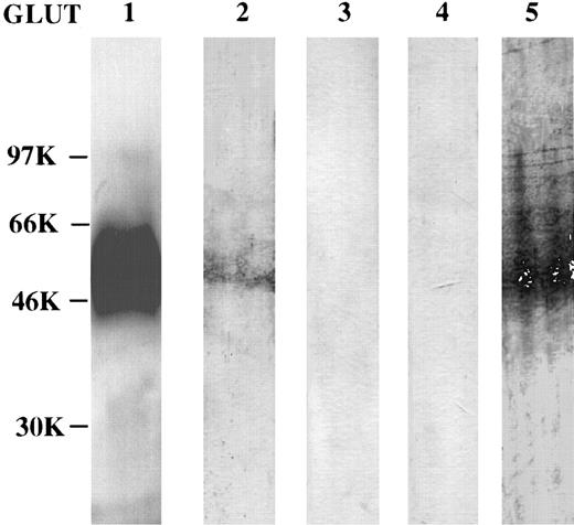
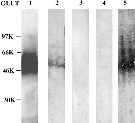
The presence of GLUT1, GLUT2, and GLUT5 in the human erythrocytes was confirmed by immunoblotting. One broad immunoreactive band, with an estimated Mr of 45,000, was observed in total membrane proteins of erythrocytes probed with anti-GLUT1 (Fig 3). A weakly reactive band with an estimated Mr of 50,000 was observed in membranes reacted with GLUT2 antibodies. As expected from the immunolocalization experiments, no immunoreactive band was observed in samples probed with GLUT3 and GLUT4 antibodies (Fig 3). On the other hand, a prominent band with an estimated Mr of 50,000, was observed in membranes tested with GLUT5 antibodies (Fig 3). Overall, the immunolocalization and the immunoblotting data are consistent with the expression of three different facilitative hexose transporters in human erythrocytes; the glucose transporter GLUT1, the low-affinity glucose-fructose transporter GLUT2, and the high-affinity fructose transporter GLUT5.
Expression of hexose transport proteins in erythrocytes. For immunoblotting, total erythrocyte membrane proteins were loaded on each lane of a sodium dodecyl sulfate-containing polyacrylamide gel, electrophoresed, transferred to Immobilon, reacted with the different anti-GLUT antibodies, and visualized using horseradish peroxidase antirabbit IgG and enhanced chemiluminescence. Sizes on the left are kD.
Expression of hexose transport proteins in erythrocytes. For immunoblotting, total erythrocyte membrane proteins were loaded on each lane of a sodium dodecyl sulfate-containing polyacrylamide gel, electrophoresed, transferred to Immobilon, reacted with the different anti-GLUT antibodies, and visualized using horseradish peroxidase antirabbit IgG and enhanced chemiluminescence. Sizes on the left are kD.
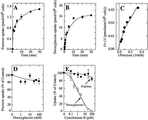
Functional studies showed that human erythrocytes had the capacity to take up fructose. Time-course experiments showed that the transport of fructose by the erythrocytes occurred slowly compared with the uptake of deoxyglucose (Fig 4A and B). Deoxyglucose uptake proceeded rapidly, approaching steady state in about 10 minutes. Considering an intracellular exchange volume for the erythrocytes of approximately 0.5 μL per 10 million cells, the deoxyglucose uptake data indicated that half the expected equilibrium concentration of deoxyglucose was reached in about 2 minutes. On the other hand, fructose uptake proceeded in a linear fashion for the first 3 minutes, followed by a second, slower uptake component that lasted for the length of the uptake assay (Fig 4A). Total fructose uptake accounted for only about 5% of the uptake of deoxyglucose under the same conditions. Because these experiments were performed at 4°C, the difference in uptake cannot be accounted for by differences in intracellular metabolism.
Kinetic analysis of the uptake of fructose by erythrocytes. (A) Time course of the uptake of 0.5 mmol/L fructose. (B) Time course of the uptake of 0.5 mmol/L deoxyglucose. (C) Double-reciprocal plot of the substrate dependence for fructose transport. Uptake was measured in the presence of concentrations of fructose ranging from 3 to 40 mmol/L. (D) Semilog plot of the concentration dependence for inhibition of fructose transport by deoxyglucose. Measurements were performed at 0.5 mmol/L fructose using 1-minute uptake assays. (E) Semilog plot of the concentration dependence for inhibition of fructose (•) and deoxyglucose transport (○) by cytochalasin B. Measurements were performed at 0.5 mmol/L fructose or deoxyglucose using 1-minute uptake assays. Cells were preincubated for 5 minutes with cytochalasin B. Where appropriate, data represent the mean ± SD of at least four samples.
Kinetic analysis of the uptake of fructose by erythrocytes. (A) Time course of the uptake of 0.5 mmol/L fructose. (B) Time course of the uptake of 0.5 mmol/L deoxyglucose. (C) Double-reciprocal plot of the substrate dependence for fructose transport. Uptake was measured in the presence of concentrations of fructose ranging from 3 to 40 mmol/L. (D) Semilog plot of the concentration dependence for inhibition of fructose transport by deoxyglucose. Measurements were performed at 0.5 mmol/L fructose using 1-minute uptake assays. (E) Semilog plot of the concentration dependence for inhibition of fructose (•) and deoxyglucose transport (○) by cytochalasin B. Measurements were performed at 0.5 mmol/L fructose or deoxyglucose using 1-minute uptake assays. Cells were preincubated for 5 minutes with cytochalasin B. Where appropriate, data represent the mean ± SD of at least four samples.
Dose-response experiments examining uptake at 4°C using incubation times of 5 minutes indicated the presence of two functional components involved in the transport of fructose in human erythrocytes. The high-affinity component presented an apparent Km for the transport of fructose of approximately 10 mmol/L (Fig 4C). The second functional component did not show evidence of saturation at concentrations of fructose of 50 mmol/L or higher. Because these fructose concentrations greatly exceeded the normal fructose content of blood even after a meal rich in fructose, no further attempt to characterize this activity was undertaken. It is known that GLUT2 transports fructose with low-affinity, with a Km for transport of about 70 mmol/L. On the other hand, the Km for the transport of fructose by GLUT5 is in the range of 10 mmol/L. Thus, our data point to GLUT5 as the main route of entry of fructose in human erythrocytes. Consistent with this notion was the finding in competition experiments that uptake of fructose was not decreased in the presence of high concentrations of deoxyglucose that completely blocked the transport of methylglucose by the human erythrocytes (Fig 4D). Similarly, cytochalasin B, a potent inhibitor of the functional activities of GLUT1 and GLUT2 that does not inhibit GLUT5, did not affect the uptake of fructose by human erythrocytes at concentrations that caused a total inhibition of the uptake of deoxyglucose by these cells (Fig 4E). The data are compatible with the concept that GLUT5 is mainly responsible for the cellular uptake of fructose in human erythrocytes.
DISCUSSION
We show here that human erythrocytes express the fructose transporter GLUT5, and that this transporter is directly involved in the uptake of fructose by these cells. This conclusion is supported by the results of immunolocalization, immunoblotting and transport assays.
The immunolocalization data indicate the presence in human erythrocytes of three members of the family of mammalian facilitative hexose transporters, the proteins GLUT1, GLUT2, and GLUT5. The glucose transporter GLUT1 present in human erythrocytes has been extensively studied using whole cells as well as reconstituted systems.1 The results of the immunofluorescent localization and the immunoblotting experiments indicating a higher degree of reactivity with GLUT1 antibodies compared with GLUT5 and GLUT2 antibodies are consistent with the concept that GLUT1 is the main hexose transporter expressed in human erythrocytes, followed by the abundant expression of GLUT5 and low level expression of GLUT2.
The transport data are consistent with the expression of GLUT5 and GLUT2 in human erythrocytes and support the concept that GLUT5 is the physiological transporter of fructose in these cells. The apparent Km for the transport of fructose by human erythrocytes is similar to the Km for transport of fructose by GLUT5.15 The properties of a second transport system that did not approach saturation at concentrations of fructose as high as 50 mmol/L are compatible with the expression of low levels of GLUT2 in human erythrocytes. GLUT2 is capable of transporting fructose, but the Km for fructose transport is greater than 70 mmol/L.3 The lack of a measurable effect of deoxyglucose on fructose transport by human erythrocytes is also consistent with a major role for GLUT5 in the uptake of fructose by these cells. Deoxyglucose is transported by GLUT1 and GLUT2, but not by GLUT5.16 Thus, it is expected that deoxyglucose would compete for the transport of fructose if GLUT2 were the main transporter involved in the transport of fructose by human erythrocytes. The alkaloid cytochalasin B strongly inhibits the functional activity of GLUT1, GLUT2, GLUT3, and GLUT4, but does not inhibit GLUT5-mediated fructose transport.2,3 16 The lack of effect of cytochalasin B on the transport of fructose by human erythrocytes is also consistent with the concept that GLUT5, and not GLUT2, is the main transporter involved in the uptake of fructose by these cells.
The time-course experiments showed that fructose entered human erythrocytes at a velocity that was only a fraction of the velocity of uptake of deoxyglucose by these cells. Although the transport data are consistent with the immunolocalization data that suggest that GLUT5 is not as abundant as GLUT1 in human erythrocytes, a side-by-side comparison of the data indicates that the large difference in transport velocity cannot be explained solely by differences in expression. A plausible explanation is that the membrane translocation of fructose mediated by GLUT5 occurs very slowly. An example of slow transport of a molecule across the cell membrane is the transport of a fluorescent glucose analog by GLUT1.17
The demonstration that human erythrocytes express the fructose transporter GLUT5 and have the capacity to transport fructose has implications for our understanding of human red blood cell metabolism. There has been an increased consumption of fructose in modern times due to the widespread use of fructose as a natural sweetener. Fructose crosses the brush border membrane of the small intestinal enterocytes through GLUT5.15 In the intact organism and under normal conditions, the liver is the major site of fructose metabolism, although there is also evidence indicating that adipose tissue, and to a lesser degree muscle tissue, may be important sites for fructose metabolism.18 In liver, the transport of fructose is mediated by GLUT2. On the other hand, adipose and muscle tissue do not express measurable amounts of GLUT2, but instead express low levels of GLUT5.19 It is conceivable then that GLUT5 is directly involved in the transport of fructose in these tissues.
A better understanding of the metabolic pathways involved in the metabolism of fructose by human erythrocytes is slowly emerging. Similar to liver, it is generally believed that the first step in the metabolism of fructose in human erythrocytes is the phosphorylation to fructose-6–phosphate by hexokinase, an enzyme that primarily phosphorylates glucose, but also has low affinity for fructose.9 In liver, fructose can be phosphorylated to fructose-1–phosphate by fructokinase, and fructose-1–phosphate is a potent activator of glucose phosphorylation to glucose-6–phosphate.20 It has been described recently that human erythrocytes accumulate measurable amounts of fructose-3–phosphate when incubated in the presence of fructose.21 Fructose-3–phosphate and its metabolic product, 3-deoxyglucosone, which is a potent glycosylating agent, have also been found in the lens of diabetic rats.22 Although phosphorylated fructose intermediaries are generated intracellularly from glucose, our results indicate that fructose of dietary origin can be transported into human erythrocytes with the direct participation of the high-affinity fructose transporter GLUT5. The data presented here are compatible with the notion that human erythrocytes may be a significant, and until now unrecognized, component of nonhepatic clearance of dietary fructose.
ACKNOWLEDGMENT
We thank Dr Luis Norambuena from the Instituto de Histologı́a y Patologı́a, Universidad Austral de Chile, for tissue samples.
Supported in part by Grant No. S-95-24 from the Universidad Austral de Chile, Grant No. 196-0485 from FONDECYT, Santiago, Chile, Grants No. R01 CA30388, R01 HL42107, and P30 CA08748 from the National Institutes of Health, and by Memorial Sloan-Kettering Institutional Funds.
Address reprint requests to Ilona I. Concha, PhD, Instituto de Bioquı́mica, Facultad de Ciencias, Universidad Austral de Chile, Casilla 567, Campus Isla Teja, Valdivia, Chile.

