Abstract
Cytogenetic analysis was performed on peripheral blood lymphocyte cultures from 19 patients with mycosis fungoides (MF )/Sézary syndrome (SS) stimulated with either phytohemagglutinin, a conventional mitogen, or a combination of interleukin-2 (IL-2) plus IL-7. The use of both PHA-stimulated and IL-2 plus IL-7–stimulated cultures enhanced the ability to identify clonal abnormalities. Clonal abnormalities were observed in 11 patients (53%) including one with monosomy for the sex chromosome as the sole abnormality. Five of the 11 patients with clonal abnormalities had normal peripheral white blood cell counts, indicating detectability of clones in the absence of frankly leukemic disease. The presence of clonal abnormalities correlated with advanced stage disease and a significantly reduced survival duration from the time of cytogenetic studies. Clonal abnormalities involving chromosomes 1 and 8 were observed in six cases. In five cases with aberrations of chromosome 1, loss of material involved the region between 1p22 and 1p36. In an additional case, a reciprocal translocation involving 1p33 was observed. Clonal abnormalities involving chromosomes 10 and 17 were observed in 5 cases, clonal abnormalities involving chromosome 2 in 4 cases, and clonal abnormalities involving chromosomes 4, 5, 6, 9, 13, 15, 19, and 20 in 3 cases. In 2 cases a der(8)t(8; 17)(p11; q11) was observed. Regions of the genome that encode T-cell receptors were not involved in abnormalities. The region between 1p22 and 1p36 is identified as a region of the genome that requires detailed analysis toward the identification of potential gene(s) involved in the process of malignant transformation and/or progression in MF/SS.
MYCOSIS FUNGOIDES (MF ) and the Sézary syndrome (SS) represent a spectrum of cutaneous T-cell lymphomas (CTCL). MF classically presents in the skin as flat patches that may evolve into raised, infiltrated plaques and tumor nodules with subsequent involvement of lymph nodes and ultimately viscera. Patients who have SS classically exhibit erythroderma, lymphadenopathy, and leukemic involvement of the peripheral blood (PB). It is not uncommon for MF patients to have small numbers of circulating tumor cells at some point in their clinical course.
The rarity of the disease, difficulty in obtaining malignant cells from skin biopsies, paucity of malignant cells in the circulation, and poor morphology of the chromosomes are limiting factors in studying the cytogenetics of MF/SS. Despite these limitations, there are numerous studies on CTCL. Early studies on unbanded chromosomal preparations1-5 detected numerical and gross structural abnormalities. Although banded preparations were subsequently studied,6-15 complete karyotypes have not been provided in a number of cases because of the complexity of the karyotype.16-24 Although these studies have revealed numerical and structural abnormalities involving many chromosomes, recurring abnormalities have not been reported.
Interpretation of data from various reports is also complicated by differences in clinical stage, source of the malignant cells (PB, bone marrow, lymph node, and skin biopsy), and tissue culture technique (particularly the kind of mitogen used). In neoplastic disorders marked by low proliferative fractions, such as B-cell chronic lymphocytic leukemia, B-cell mitogens (phorbol 12-myristate, 13-acetate [TPA], phorbol 12, 13-dibutyrate, pokeweed mitogen [PWM], and lipopolysaccharide) have been useful in enhancing the number of malignant cells in division. In addition to phytohemagglutinin (PHA), the most frequently used agent, other mitogens used in the study of CTCL are concanavalin A,18 calcium ionophore,18 pokeweed,19 anti–β2-microglobulin,19 Cowan, protein A,19 staphylococcal protein A,25 TPA,9 interleukin-2 (IL-2),9 and A23187.9
Recent studies have shown that IL-2 and IL-7 act synergistically to induce proliferation of Sézary cells in vitro.26 This method has not been used previously in cytogenetic studies. The objectives of this study were: to evaluate the use of IL-2 and IL-7 in the cytogenetic study of circulating malignant cells in MF/SS; to identify recurring chromosomal abnormalities; and to correlate clonal chromosome abnormalities with clinical stage and survival. The current report is the largest series of MF/SS patients studied clinically and cytogenetically at a single institution who had PB used as the source of malignant cells, and PHA and combined IL-2 and IL-7 (henceforth referred to as IL-2/IL-7) as mitogens.
MATERIALS AND METHODS
Patients.Nineteen patients (10 men, 9 women) with a mean age of 61 years, and who were diagnosed with MF/SS at Northwestern Memorial Hospital or the Veterans Affairs Lakeside Medical Center between December 1992 and January 1995 were studied. Diagnosis was based on clinical evaluation, with skin biopsy confirmation. All the cases were clinically evaluated by a multidisciplinary CTCL clinic at Northwestern University to confirm the diagnosis. At the time of cytogenetic evaluation, a PB sample from each patient was analyzed for complete blood count and differential leukocyte count. Quantitation of Sézary cells was performed on Wright's stained blood film from each patient. Staging was based on TNM criteria.27 Table 1 summarizes clinical stage, white blood cell (WBC) count and Sézary cell count in each patient. Seventeen of the 19 patients had been treated before being studied. Some patients received numerous therapeutic modalities, including systemic chemotherapy and immunomodulation therapy. Three healthy volunteers were analyzed as controls.
Cell culture and mitogens.Twenty-three PB specimens from 19 patients and samples from three healthy volunteers were studied. Whole blood, 0.5 mL, was cultured in 10 mL of RPMI 1640 with 10% fetal bovine serum (FBS) for 72 hours. Cultures stimulated with PHA and those stimulated with IL-2 (10 Biological Response Modifier Program [BRMP] U/mL) plus IL-7 (25 ng/mL) were studied in specimens from 16 patients. Specimens from three patients (nos. 7, 11, and 16) were cultured only with PHA. IL-2 and IL-7 were used in two cultures separately for specimen from one patient (no. 15). PB specimens from the three normal individuals were also stimulated with IL-2/IL-7.
Cytogenetic analysis.Photomicrographs of trypsin-Giemsa method stained metaphase cells were karyotyped. Karyotypes were described according to the ISCN nomenclature.28
Statistical analyses.Statistical analyses were performed using SPSS software (Chicago, IL), version 6.1.2 for Microsoft Windows (Microsoft Corp, Bellevue, WA). Survival data were generated by the method of Kaplan and Meier.28a Differences in survival were analyzed by the log-rank method. The relationship between karyotype and disease stage was evaluated using Fisher's exact test using 2 × 2 contingency tables.
RESULTS
Normal karyotypes were observed in the three specimens from normal individuals. Metaphase cells were available for analysis from all 22 specimens stimulated with PHA and from 16 of the 17 specimens stimulated with IL-2/IL-7 (Table 2). Metaphase cells from patient 19 could not be analyzed completely because of poor morphology. Metaphase cells were also obtained from the one patient (no. 15) whose cells were stimulated with IL-2 and IL-7 in separate cultures. In two patients (nos. 12 and 13) the abnormal clones were observed in the cultures stimulated with IL-2/IL-7, but not in the cultures stimulated with PHA. In two patients (nos. 14 and 19), the cultures stimulated with PHA were informative; cultures stimulated with IL-2/IL-7 either did not yield metaphase cells (no. 14) or yielded metaphase cells that could not be analyzed because of poor morphology (no. 19).
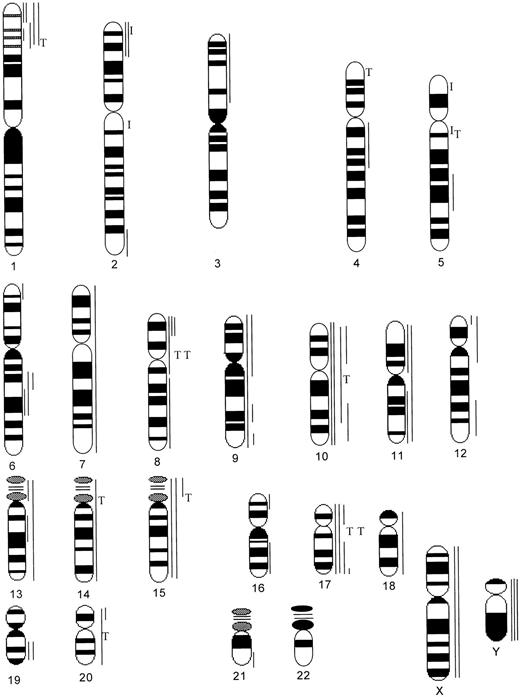
In eight patients (42%), all of the cells analyzed had either a normal karyotype (nos. 1 to 4) or a normal karyotype with nonclonal abnormalities (nos. 5 to 8). In one woman (no. 11) monosomy for the sex chromosome was observed as the sole abnormality. Clonal abnormalities were observed in the remaining 10 patients (53%). Sex chromosomes and all autosomes, with the exception of chromosome 22, were involved in clonal abnormalities (Fig 1). Patients with advanced-stage disease were more likely to have clonal cytogenetic abnormalities detectable in PB lymphocytes (10 of 15 patients with clinical stage III or IV) than patients with early-stage disease (0 of 4 patients with clinical stages I and II) (P = .03). Clonal abnormalities were detected in 4 of 11 patients (36%) with WBC within normal reference range for our laboratory (3.5 to 10.5 × 10⁹/L).
Schematic diagram illustrating the banding pattern of human chromosomes and the distribution of monosomy and breakpoints in structural rearrangements observed in 18 patients with MF/SS. ( — ), Monosomy, deletion, and translocation with unidentified segment; T, translocation; I, inversion.
Schematic diagram illustrating the banding pattern of human chromosomes and the distribution of monosomy and breakpoints in structural rearrangements observed in 18 patients with MF/SS. ( — ), Monosomy, deletion, and translocation with unidentified segment; T, translocation; I, inversion.
Single clones with chromosome number close to diploidy (43 to 48 chromosomes) were observed in six patients (nos. 9, 10, 11, 12, 13, and 14). Multiple near diploid clones were observed in two patients (nos. 15 and 16) and near tetraploid clones in three cases (nos. 17, 18, and 19). Loss of the sex chromosome was the most frequent abnormality (seven cases). As mentioned previously, in one patient (no. 11) monosomy for the X chromosome was the sole abnormality. In two patients (nos. 9 and 18) an apparently identical rearrangement, der(8)t(8; 17)(p11; q11), was observed (Fig 2).
Partial karyotype of metaphase cells showing the der(8)t(8; 17)(p11; q11). The der(8) chromosomes are identified with arrows.
Partial karyotype of metaphase cells showing the der(8)t(8; 17)(p11; q11). The der(8) chromosomes are identified with arrows.
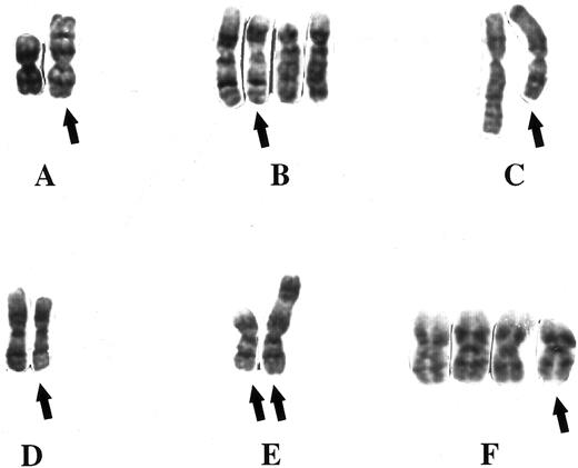
Clonal abnormalities involving chromosomes 1 and 8 were observed in six patients. In five patients with chromosome 1 abnormalities, loss of material involved the region between 1p22 and 1p36 (Figs 1 and 3). In one patient (no. 16), rearrangements resulted in loss of material from the short arm of both chromosome 1 homologs (Fig 3). In an additional patient (no. 10), a reciprocal translocation involved the short arm of chromosome 1 at 1p33, t(1; 4)(p33; p16) (Fig 3). In five patients, rearrangements resulted in complete or partial monosomy for the short arm of chromosome 8. Clonal abnormalities involved chromosomes 10 and 17 in five patients. Monosomy for chromosome 10 was observed in three patients. Complete monosomy for one chromosome 10 homlog and monosomy for the region between p11 and p15 of the homologous chromosome was observed in one patient (no. 9). Rearrangements resulted in complete or partial monosomy for the short arm of chromosome 17 in five patients. Chromosome 2 was rearranged in four patients, and clonal abnormalities involving chromosomes 4, 5, 9, 13, 15, 19, and 20 were observed in three patients. Rearrangements resulted in partial monosomy for the long arm of chromosome 6 in two patients.
Partial karyotypes of metaphase cells showing abnormalities involving the short arm of chromosome 1. The rearranged chromosome 1 homologs are identified with arrows. (A) Case 9, add(1)(p36); (B) case 10, t(1; 4)(p33; p16); (C) case 14, add(1)(p36) and ins(1:?)(q?25; ?); (D) case 15, del(1)((p34p35)); (E) case 16, del(1)(p?32p35) and add(1)(p33); (F ) case 18, del(1)(p33).
Partial karyotypes of metaphase cells showing abnormalities involving the short arm of chromosome 1. The rearranged chromosome 1 homologs are identified with arrows. (A) Case 9, add(1)(p36); (B) case 10, t(1; 4)(p33; p16); (C) case 14, add(1)(p36) and ins(1:?)(q?25; ?); (D) case 15, del(1)((p34p35)); (E) case 16, del(1)(p?32p35) and add(1)(p33); (F ) case 18, del(1)(p33).
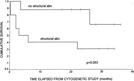
Patients with advanced-stage disease had shortened survival in comparison to patients with earlier-stage disease, although this difference was not statistically significant (P = .47). The 10 patients with clonal structural abnormalities had shorter survival from the time of cytogenetic study than patients without structural chromosome abnormalities (Fig 4). This difference approached statistical significance (P = .053 by log-rank test). Median survival for patients with clonal structural chromosome abnormalities was 3 months from cytogenetic study. Median survival for patients without clonal structural chromosome abnormalities has not yet been reached, with seven of nine patients still alive.
Kaplan-Meier product limit curve illustrating survival duration in 19 patients with MF/SS. Survival is illustrated from time of cytogenetic study and is stratified by cytogenetic status. Patients with clonal structural chromosome abnormalities (abn) display shorter survival duration than patients without structural chromosome abnormalities. +, Patients alive at last follow-up.
Kaplan-Meier product limit curve illustrating survival duration in 19 patients with MF/SS. Survival is illustrated from time of cytogenetic study and is stratified by cytogenetic status. Patients with clonal structural chromosome abnormalities (abn) display shorter survival duration than patients without structural chromosome abnormalities. +, Patients alive at last follow-up.
DISCUSSION
Compared with other hematologic malignant disorders, there is limited information on the cytogenetic characteristics of malignant cells in MF/SS. This study substantiates previous reports of hyperdiploidy and complex karyotypes in MF/SS. In the current series, although both numerical and structural abnormalities involving most chromosomes were observed, structural abnormalities were more frequent. Furthermore, the most frequent abnormalities resulted in loss of material from the short arms of chromosomes 1 and 8. Regions of the genome to which genes encoding T-cell receptors have been mapped (14q11, 14q32, 7p15-14, 7q35) were not involved, suggesting that the genetic basis for malignant transformation in primary cutaneous T-cell lymphomas appears to be different from that involved in other T-cell malignant disorders.
Among the abnormalities observed in this series, rearrangements resulting in loss of material from the short arm of chromosome 1 (1p33-36) are most impressive (5 of 10 patients). Nonrandom involvement of chromosome 1 in structural abnormalities in MF/SS has been previously reported.10,15,21,22 Loss of material from the short arm of chromosome 1 has been observed in a variety of tumors,29-38 suggesting the presence of one or more tumor suppressor gene(s) in this region. Loss of heterozygosity studies of alleles between 1p33 and p36 will help determine the significance of this abnormality.
A number of recurring balanced translocations involving 1p have also been observed in other neoplasms. These include t(1; 17)(p36; q21), t(1; 3)(p36; q21), t(1; 22)(p13; q13), and t(1; 7)(p11; p11) in AML; t(1; 9)(p11; p11) in PV; t(1; 14)(p32-34; q11) in T-ALL; t(1; 11)(p32; q23) in ALL; t(1; 8)(p22; q12) in pleomorphic adenoma of the salivary gland; and t(1; 12)(p33-34; q13-15) in lipoma.39 The band 1p33 involved in the apparently balanced translocation, t(1; 4)(p33; p16), in patient 10 is interesting because the breakpoint in this rearrangement may be similar to that observed in the t(1; 14) in T-ALL, the t(1; 11) in ALL, and t(1; 12) in lipoma. Genes on the short arm of chromosome 1 involved in malignant transformation include the TAL1 (TCL5, SCL) gene [involved in the t(1; 14)(p32-34; q11)]40 and proto-oncogenes MYCL-1 (1p32)41 and JUN (1p32-31).42 Another gene of interest in the context of MF/SS is VCAM-1, a member of the vascular cell adhesion molecules (VCAM), mapped to 1p32-p31.43 It is speculated that VCAMs may play a role in tumor invasion and metastasis.
Partial monosomy for the long arm of chromosome 6 in two patients in the current series is of interest because the presence of at least three tumor-suppressor genes on the long arm of chromosome 6 has been suggested based on three regions of minimal molecular deletion (RMD), RMD1 at 6q25-27, RMD2 at 6q21-23, and RMD3 at 6q23 in non-Hodgkin's lymphoma.44 Nonrandom involvement of chromosome 6 in structural aberrations in MF/SS has been previously reported.10,14,21 24 Detailed analysis of the region between q15 and q23 in patients with MF/SS is required to determine if other non-Hodgkin's lymphomas and MF/SS have involvement of the same tumor-suppressor genes.
Loss of material from the short arms of chromosomes 10 and 8 and from the short and long arms of chromosome 17 are also observations that require detailed analysis at the molecular level. Nonrandom involvement of chromosomes 10 and 17 has been previously reported.15,20 45
Recurring balanced rearrangements have not been reported in MF/SS. The der(8)t(8; 17)(q11; p11) is the only translocation observed in two cases in this series. We are unable to confirm if the rearrangement in the two cases (nos. 9 and 18) are identical by additional studies because of nonavailability of cells from one of the patients (no. 18). The breakpoint at 8q11 is of interest because the gene for IL-7 is located at 8q12-13.46
This study shows that clonal chromosomal abnormalities can be detected in PB samplings even in the absence of frankly leukemic disease. The use of a combination of IL-2/IL-7 in addition to conventional mitogens such as PHA appears to enhance detection of clonal abnormalities in MF/SS. The mitogen used may be important in obtaining sufficient numbers of metaphase cells in MF/SS. Differences in response to mitogens between different clones even in a single patient have been reported.9 A variety of mitogens have been used in previous studies. The synergistic effect of combined IL-2/IL-7 in inducing division of Sézary cells in culture, described recently,26 has not been previously used in cytogenetic studies. The numbers of patients in this series are too small to determine the relative efficacy of PHA or IL-2/IL-7 in cytogenetic studies of MF/SS. However, observations in this study suggest that the combination of IL-2/IL-7 offers a viable alternative methodology, and its use along with other mitogens enhances detection of abnormal clones in cytogenetic studies of circulating malignant cells in MF/SS.
A correlation between the level of Sézary cells in circulation and cytogenetic abnormalities has been previously reported.24 In the current series of patients there was no correlation between the number of circulating Sézary cells and the presence or absence of cytogenetic abnormalities in patients with Sézary cell counts above zero. However, clonal abnormalities were not detected when Sézary count was zero. Although abnormal clones were observed in one patient (no. 15) with only 0.3 × 10⁹/L Sézary cells, clonal abnormalities were not observed in the specimen from another patient (no. 8) with 3.3 × 10⁹/L Sézary cells. These observations may be because of differences in response to mitogens between patients. The ability to detect abnormal clones in the absence of frankly leukemic disease suggests that PB cells may be used in cytogenetic studies of primary cutaneous T-cell lymphomas, diminishing the need for solid tumor biopsies.
In the current study, survival duration from the time of cytogenetic study was shorter in patients with clonal structural chromosome abnormalities (P = .053). This may reflect the fact that patients with structural chromosome abnormalities were significantly more likely to have advanced-stage disease (P = .03). Clinical stage in MF/SS has been shown to correlate well with survival.47 Patients in the current study with advanced-stage disease had shorter survival times than patients with earlier-stage disease. The fact that this difference did not reach statistical significance is likely because of limited sample size. Regardless, the presence of clonal cytogenetic abnormalities in the current study was more effective than stage in predicting survival.
Based on T-cell receptor chain gene rearrangements by Southern blot analysis, it has been suggested that circulating Sézary cells may be reactive in nature.48 The presence of clonal chromosomal abnormalities in circulating PB lymphocytes in this study suggests that Sézary cells may indeed be malignant in nature. The malignant nature of these cells is underscored by the complexity of clonal abnormalities in MF/SS patients, and by the lack of cytogenetic abnormalities in normal controls.
Our data indicate that, with the use of novel mitogen combinations, cytogenetic studies can be carried out in MF/SS patients using PB samples, even in the absence of frankly leukemic disease. Furthermore, the presence of clonal chromosomal abnormalities correlates with a relatively poor prognosis. The detection of recurring structural chromosome abnormalities in MF/SS, particularly that involving the region 1p32-36, is a novel finding worthy of further investigation. Additional studies at the cytogenetic and molecular levels should help in determining the significance of this region in the etiology of MF/SS. Cytogenetic studies on sorted subpopulations will be critical towards determining if the clonal abnormalities are characteristic of circulating malignant cells in MF/SS.
Supported in part by a generous contribution from Jaunine C. Clark Leukemia Research Fund.
Address reprint requests to Maya Thangavelu, PhD, Northwestern Memorial Hospital, Room 1564, Prentice Pavilion, 333 E Superior, Chicago, IL 60611.


