Abstract
P-selectin (CD62P) is a Ca2+-dependent endogenous lectin that can be expressed by vascular endothelium and platelets. The major ligand for P-selectin on leukocytes is P-selectin glycoprotein ligand-1 (PSGL-1). P-selectin can also bind to carcinoma cells, but the nature of the ligand(s) on these cells is unknown. Here we investigated the P-selectin binding to a breast and a small cell lung carcinoma cell line that are negative for PSGL-1. We report that CD24, a mucin-type glycosylphosphatidylinositol-linked cell surface molecule on human neutrophils, pre B lymphocytes, and many tumors can promote binding to P-selectin. Latex beads coated with purified CD24 from the two carcinoma cell lines but also neutrophils could bind specifically to P-selectin-IgG. The binding was dependent on divalent cations and was abolished by treatment with O-sialoglycoprotein endopeptidase but not endoglycosidase F or sialidase. The beads were stained with a monoclonal antibody (MoAb) to CD57 (HNK-1 carbohydrate epitope) but did not react with MoAbs against the sialylLex/a epitope. The carcinoma cells and CD24-beads derived from these cells could bind to activated platelets or P-selectin transfected Chinese hamster ovary cells (P-CHO) in a P-selectin–dependent manner and this binding was blocked by soluble CD24. Transfection of human adenocarcinoma cells with CD24 enhanced the P-selectin–dependent binding to activated platelets. Treatment of the carcinoma cells or the CD24 transfectant with phosphatidylinositol-specific phospholipase C reduced CD24 expression and P-selectin–IgG binding concomitantly. These results establish a role of CD24 as a novel ligand for P-selectin on tumor cells. The CD24/P-selectin binding pathway could be important in the dissimination of tumor cells by facilitating the interaction with platelets or endothelial cells.
AMONG THE CELL adhesion molecules, the selectins comprise a family of molecules which are able to bind to specific sugar determinants on the surface of adjacent cells.1-4 Selectins are involved in the rolling adhesion of leukocytes to endothelial cells and platelets under the shear forces present in the blood circulation. Three members of this family, E-selectin (CD62E), L-selectin (CD62L), and P-selectin (CD62P), have been identified and characterized. P-selectin can be found on the cell surface of activated platelets and endothelial cells.5,6 It is stored in intracellular granula and is rapidly mobilized to the cell surface within minutes after stimulation with pro-inflammatory agents like histamine or thrombin.7 P-selectin, expressed by thrombin-activated platelets and endothelial cells, binds to ligands on myeloid cells and subsets of lymphocytes. Recent studies have provided evidence that certain tumor cells can also bind to activated platelets via P-selectin8 and that human carcinomas can react with P-selectin– or E-selectin–IgG constructs.9-12
The nature of the high-affinity binding between selectins and their ligands is under intense study. Earlier investigations had shown that all selectins could recognize the blood group determinants sialylLex and sialylLea or related sulfated structures.3 P-selectin can also bind to other sulfated moieties like the SGNL (3-sulfo-glucoronyl-neolactose) glycolipid13 or sulfatides.14 A major glycoprotein ligand for P-selectin was then identified and termed P-selectin glycoprotein ligand-1 (PSGL-1).15 PSGL-1 is a 220-kD dimeric mucin-type glycoprotein that carries O-linked sialylLex glycans16,17 and a sulfated Tyr residue at the N-terminus of the molecule which appears to be critical for its function.18,19 It is expressed on human myeloid HL-60 cells, neutrophils, lymphocytes,20 CD34+ early progenitor cells,21 and other hematopoietic cells, though not always in an active form.22 On leukocytes PSGL-1 appears to account for most high-affinity binding sites for P-selectin and is essential for neutrophils to roll on P-selectin at physiological shear.20 23
CD24 consists of a small protein core comprising 27 amino acids which is extensively glycosylated and is bound to the membrane via a phosphatidyl-inositol anchor.24-26 Nearly half of the amino acids in CD24 are composed of Ser and Thr residues that are potential sites for O-linked glycans. CD24 is expressed at the early stages of B-cell development, it is highly expressed on neutrophils, but it is absent on normal T cells or monocytes.24 Although CD24 is not present in adult human tissues, it is expressed in many human carcinomas.27,28 The homologous murine molecule is HSA (heat-stable antigen), a 28 to 70-kD cell-surface glycoprotein expressed by many cell types in the mouse.29-31 In a previous study we have presented evidence that certain glycoforms of HSA could act as a specific ligand for P-selectin.32 33 We have now examined whether CD24 can serve as a P-selectin ligand too and, if so, whether the expression on tumor cells is related to the ability to bind to P-selectin. Our results suggest that CD24 is a major ligand for P-selectin on carcinoma cells.
MATERIALS AND METHODS
Cells.The small cell lung carcinoma line SW2 was described before.27 The human breast carcinoma cell line KS34 was provided by B. Gückel (University of Heidelberg). Cells were cultivated in Dulbecco's modified Eagle medium (DMEM) supplemented with 10% fetal calf serum (FCS) at 37°C, 5% CO2 , and 100% humidity. Human platelets were isolated from platelet-rich plasma by Sepharose 2B chromatography. Neutrophils were isolated from human blood using Dextran-sedimentation of erythrocytes followed by Ficoll-Hypaque (Pharmacia, Freiburg, Germany) separation as described.35 Residual erythrocytes were lysed by brief incubation in 155 mmol/L NH4Cl, 0.1 mmol/L EDTA, 10 mmol/L KHCO3 solution followed by washing of the cells. Chinese hamster ovary (CHO) cells expressing human P-selectin were established by cotransfection a P-selectin cDNA in pCDM8 (obtained from Dr E.C. Butcher, Stanford University, Stanford, CA) with a neomycin-resistence gene using Lipofectamin (GIBCO-BRL Eggenstein, Germany). After neomycin selection P-selectin expressing transfectants were selected by fluorescence-activated cell sorting (FACS) using a P-selectin specific monoclonal antibody (MoAb) (see below). The isolation of CD24 transfected A125 adenocarcinoma cells has been described.36
Antibodies.Anti-CD24 MoAbs ML-5 and 32D12 were obtained from S. Funderud (Radiumhuset, Oslo, Norway) and the MoAb SWA11 has been described.27 MoAbs WAPS 12.2 against human P-selectin, HECA 452 (anti sialylLex and sialylLea 37), and CSLEX-1 (anti sialyl-Lex) and DREG 56 against L-selectin were a kind gift from Dr E.C. Butcher. MoAb HNK-1 (CD57) and fluorescein isothiocyanate (FITC)-conjugated limax flavus agglutinin were obtained from Dianova (Hamburg, Germany). MoAb W 6/32 (major histocompatibility complex [MHC] class I) was obtained from G. Moldenhauer (DKFZ Heidelberg). Human P-selectin-IgG was a gift from Genetics Institute (Boston, MA). The murine P-selectin–IgG fusion protein has been described.38
Affinity purification of CD24.CD24 was purified by affinity chromatography on an MoAb SWA11 column as described for HSA.32 Briefly, human neutrophils, KS or SW2 tumor cells were lysed in the presence of NP-40, cell lysates were prepared by centrifugation, and passed over the antibody column. After washing the bound antigen was eluted at pH 11.5 in the presence of β-octylglucoside (BOG) as detergent. Eluted antigen was neutralized and analyzed by enzyme-linked immunosorbent assay (ELISA) and sodium dodecyl sulfate-polyacrylamide gel electrophoresis (SDS-PAGE). Routine staining of the gel with Coomassie brillant blue or silver nitrate did not show any distinct band on the gel. Because of its small protein core, CD24 cannot be detected under these conditions and requires modification with digoxigenin-3-O-succinyl-ε-amino-caproic-acid-hydrazide (see below). Lens culinaris binding glycoproteins were isolated from SW2 cell lysate on a lens culinaris agglutinin affinity column. After washing of the column bound glycoproteins were eluted with 0.5 mol/L α-methylmannoside in 50 mmol/L BOG. Antigens were dialyzed and stored at −70°C.
Coating of antigen to polystyrene beads.CD24 antigen (∼150 μg/mL) in BOG were incubated with polystyrene beads (Serva, Heidelberg, Germany) and the detergent was removed by dialysis as described.39 Remaining binding sites were blocked by incubation with 1% bovine serum albumin (BSA) in phosphate-buffered saline (PBS) for 1 hour. The density of coated CD24 was determined by FACS analysis using MoAb ML-5 as described below. For control purposes purified MHC class I antigen (a kind gift of Dr H. Kropshofer, DKFZ) or lens culinaris binding glycoproteins were adsorbed under similar conditions. The control beads could be stained either with MoAb W6/32 against MHC class I or FITC-conjugated lens culinaris agglutinin, respectively.
Cytofluorography.The staining of cells or antigen-coated beads with MoAbs (hybridoma supernatant or purified antibody) and phycoerythrin-conjugated goat antibodies to mouse Igs (SERVA, Heidelberg, Germany) has been described.40 Stained cells or beads were analyzed with a FACScan fluorescence activated cell analyzer (Becton Dickinson, Heidelberg, Germany). The staining with P-selectin–IgG was as described previously.33
Isolation of RNA and reverse transcriptase-polymerase chain reaction (RT-PCR) analysis.The isolation of total RNA from cells has been described in detail elsewhere.41 Total RNA (6 μg) was transcribed into cDNA using 300 U of M-MLV RT (Promega, Heidelberg, Germany) and oligo(dT)20 for priming. cDNA synthesis proceeded for 2 hours at 37°C. After heat-inactivation of the enzyme the RNA/DNA hybrid was treated with RNAse H for 20 minutes at 37°C. The cDNA was used as template for PCR using an annealing temperature of 60°C and 40 cycles of 80 seconds. The following primers for PSGL-1 were used: forward, GCCCTCGAGGAAGCTTTCCCATGCTCTGCTG; reverse, AGCGGATCCGAGGTGGGGTCTTGCCAAAACAG. For the detection of CD24 the following primers were used: forward, TCCAAGGCACCCAGCATCCTGCTAGA; reverse, TAGAAGACGTTTCTTGGCCTGAGTCT. PCR products were separated on a 1% agarose gel containing 0.5 μg/mL ethidium bromide.
Enzyme treatment.Sialic acid residues were cleaved from CD24-coated beads by incubation with 0.1 U/mL of Vibrio cholerae sialidase (Behringwerke, Marburg, Germany) in Hanks' balanced saline solution (HBSS) containing 2 mmol/L CaCl2 , 2 mmol/L MgCl2 , 10 mmol/L HEPES for overnight at 37°C. To control for sialidase cleavage the beads were stained before and after treatment with FITC-conjugated limax flavus agglutinin (LFA), which detects sialic acids. Treatment of beads with endoglycosidase F to remove N-linked glycans was done in 50 mmol/L sodium-phosphate/35 mmol/L EDTA pH 6.1 for overnight at 37°C according to the instructions given by the manufacturer (Boehringer, Mannheim, Germany). These assay conditions have been previously found to reduce P-selectin binding to HSA.33 Treatment of beads with O-sialoglycoprotein endopeptidase (OSGE) from Pastorella hemolytica (Camon, Wiesbaden, Germany) was done with 20 μL enzyme in HBSS supplemented with 2 mmol/L Ca2+ for 2 hours at 37°C or as otherwise indicated. After treatment the beads were washed, stained, and analyzed by FACS as described above. Treatment of cells with phosphatidylinositol-specific phospholipase-C (PIPL-C) was done as described previously.42
Cell-binding assay.Binding of tumor cells to platelets or P-selectin–CHO monolayers was done using Eur3+-labeled cells.43 Briefly, cells were washed with cold Dulbecco's-PBS and 1 × 10⁷ cells were suspended in 1.5 mL labeling buffer (pH 7.3) containing 50 mmol/L HEPES, 93 mmol/L NaCl, 5 mmol/L KCl, 2 mmol/L MgCl2 , and 2 mmol/L EuCl3 (Sigma, Taufkirchen, Germany) and 5 mmol/L diethyleneaminopentaacetate (DPTA Titriplex V; Merck, Darmstadt, Germany). Cells were incubated for 1 hour at 4°C with occasional shaking. Cells were then washed with RPMI 1640 containing 5% FCS and resuspended in HBSS plus 2 mmol/L Ca2+ and Mg2+. One hundred microliters of the cell suspension (5 × 10⁵ cells/mL) was allowed to bind to the monolayer of platelets or P-selectin–CHO cells in a 96-well plate for 30 minutes at room temperature on a rotary platform (70 to 80 rpm). Before the assay the platelets had been activated by phorbolmyristate acetate (PMA) treatment (50 ng/mL for 10 minutes) and adsorbed to the microtiter plate. PMA-treated platelets stained positive with MoAbs to P-selectin and FITC-conjugated antibodies to von Willebrand factor. After the binding assay bound and nonbound cells were separated on the basis of buoyant density using Percoll as described.44 For detection of the bound Eu3+-labeled cells, the plates were inverted to remove the Percoll and fixative, then washed once in PBS, refilled with 50 μL PBS and 50 μL Europium-enhancement solution (Delfia; LKB Wallac, Turku, Finland). The fluorescence was measured in a time-resolved fluorometer (Arcus 1230; LKB-Wallac) after 5 minutes. Binding of CD24-coated polystyrene beads was done with platelets adsorbed to LABTEK glass chamberslides (Nunc, Wiesbaden, Germany). Beads at 2 × 10⁶/mL in HBSS containing 10 mmol/L HEPES, 2 mmol/L Ca2+ and Mg2+ were allowed to bind for 30 minutes at room temperature on a rotary platform to the platelet monolayer. For antibody-mediated blocking the cells or beads were preincubated with saturating amounts of the indicated antibodies for 10 minutes and transferred to the slides. After the assay the slides were dipped in PBS to remove unbound beads, fixed with 2% glutaraldehyde, and counted.
Biochemical analysis.Purified CD24 was separated by SDS-PAGE on a 10% slab gel under reducing conditions and transferred to Nitrocellulose (Schleicher & Schüll, Dassel, Germany). The blot was developed with CD24-specific MoAbs followed by peroxidase-conjugated goat-antimouse IgG and ECL detection (Amersham, Braunschweig, Germany). Glycoproteins on the membrane were also reacted with digoxigenin-3-O-succinyl-ε-amino-caproic-acid-hydrazide following periodate oxidation of carbohydrates as described by the manufacturer (Boehringer, Mannheim, Germany). Incorporated digoxigenin was detected with specific antibodies (Boehringer).
RESULTS
P-selectin ligands on human carcinoma cells.Human carcinomas in situ and established tumor cell lines can bind P-selectin.9 From a panel of CD24+ tumor lines we selected the breast carcinoma KS and the small cell lung carcinoma SW2 to investigate the nature of these ligands. Figure 1 shows that both carcinoma cell lines similar to human neutrophils and the monocytic cell line HL-60 were stained with human P-selectin–IgG. The binding was specific because it was blocked in the presence of an MoAb to human P-selectin.
Staining of human carcinoma cells with P-selectin-IgG. Human neutrophils (PMNs), the myeloid cell line HL-60 cells, KS breast cancer cells, and SW2 small cell lung cancer cells were stained with P-selectin–IgG and FITC-conjugated goat antibodies directed against the Fc part of human IgG. The staining was done in the presence (a) or absence (b) of a blocking MoAb to human P-selectin.
Staining of human carcinoma cells with P-selectin-IgG. Human neutrophils (PMNs), the myeloid cell line HL-60 cells, KS breast cancer cells, and SW2 small cell lung cancer cells were stained with P-selectin–IgG and FITC-conjugated goat antibodies directed against the Fc part of human IgG. The staining was done in the presence (a) or absence (b) of a blocking MoAb to human P-selectin.
PSGL-1 has been shown to be a major ligand for P- and E-selectin leukocytes.20,23 45 Therefore, we investigated the expression of PSGL-1 in KS and SW2 tumor cells by RT-PCR analysis. As shown in Fig 2 the PSGL-1–specific primers could amplify an expected band of 1,200 bp from HL-60 cells; however, both KS and SW2 cells did not express detectable levels of PSGL-1. These findings suggested that in KS and SW2 cells the binding of P-selectin was independent of PSGL-1.
RT-PCR analysis for PSGL-1 and CD24 expression. cDNAs derived from HL-60, KS and SW2 cells were amplified with sequence-specific oligonucleotide primers specific for PSGL-1 or CD24. For internal control β-actin specific primers were used. The size of the expected bands for each product are indicated. Lane 1, HL-60 cells β-actin primer; lane 2, HL-60 cells CD24 primer; lane 3, HL-60 cells PSGL-1 primer; lane 4, KS cells β-actin primer; lane 5, KS cells CD24 primer; lane 6, KS cells PSGL-1 primer; lane 7, SW2 cells β-actin primer; lane 8, SW2 cells CD24 primer; lane 9, SW2 cells PSGL-1 primer.
RT-PCR analysis for PSGL-1 and CD24 expression. cDNAs derived from HL-60, KS and SW2 cells were amplified with sequence-specific oligonucleotide primers specific for PSGL-1 or CD24. For internal control β-actin specific primers were used. The size of the expected bands for each product are indicated. Lane 1, HL-60 cells β-actin primer; lane 2, HL-60 cells CD24 primer; lane 3, HL-60 cells PSGL-1 primer; lane 4, KS cells β-actin primer; lane 5, KS cells CD24 primer; lane 6, KS cells PSGL-1 primer; lane 7, SW2 cells β-actin primer; lane 8, SW2 cells CD24 primer; lane 9, SW2 cells PSGL-1 primer.
Neutrophils, HL-60 cells, as well as the two carcinoma lines react with MoAbs to CD24 (not shown). As shown in Fig 2 the RT-PCR analysis confirmed the expression of CD24 in the tumor cell lines because an expected band of ∼320 bp was amplified from the respective cDNAs.
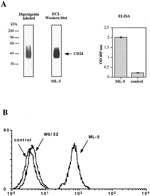
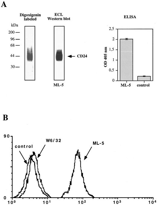
Isolated CD24 binds P-selectin.Previous analysis in the mouse has shown that HSA, the murine homologue to CD24,26 is a ligand for mouse P-selectin.32 33 Therefore, we set out to test whether CD24 on human cells had a similar role. For functional analysis CD24 was first isolated by affinity chromatography from lysate of neutrophils as well as from KS and SW2 tumor cells. As shown in Fig 3A the purified CD24 reacted positive in ELISA and showed the expected bands of 38 to 68 kD in SDS-PAGE. The affinity-purified CD24 could be adsorbed to polystyrene beads. As shown in Fig 3B the beads were stained with the CD24-specific MoAb ML-5 but not with control MoAbs against human MHC class I or DREG 56 (anti–L-selectin, not shown).
Characterization of purified CD24. (A) Affinity-purified CD24 antigen isolated from human neutrophils was analyzed by Western blotting. Antigen was detected by MoAbs to CD24 (mixture of ML-5, SWA11, 32D12) followed by peroxidase-conjugated secondary antibody and ECL detection. Antigen was also detected on the membrane after treatment with digoxigenin-3-O-succinyl-ε-amino-caproic acidhydrazide followed by Digoxigenin-specific antibodies. Antigen was also coated to microtiter plates and analyzed by ELISA using MoAb ML-5 hybridoma supernatant or tissue culture medium for control. (B) Detection of CD24 after coating to polystyrene beads. Beads were stained using MoAb ML-5 specific for CD24 or control MoAbs W 6/32 to MHC class I followed by PE-conjugated rabbit-antimouse IgG. Background control was the PE-conjugated antibody alone. Stained beads were analyzed by FACScan.
Characterization of purified CD24. (A) Affinity-purified CD24 antigen isolated from human neutrophils was analyzed by Western blotting. Antigen was detected by MoAbs to CD24 (mixture of ML-5, SWA11, 32D12) followed by peroxidase-conjugated secondary antibody and ECL detection. Antigen was also detected on the membrane after treatment with digoxigenin-3-O-succinyl-ε-amino-caproic acidhydrazide followed by Digoxigenin-specific antibodies. Antigen was also coated to microtiter plates and analyzed by ELISA using MoAb ML-5 hybridoma supernatant or tissue culture medium for control. (B) Detection of CD24 after coating to polystyrene beads. Beads were stained using MoAb ML-5 specific for CD24 or control MoAbs W 6/32 to MHC class I followed by PE-conjugated rabbit-antimouse IgG. Background control was the PE-conjugated antibody alone. Stained beads were analyzed by FACScan.
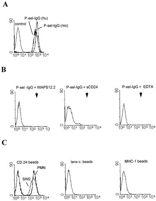
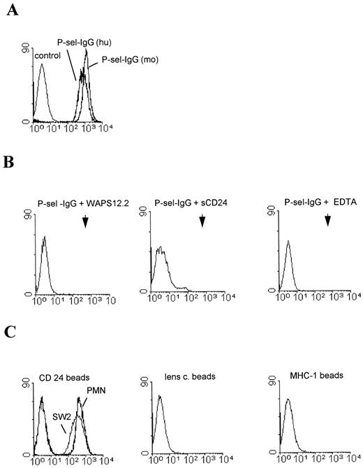
To study whether isolated CD24 could bind to P-selectin we used FACS staining with P-selectin-IgG. Figure 4A shows that the beads coated with CD24 from KS tumor cells could be stained with human or mouse P-selectin–IgG. The reactivity of the human P-selectin–IgG was blocked by an MoAb to human P-selectin, and was abolished in the presence of 5 mmol/L EDTA or soluble CD24 (Fig 4B). CD24 isolated from SW2 or neutrophils and coated to beads showed a similar reactivity. Again the beads were stained with human (Fig 4C) or mouse P-selectin–IgG (not shown). Control beads coated with lens culinaris binding glycoproteins from SW2 tumor cells or MHC class I did not bind to P-selectin–IgG (Fig 4C).
Fluorescent staining of CD24-coated beads with selectin-IgGs. (A) CD24-coated polysterene beads derived from KS tumor cells were stained with human or mouse P-selectin–IgG chimeric proteins and FITC-conjugated goat antibodies directed against the Fc part of human IgG. (B) Staining of CD24 beads (KS cells) with human P-selectin–IgG in the presence of 2 mmol/L EDTA, an MoAb to P-selectin or soluble CD24. Arrowheads indicate the position of the positive control (see A). (C) CD24-coated latex beads derived from SW2 tumor cells or neutrophils, control beads coated with lens culinaris glycoproteins from SW2 cells, or MHC class I–coated beads were stained with human P-selectin–IgG as described.
Fluorescent staining of CD24-coated beads with selectin-IgGs. (A) CD24-coated polysterene beads derived from KS tumor cells were stained with human or mouse P-selectin–IgG chimeric proteins and FITC-conjugated goat antibodies directed against the Fc part of human IgG. (B) Staining of CD24 beads (KS cells) with human P-selectin–IgG in the presence of 2 mmol/L EDTA, an MoAb to P-selectin or soluble CD24. Arrowheads indicate the position of the positive control (see A). (C) CD24-coated latex beads derived from SW2 tumor cells or neutrophils, control beads coated with lens culinaris glycoproteins from SW2 cells, or MHC class I–coated beads were stained with human P-selectin–IgG as described.
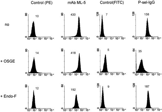
CD24 carries O-linked glycans and the HNK-1 epitope (CD57).We investigated the type of glycan involved in the P-selectin–IgG binding to CD24. As shown in Fig 5 for neutrophil CD24, treatment with endoglycosidase F, which removes N-linked glycans from the protein, caused a reduction in MoAb ML-5 staining (∼50%) yet left the reactivity with P-selectin–IgG intact. In contrast, short treatment (30 minutes) of CD24 with OSGE, which cleaves the protein at sialylated O-linked glycans, significantly reduced P-selectin–IgG staining. The reactivity with ML-5 was retained or was only weakly decreased. Treatment of the beads for prolonged time (2 to 3 hours) abolished also MoAb ML-5 reactivity (see below).
Effect of enzymatic treatment on P-selectin binding to CD24-coated beads. CD24-coated beads derived from PMNs were stained with human P-selectin–IgG and MoAb ML-5 against CD24 after treatment of the beads with endoglycosidase F to remove N-linked glycans or with OSGE from Pastorella hemolytica to cleave the protein at O-linked glycans. Samples treated with enzyme buffer alone did not show any change (not shown). Note that similar results were obtained with beads coated with CD24 antigen derived from SW2 or KS tumor cells.
Effect of enzymatic treatment on P-selectin binding to CD24-coated beads. CD24-coated beads derived from PMNs were stained with human P-selectin–IgG and MoAb ML-5 against CD24 after treatment of the beads with endoglycosidase F to remove N-linked glycans or with OSGE from Pastorella hemolytica to cleave the protein at O-linked glycans. Samples treated with enzyme buffer alone did not show any change (not shown). Note that similar results were obtained with beads coated with CD24 antigen derived from SW2 or KS tumor cells.
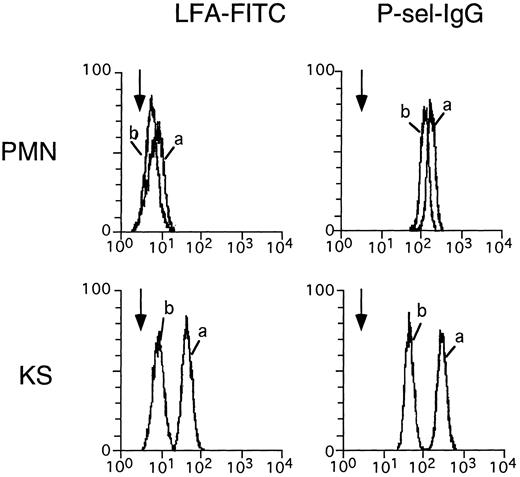
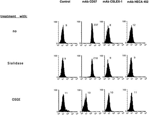
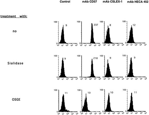
We investigated the presence of sialic acid on CD24 by using the sialic-acid–specific lectin LFA and compared both neutrophilic and KS-tumor–derived CD24. The amount of CD24 associated with each sort of beads was approximately equal as measured by cytofluorographic analysis with MoAb ML-5 (not shown). Staining of the beads with FITC-conjugated LFA showed a low level of staining with neutrophilic CD24 and a much higher level with KS tumor CD24 beads. (Fig 6). Treatment with Vibrio cholerae sialidase reduced the lectin staining strongly for tumor but only weakly for neutrophil CD24 (Fig 6). The lower level of expression as well as the resistance to sialidase treatment suggested that CD24 from neutrophils contained a higher degree of glycans in which the sialic acid was substituted by sulfate groups. Such forms are known to be resistant to sialidase treatment. Sialidase treatment also affected the staining with P-selectin–IgG to different extents. Whereas the tumor-derived CD24 was clearly decreased, the neutrophil CD24 was not (Fig 6). The MoAb ML-5 reactivity was not influenced by sialidase treatment. HSA can be modified with the HNK-1/L2 epitope.32 Therefore, we analyzed the CD24-associated glycans using carbohydrate-specific MoAbs, including an MoAb against CD57 (HNK-1). As shown in Fig 7, the CD24 from neutrophils stained strongly with the CD57-specific MoAb. The staining was completely abolished after OSGE treatment of the beads. Staining with the MoAb to CD57 was not affected after sialidase treatment (Fig 7) or by endoglycosidase F treatment (not shown). As shown in Fig 7 the CD24 beads did not react with MoAbs HECA 452 or CSLEX-1 against sialylLex and sialylLea, although both antibodies could bind to human neutrophils (not shown).
Effect of sialidase treatment. CD24-coated beads were stained with FITC-conjugated LFA before (a) or after (b) sialidase treatment. Background staining of the beads was done in the presence of 5 mmol/L ETDA which prevents lectin and P-selectin–IgG specific staining and is indicated by arrowheads.
Effect of sialidase treatment. CD24-coated beads were stained with FITC-conjugated LFA before (a) or after (b) sialidase treatment. Background staining of the beads was done in the presence of 5 mmol/L ETDA which prevents lectin and P-selectin–IgG specific staining and is indicated by arrowheads.
Analysis of CD24 associated carbohydrates. CD24 beads derived from PMNs were stained with MoAb HECA-452 (sialylLex/a) and MoAb CSLEX (sialylLex) followed by phycoerythrin (PE)-conjugated secondary antibodies goat-antirat-IgG or antimouse-IgG, respectively, or biotinylated CD57 MoAb followed by streptavidin-PE. Beads were stained also after treatment with OSGE from P hemolytica to cleave the protein at O-linked glycans. Samples treated with enzyme buffer alone did not show any change (not shown). Note that similar results were obtained with beads coated with CD24 antigen derived from SW2 tumor cells.
Analysis of CD24 associated carbohydrates. CD24 beads derived from PMNs were stained with MoAb HECA-452 (sialylLex/a) and MoAb CSLEX (sialylLex) followed by phycoerythrin (PE)-conjugated secondary antibodies goat-antirat-IgG or antimouse-IgG, respectively, or biotinylated CD57 MoAb followed by streptavidin-PE. Beads were stained also after treatment with OSGE from P hemolytica to cleave the protein at O-linked glycans. Samples treated with enzyme buffer alone did not show any change (not shown). Note that similar results were obtained with beads coated with CD24 antigen derived from SW2 tumor cells.
Mapping of the CD24-associated P-selectin binding site.To analyze the spatial relationship of the MoAb ML-5 in relation to the P-selectin binding site, time-course analyses of OSGE treatment were performed on neutrophil CD24 beads. We assumed that OSGE cleavage procedes from the N-terminus and that the adsorbtion of isolated CD24 to the beads occurs via the hydrophobic lipid tail, thereby exposing the N-terminus similar to the cell surface. OSGE treatment was performed for variable length of time and the binding of P-selectin–IgG, CD57, and ML-5 was tested at each timepoint. As shown in Table 1, the P-selectin–IgG and CD57 (HNK-1) binding declined concomitantly after a short period of time (10 to 30 minutes) whereas a significant loss of ML-5 reactivity occurred much later.
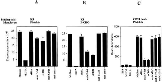
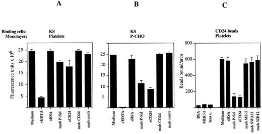
Involvement of CD24 in tumor-cell binding to platelets.We next studied the role of CD24 in the interaction of tumor cells with P-selectin using functional binding assays. The binding of CD24 beads to human platelet monolayers expressing P-selectin was investigated. As shown in Fig 8C the CD24 beads were able to bind, whereas the control beads coated with BSA, lens culinaris glycoproteins, or MHC class I antigen could not bind (Fig 8C). The binding of the CD24 beads was P-selectin dependent because it was blocked with MoAb WAPS 12.2 against P-selectin. It was also inhibited in the presence of soluble CD24 (2 μg/mL) but not by HSA from ESb that cannot bind to P-selectin.32 The available MoAbs to CD24 (ML-5, SWA11, 32D12) were unable to inhibit the binding of the CD24 beads, suggesting that they were functionally nonblocking. The binding of KS tumor cells to the platelets or P-selectin–transfected CHO cells was also studied. As shown in Fig 8A and B the KS cells could bind to the platelet monolayer as well as to the P-selectin–CHO cells in a Ca- and Mg-dependent manner since it was blocked in the presence of 5 mmol/L EDTA. The binding was mediated in part by P-selectin because the MoAb to P-selectin could block the interaction by ∼20% (platelets) or ∼50% (P-selectin–CHO), respectively. Purified CD24 at 2 μg/mL blocked the binding of KS cells to both substrates to a similar extend as the P-selectin MoAb. HSA from ESb or the MoAb ML-5 to CD24 failed to block the binding of KS cells to both substrates. It is conceivable that the non–P-selectin–mediated binding in both types of cell-cell binding assays was largely caused by integrins.
Binding of CD24-coated beads and KS cells to platelets and P-CHO. PMA-activated platelets were adsorbed to 96-well tissue culture plates at 37°C for 1 hour (A). P-CHO cells were grown to confluency in 96-well plates (B). Europium-labeled KS tumor cells were allowed to bind in the absence or presence of the indicated antibodies or 2 μg/mL soluble CD24 or HSA, respectively. Bound and nonbound cells were separated by buoyant density as described in Materials and Methods. After fixation of bound cells the Europium fluorescence was measured. (C) PMA-activated platelets were adsorbed to glass slides. CD24 beads and control beads were allowed to bind in the presence or absence of soluble glycoproteins or the indicated MoAbs. The binding assay was performed for 30 minutes at room temperature. The slides were washed and fixed in 2% glutaraldehyde before counting.
Binding of CD24-coated beads and KS cells to platelets and P-CHO. PMA-activated platelets were adsorbed to 96-well tissue culture plates at 37°C for 1 hour (A). P-CHO cells were grown to confluency in 96-well plates (B). Europium-labeled KS tumor cells were allowed to bind in the absence or presence of the indicated antibodies or 2 μg/mL soluble CD24 or HSA, respectively. Bound and nonbound cells were separated by buoyant density as described in Materials and Methods. After fixation of bound cells the Europium fluorescence was measured. (C) PMA-activated platelets were adsorbed to glass slides. CD24 beads and control beads were allowed to bind in the presence or absence of soluble glycoproteins or the indicated MoAbs. The binding assay was performed for 30 minutes at room temperature. The slides were washed and fixed in 2% glutaraldehyde before counting.
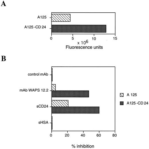
SW2 cells also bound to platelets and P-CHO in a P-selectin–dependent manner. However, these cells formed homotypic aggregates during the assay which did not permit an accurate quantification (data not shown). To further assess the role of CD24 we compared the binding of CD24 transfected A125-7/4 adenocarcinoma cells versus nontransfected cells. Initial experiments indicated that the CD24-transfected cells could be stained much better with the P-selectin–IgG compared with the parental cells (not shown). As indicated in Fig 9A the CD24-transfected cell line showed also an increased binding to the platelet monolayer. In addition, the relative blocking ability of the P-selectin–specific MoAb and the soluble CD24 was much higher for CD24 transfected A125-7/4 cells (50% to 60%) as compared with the nontransfected A125 cells (8% to 22%). The ability of purified CD24 to block the binding of nontransfected cells, though to a much lesser extent, suggested that the CD24-associated carbohydrates could compete for other nondefined ligands. However, in the presence of CD24 the P-selectin–mediated binding was clearly enhanced.
Binding and inhibition of CD24-transfected A125 adenocarcinoma cell versus nontransfected cells to human platelets. (A) PMA-activated platelets were adsorbed to 96-well tissue culture plates at 37°C for 1 hour. Europium-labeled tumor cells (A125 v A125-CD24) were adjusted to 106/mL and equal numbers of cells were allowed to bind. Note that the Eur3+ labeling of both types of cells was similar. (B) PMA-activated platelets were adsorbed to 96-well tissue culture plates at 37°C for 1 hour. Europium-labeled tumor cells A125 and A125-CD24 were allowed to bind in the absence or presence of the indicated antibodies or 2 μg/mL soluble CD24 or HSA, respectively. Bound and nonbound cells were separated by buoyant density as described in Materials and Methods. After fixation of bound cells the Europium fluorescence was measured and the results are expressed in percent inhibition of the added compound relative to medium control. Three independent experiments with similar results were performed for (A) and (B) and one representative experiment is shown.
Binding and inhibition of CD24-transfected A125 adenocarcinoma cell versus nontransfected cells to human platelets. (A) PMA-activated platelets were adsorbed to 96-well tissue culture plates at 37°C for 1 hour. Europium-labeled tumor cells (A125 v A125-CD24) were adjusted to 106/mL and equal numbers of cells were allowed to bind. Note that the Eur3+ labeling of both types of cells was similar. (B) PMA-activated platelets were adsorbed to 96-well tissue culture plates at 37°C for 1 hour. Europium-labeled tumor cells A125 and A125-CD24 were allowed to bind in the absence or presence of the indicated antibodies or 2 μg/mL soluble CD24 or HSA, respectively. Bound and nonbound cells were separated by buoyant density as described in Materials and Methods. After fixation of bound cells the Europium fluorescence was measured and the results are expressed in percent inhibition of the added compound relative to medium control. Three independent experiments with similar results were performed for (A) and (B) and one representative experiment is shown.
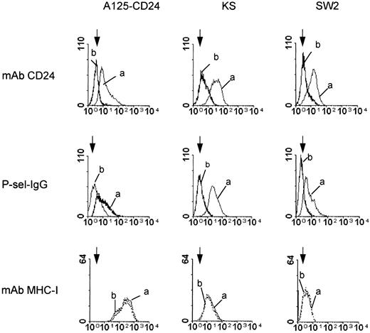
PIPL-C treatment of carcinoma cells affects P-selectin–IgG staining.CD24 is a GPI-anchored cell surface molecule which can be removed by PIPL-C treatment. We tested whether the treatment would also influence the staining of carcinoma cells and the CD24-transfected cell line. As shown in Fig 10, the PIPL-C treatment removed CD24 as demonstrated by reduced cell-surface staining with MoAb ML-5 to CD24 and concomitantly reduced the staining of the cells with the P-selectin–IgG. As expected, the staining of the transmembranal MHC class-1 antigen was not effected by the PIPL-C treatment.
Effect of PIPL-C treatment on CD24 or P-selectin–IgG staining. CD24-transfected adenocarcinoma cells (A125-CD24), KS cells, or SW2 cells were stained before (a) or after PIPL-C treatment (b) with MoAb ML-5 to CD24, human P-selectin–IgG, or MoAb W6/32 to MHC class I for control. The arrowheads indicate the negative control using the secondary antibody only.
Effect of PIPL-C treatment on CD24 or P-selectin–IgG staining. CD24-transfected adenocarcinoma cells (A125-CD24), KS cells, or SW2 cells were stained before (a) or after PIPL-C treatment (b) with MoAb ML-5 to CD24, human P-selectin–IgG, or MoAb W6/32 to MHC class I for control. The arrowheads indicate the negative control using the secondary antibody only.
DISCUSSION
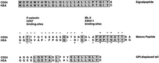
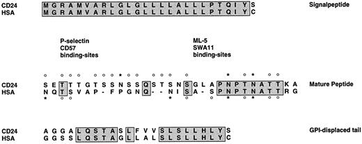
The present work was initiated by our observation that some forms of HSA, the murine homologue of CD24, could serve as a specific ligand for P-selectin.32,33 HSA forms that were modified with the HNK-1/L2 sulfate-containing carbohydrate epitope could react with P-selectin–IgG in biochemical assays32 as well as support the binding of myeloid cells to P-selectin on platelets and endothelial cells in functional assays.33 We now tested the hypothesis of whether CD24 could act as a ligand for human P-selectin in a similar way. This is not trivial because during evolution the sequence of the HSA and CD24 coreproteins have significantly diverged (see Fig 11). We were particulary interested in the role of CD24 on human tumor cells where it is abundantly expressed. Here we present evidence that CD24 from carcinoma cells can bind to P-selectin and that CD24 is a major cell-surface glycoprotein that supports the binding of tumor cells to P-selectin.
Sequence comparison of murine HSA and human CD24. Primary amino acid sequences for HSA and human CD24 were aligned. Boxes indicate identity of sequence. Potential N-linked glycosylation sites are marked with an asterisk, and potential O-linked glycosylation sites are marked with a circle above the amino acid. Putative binding sites for P-selectin, MoAb CD57, and the CD24-specific MoAbs SWA11 and ML-5 are indicated.
Sequence comparison of murine HSA and human CD24. Primary amino acid sequences for HSA and human CD24 were aligned. Boxes indicate identity of sequence. Potential N-linked glycosylation sites are marked with an asterisk, and potential O-linked glycosylation sites are marked with a circle above the amino acid. Putative binding sites for P-selectin, MoAb CD57, and the CD24-specific MoAbs SWA11 and ML-5 are indicated.
Our results are based on the analysis of purified CD24 that was isolated from tumor cells or neutrophils. Both normal and tumor-cell–derived CD24 forms were able to bind to P-selectin. The binding appeared to involve the selectin-portion of P-selectin because it was divalent cation dependent and was inhibited by a functionally blocking MoAb against this domain. Control proteins coated to beads, ie, MHC class I or a mixture of lens culinaris binding glycoproteins could not bind to P-selectin. Importantly, the tumor cells from which CD24 was isolated did not express PSGL-1, excluding the possibility of contamination with the already known P-selectin ligand. We conclude that CD24 can act as a P-selectin ligand in its own right and is functionally similar to HSA. However, in contrast to HSA which did not bind to murine E-selectin, CD24 also bound to human E-selectin (S.A. and P.A., unpublished observations). Additional carbohydrate analysis showed that the binding of P-selectin to CD24 was sensitive to OSGE treatment but not endoglycosidase F, suggesting the presence of O-linked mucin-type glycans. Unexpectedly, CD24 did not carry detectable sialylLex/a carbohydrate epitopes. Instead, it was modified with the CD57/HNK-1 epitope, a sulfate containing carbohydrate moiety related in structure to the SGNL (3-sulfo-glucoronyl-neolactose) glycolipid that has been found previously to be associated with P-selectin binding.13 Thus, CD24 constitutes a novel member of the group of cell-surface mucins that can serve as selectin ligands.
It is interesting to compare CD24 to PSGL-1, another mucin and major ligand for P-selectin and E-selectin on leukocytes.15-19,45 PSGL-1 carries sLex containing O-linked glycans that appear to be necessary for P-selectin binding.15-19,21 A Tyr-sulfation site within the N-terminal 19 amino acids of the mature PSGL-1 polypeptide was described that seems to be particularily important in rendering the molecule a high-affinity ligand.18,19 MoAb PL1 blocks the binding of purified PSGL-1, the binding of neutrophils to immobilized P-selectin, and the rolling of neutrophils on P-selectin–transfected CHO cells.20 Recently, epitope mapping has identified a linear sequence LDYDFLPETEPPEM to which MoAb PL1 could bind.46 This site is close to the N-terminus of PSGL-1 and includes the putative Tyr-sulfation site.18,19 It is conceivable that under physiologic conditions this site is indeed sulfated. The sulfate group together with several surrounding acidic amino acids could create a negatively charged patch at the N-terminus of PSGL-1. The CD24 glycoprotein does not contain a sulfated tyrosine residue, because no tyrosine is present in the primary sequence. However, the association of both mouse and human CD24 with the HNK-1 sulfate-containing epitope, the observed correlation in the mouse between HNK-1 expression and P-selectin binding, and the previous finding that the SGNL lipid, which carries the HNK-1 epitope, is recognized by P-selectin13 may suggest that this epitope could play a similar role as the sulfated Tyr residue in PSGL-1.
A sequence comparison of human and mouse CD24 shows that during the evolution the human CD24 molecule has acquired additional serine and threonine residues rendering the molecule more like a typical mucine (see Fig 11). Putative O-linked glycosylation sites emerged in the N-terminal half of the molecule, creating a densely packed stretch of the glycans at the tip of the molecule. Additional glycosylation sites are present juxtaposed to the membrane surface, giving the molecule a dumbbell-like shape. The epitope for the ML-5 MoAb (and several other CD24 MoAbs) has been located in the center of the molecule around the LAP motif.47 In agreement with the OSGE kinetic experiment in Table 1 it is conceivable that P-selectin–IgG and CD57 binding sites are located at the tip of the molecule as opposed to the ML-5 epitope and are therefore removed consecutively. The spatial separation of the P-selectin binding site and the ML-5 epitope is also supported by the lack of CD24 MoAbs to inhibit the binding of beads to the platelet monolayer. Thus, the P-selectin binding site is distinct from the epitope for the CD24-specific MoAb ML-5.
The available MoAbs to CD24 also did not block in cell-cell binding assays in which the binding of KS cells to platelets or P-selectin–transfected CHO cells was tested. However, soluble CD24 was able to block this binding to a similar extent as the P-selectin–specific MoAb. Soluble HSA from mouse ESb cells, that cannot bind to P-selectin, could not block. This suggested that the CD24-associated glycans were important in mediating the blocking effect. Further support for an important role of CD24 in the binding to P-selectin came from experiments with the A125-7/4 adenocarcinoma cells. The CD24 transfected subline bound much better to P-selectin and this increased binding appeared to be due to the expression of CD24 as indicated by competition experiments. Importantly, PIPL-C treatment of the transfected adenocarcinoma cells as well as the two carcinom cells studied removed CD24 from the cell surface and at the same time decreased P-selectin binding. Interestingly, in neutrophils DeBruijne-Admiraal et al48 have failed to detect a significant role of GPI-anchored molecules, including CD24, in the initial binding to P-selectin. It may well be that PSGL-1 is the dominant ligand in this process. PSGL-1 is a large molecule that has been found to localize on the tip of microvilli on neutrophils and it has been argued that this localization may facilitate the interaction with P-selectin under high shear forces.20,23,49 In contrast, GPI-anchored molecules, like CD24, typically form dense clusters at the cell surface50 51 and therefore could present carbohydrates at high density. It is indeed possible that different classes of P-selectin ligands exist which only become apparent if the dominant molecule PSGL-1 is missing.
Finally, it is interesting to address the potential role of CD24 on tumor cells. CD24 has been found to be expressed on leukemias of the B-cell lineage such as acute lymphoblastic leukemia (ALL), chronic lymphocytic leukemia (CLL), and non-Hodgkin's B-cell lymphoma.52 The reduction or absence of CD24 expression on ALL cells predicts a better prognosis.53 Expression of CD24 was also shown on several solid (nonhematopoietic) tumors such as small cell lung carcinoma, neuroblastoma, rhabdomyosarcoma, and renal cell carcinoma.28,54 Moreover, recent studies have suggested that CD24 is a potential early tumor marker gene which correlates with p53 mutation in hepatocellular carcinoma.55 CD24 was also found to be associated with c-fgr or lyn in human tumor cell lines.56 The significance of CD24 expression on so many tumors is not known. As shown in this study, CD24 from two tumors were able to bind to P-selectin and additional screening of human tumor cell lines have shown a good correlation between CD24 expression and P-selectin binding (Z.M.S. and P.A., unpublished observations). Studies by Stone and Wagner8 have provided evidence that many tumor cell lines can bind to platelets via P-selectin. The ability of tumor cells to bind to platelets in the blood is considered to be important because stabilized platelets-tumor thrombi can physically protect tumor cells from destruction as well as help and promote tumor extravasation and tissue penetration.57 58 Thus, CD24 expression might be of advantage for tumor cells. It will be important to investigate whether the CD24/P-selectin binding pathway as described here plays a role in the interaction of tumor cells with platelets or endothelial cells in vivo and has significance in tumor metastasis.
ACKNOWLEDGMENT
We thank Claudia Geiger for excellent technical assistance and Dr B. Gückel for tumor cell lines and helpful discussions. We thank K.L. Moore for comments on the manuscript.
Supported by a grant from Deutsche Forschungsgemeinschaft to P.A. (Al 170/4-1 and Al 170/4-2) and by a guest scientist stipend from DKFZ to Z.M.S. R.S. was supported by a grant from the Swiss National Science Foundation 31-37258.93.
Address reprint requests to Peter Altevogt, PhD, Tumor Immunology Programme, 0710, German Cancer Research Center, Im Neuenheimer Feld 280, D-69120 Heidelberg, Germany.


