Abstract
We have identified a small molecular weight compound, SCH 14988, which specifically stimulates in vitro granulocyte-colony stimulating factor (G-CSF ) production from activated human peripheral blood mononuclear cells and monocytes but not other cytokines or CSFs with hematoregulatory activity. In vivo administration of SCH 14988 to mice rendered neutropenic by cyclophosphamide treatment resulted in the accelerated recovery of the peripheral neutrophil compartment. This activity correlated with increased in vivo G-CSF levels and stimulation of marrow granulopoiesis, and was comparable to that of exogenously administered recombinant human G-CSF. No alterations to other leukocyte populations in peripheral blood, spleen, or the peritoneal cavity were observed. These findings suggest that SCH 14988 may be clinically useful to enhance neutrophil granulopoiesis, as well as to study the mechanisms involved in G-CSF gene regulation.
GRANULOCYTE COLONY-stimulating factor (G-CSF ) is produced by monocytes, T lymphocytes, fibroblasts, and endothelial cells and stimulates the production of segmented neutrophils from the bone marrow.1-3 Administration of recombinant murine or recombinant human (rh) G-CSF has been shown to induce neutrophil granulocytosis in normal animals4-8 and to accelerate the recovery of neutrophil numbers and reduce neutrophil nadir times after chemotherapy9-12 and radiation therapy13-17 in animal models. Rh G-CSF (Neupogen, filgastrim) has shown marked efficacy in cancer patients and after bone marrow transplantation, thereby permitting more rigorous chemotherapy regimens.3,18,19 However, the high cost of Neupogen therapy, the requirement for subcutaneous or intravenous administration, and its potential side effects (eg, medullary bone pain)1-3,18,19 suggest that small molecular weight compounds that can be delivered at low cost would be of significant clinical use.
We have identified a small molecular weight compound, SCH 14988, which significantly and selectively stimulates G-CSF production, but not a variety of other cytokines, from in vitro phytohemagglutin (PHA)-stimulated human peripheral blood mononuclear cells (PBMNCs) and from lipopolysaccharide (LPS)-stimulated human peripheral blood monocytes (PBMNs). Administration of SCH 14988 to cyclophosphamide (CY)-treated mice led to enhanced recovery of the peripheral neutrophil compartment and possessed comparable activity with exogenous rhG-CSF treatment. No alterations to other leukocyte populations in peripheral blood, spleen, or peritoneal cavity were seen. The activity of SCH 14988 correlated with an increase in steady-state bone marrow G-CSF mRNA levels as well as serum G-CSF protein levels. These results indicate that SCH 14988 may be of use in the clinic for enhancing neutrophil granulopoiesis. Moreover, these data suggest that SCH 14988 may be a useful tool for investigating the mechanisms involved in G-CSF gene regulation.
MATERIALS AND METHODS
In vitro cell stimulation and cytokine measurements.Human PBMNCs were prepared from normal donors by standard techniques using Ficoll-Hypaque (Pharmacia, Piscataway, NJ). For determination of the effect of SCH 14988 on cytokine production by PBMNCs, cells were cultured at 1 × 10⁶ cells/mL with 5 μg/mL PHA (Murex Diagnostics, Dartford, UK) in the presence of SCH 14988 or vehicle (0.1% dimethylsulfoxide). Human PBMNs were prepared from normal donors by centrifugal elutriation (Beckman Instruments, Somerset, NJ) as described.20 Monocyte purity was greater than 88%, as determined by flow cytometry. Monocytes were cultured at 1 × 10⁶ cells/mL with 0.1 to 1 ng/mL LPS (Sigma, St Louis, MO) in the presence or absence of SCH 14988. Supernatants were collected after 40 hours and cytokine levels determined by enzyme-linked immunosorbent assay (R & D Systems, Minneapolis, MN). All data is expressed as percent of control; absolute cytokine values in the control group are listed in the appropriate figure legends. Cell viability was unaffected by any of the treatment conditions, and was always greater than 90%.
Determination of steady-state human G-CSF mRNA levels.Human PBMNCs were prepared as described and stimulated with PHA plus SCH 14988 or vehicle for 24 hours. Cells were collected, washed, and total cellular RNA prepared using Trisolv (Biotex Laboratories, Edmonton, Alberta, Canada). RNA was reverse transcribed (GIBCO-BRL, Grand Island, NY) and subjected to polymerase chain reaction (PCR) for 25 cycles at 94°C for 0.5 minutes, 50°C for 0.5 minutes and 72°C for 1 minute. For G-CSF, a 560-bp product was obtained using the following primers: forward, 5′-ACTCCATAGCGGCCTTTTCC-3′; and reverse, 5′-TCATCCCAGTGCCCATTGCA-3′. For glyceraldehyde 3-phosphate dehydrogenase (GAPDH), a 983-bp product was obtained using these primers: forward, 5′-TGAAGGTCGGAGTCAACGGATTTGGT-3′; and reverse, 5′-CATGTGGGCCATGAGGTCCACCAC-3′. For photographic reproduction, a Polaroid photograph (Polaroid Corp, Cambridge, MA) was optically scanned as a Tagged Image File Format (TIFF) file using Adobe Photoshop (Adobe Systems, San Jose, CA). Quantitation of G-CSF levels for each treatment group was performed by normalizing relative gray-scale units for G-CSF to that of the respective GAPDH using Scan Analysis software (Biosoft, Cambridge, UK). Specificity of the G-CSF band was confirmed by Southern blotting with a 32P-labeled internal 120-bp probe prepared by PCR.
In vivo neutropenia model.Eight- to 12-week old female BALB/cJ mice were dosed with 250 mg/kg CY on day 0 by intraperitoneal (IP) injection and then treated daily, starting on day 1, with SCH 14988 at 40 mg/kg or carboxymethylcellulose (CMC) vehicle IP. In some experiments, rhG-CSF (R & D Systems) or the appropriate vehicle (phosphate-buffered saline [PBS] containing 1% normal mouse serum) was injected daily by subcutaneous injection. A separate group of normal age-matched untreated control mice were used for comparison in each experiment. At various timepoints, mice from each treatment group were sacrificed, and peripheral blood cells, spleen cells, or peritoneal cells were collected, washed, enumerated, and subjected to further analysis as described below. All animal care was in accordance with guidelines established by our institution's Animal Care and Use Committee.
Flow cytometry.All cells were washed extensively in Dulbecco's PBS containing 1% bovine serum albumin and 0.1% sodium azide and then stained with optimal amounts of fluorochrome-conjugated antibodies against Gr-1, B220, CD4, CD8 (Pharmingen, San Diego, CA), Mac-1 (Boehringer Mannheim, Indianapolis, IN), or IgM (Southern Biotechnology, Birmingham, AL). Cells were analyzed on a FACScan using LYSIS II or CellQuest software (Becton Dickinson Immunocytometry Systems, Mountain View, CA). Isotype-matched fluorochrome-conjugated antibodies were used as negative staining controls. The number of cells positive for a specific marker in a given tissue was calculated by multiplying the absolute number of leukocytes in that tissue by the percentage of positive cells for the marker.
Cytological analysis of bone marrow cells.Femoral bone marrow cells were prepared on day 6 after CY treatment, placed on glass slides, and stained with Wright-Giemsa for staging of granulopoiesis. A total of 300 cells per sample were counted.
Determination of steady-state G-CSF mRNA levels in murine bone marrow and peritoneum.CY-treated mice were administered SCH 14988 or vehicle as described above for 4 days. On day 5 post-CY, 24 hours after the last drug injection, bone marrow or peritoneal cells from each treatment groups were pooled and the RNA was isolated, reverse-transcribed, and subjected to PCR for 40 cycles using the following primers: murine G-CSF (mG-CSF), forward, 5′-GCTGTGGCAAACTGCACTATGGTC-3′; reverse, 5′-GAAGTGAAGGCTGGCATGGCGCTC-3′ (a 480-bp product); and β-actin, forward, 5′-ATGGTGGGAATGGGTCAGA-3′; reverse, 5′-GCACGATTTCCCTCTCAGCT-3′ (a 464-bp product). Specificity of the G-CSF band was confirmed by Southern blotting with a 32P-labeled EcoRI fragment of an mG-CSF–containing cDNA clone (provided by Dr S. Nagata, Osaka Bioscience Institute, Okaka, Japan). Quantitation was performed as described above by normalizing to β-actin levels.
Measurement of serum G-CSF levels.Serial dilutions of serum from CY-treated mice that received vehicle or SCH 14988 were harvested on day 5 or 6 and were added to the NFS-60 cell line as described.21 Serum from age-matched control mice was used for comparison. Proliferation was determined by assessing (3-(4,5-dimethylthiazol-2-yl)-2,5-diphenyltetrazolium bromide (MTT) reduction.
RESULTS
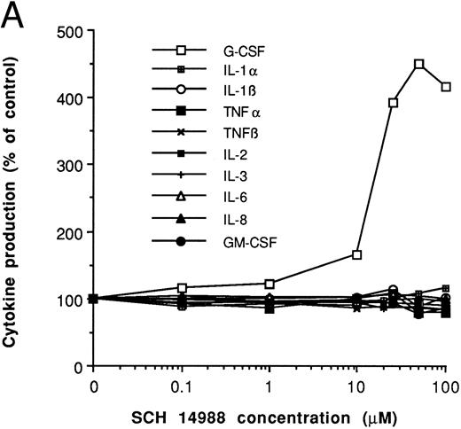
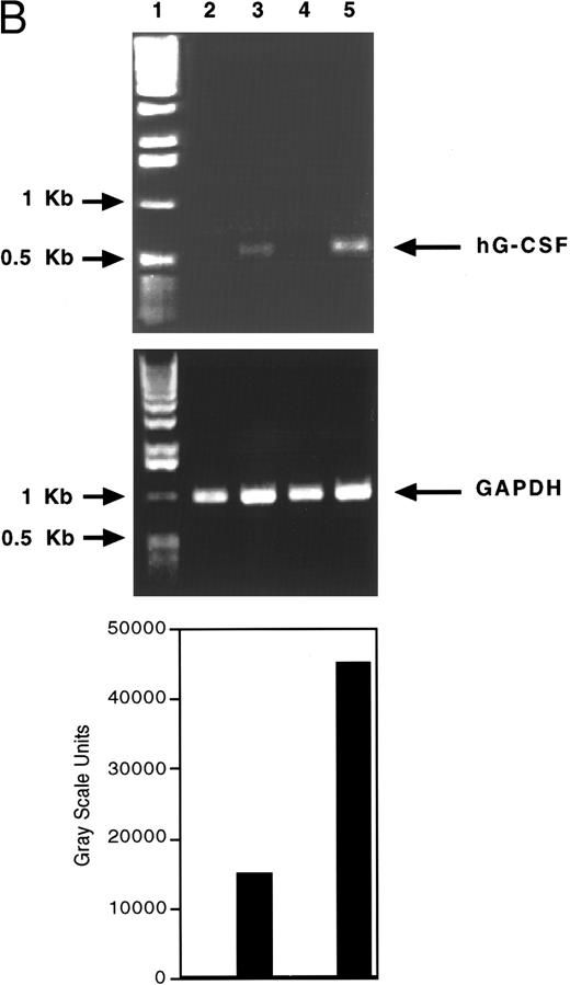
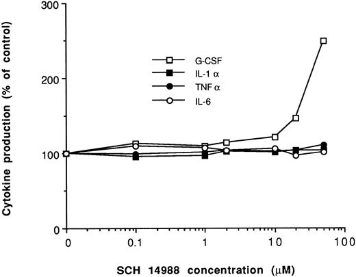
The molecule SCH 14988 is a pyridinyl-naphthyridinylsulfonyl acetamide originally synthesized as an immunomodulator (Fig 1). Examination of its activity in cultures of human PBMNCs showed that SCH 14988 was able to augment G-CSF production from PHA-stimulated cells by up to fourfold to fivefold at 25 to 50 μmol/L with an effective concentration50 of 15 μmol/L (Fig 2A). Similar activity was observed after concanavalin A treatment (data not shown). The effect of this compound was specific for G-CSF, because no change in the production of tumor necrosis factor α (TNFα), TNFβ, interleukin-1α (IL-1α), IL-1β, IL-2, IL-3, IL-6, IL-8 or granulocyte-macrophage colony-stimulating factor (GM-CSF ) was seen (Fig 2A). As depicted in Fig 2B, steady-state G-CSF mRNA levels also increased in these cultures by twofold to threefold, suggesting modulation by SCH 14988 of G-CSF gene expression by transcriptional or post-transcriptional mechanisms. Similar to the protein data, no change in steady-state TNFα or GM-CSF mRNA levels were observed (data not shown). Specific enhancement of G-CSF production was also observed after treatment of LPS-treated human peripheral blood elutriated monocytes (Fig 3). Moreover, SCH 14988 was able to significantly enhance G-CSF mRNA levels in cultures of pokeweed mitogen-stimulated murine bone marrow macrophages (data not shown). We did not observe any effect on G-CSF mRNA levels or protein production in any of the above systems when cells were treated with SCH 14988 alone or with similar concentrations of its two likely metabolic products, 6-(3-methyl- 2-pyridnyl)-1,7- naphthyridin-8-amine and N-[4-(aminosulfonyl)phenyl]acetamide.
Chemical structure of SCH 14988, N-[4-[[[6-(3-methyl-2-pyridinyl)- 1,7-naphthyridin-8-yl]amino]sulfonyl]phenyl]acetamide.
Chemical structure of SCH 14988, N-[4-[[[6-(3-methyl-2-pyridinyl)- 1,7-naphthyridin-8-yl]amino]sulfonyl]phenyl]acetamide.
Specific effect of SCH 14988 on G-CSF production by human PBMNCs. (A) Cells were cultured with PHA in the presence of varying concentrations of SCH 14988 or vehicle (0.1% DMSO). Data, expressed as percent of control, is normalized to the PHA plus vehicle (0 μmol/L SCH 14988) value, which was arbitrarily set at 100%. Each data point represents 4 to 12 donors; standard error was less than 20%. Absolute levels of each cytokine (in pg/mL) in the PHA plus vehicle group were IL-1α, 412 ± 113; IL-1β, 1,662 ± 304; IL-2, 781 ± 169; IL-3, 257 ± 36; IL-6, 10,367 ± 2,268; IL-8, 4,600 ± 246; TNFα, 2,225 ± 297; TNFβ, 1,890 ± 604; GM-CSF, 758 ± 205; and G-CSF, 3,018 ± 231. In the presence of PHA plus 25 μmol/L SCH 14988, G-CSF levels were increased to 14,185 ± 509 pg/mL. This increase in G-CSF levels was statistically significant relative to the PHA plus vehicle group, as determined by the Students' t-test (P < .01). For each cytokine, production was always less than 65 pg/mL in unstimulated cultures. (B) Steady-state G-CSF mRNA levels in PBMNCs cultured with vehicle (lane 2), PHA (lane 3), SCH 14988 alone at 25 μmol/L (lane 4), or PHA plus SCH 14988 (lane 5) for 24 hours. One kilobase ladder is shown in lane 1. Ethidium bromide-stained 1% agarose/Tris-acetate-EDTA gels are shown.
Specific effect of SCH 14988 on G-CSF production by human PBMNCs. (A) Cells were cultured with PHA in the presence of varying concentrations of SCH 14988 or vehicle (0.1% DMSO). Data, expressed as percent of control, is normalized to the PHA plus vehicle (0 μmol/L SCH 14988) value, which was arbitrarily set at 100%. Each data point represents 4 to 12 donors; standard error was less than 20%. Absolute levels of each cytokine (in pg/mL) in the PHA plus vehicle group were IL-1α, 412 ± 113; IL-1β, 1,662 ± 304; IL-2, 781 ± 169; IL-3, 257 ± 36; IL-6, 10,367 ± 2,268; IL-8, 4,600 ± 246; TNFα, 2,225 ± 297; TNFβ, 1,890 ± 604; GM-CSF, 758 ± 205; and G-CSF, 3,018 ± 231. In the presence of PHA plus 25 μmol/L SCH 14988, G-CSF levels were increased to 14,185 ± 509 pg/mL. This increase in G-CSF levels was statistically significant relative to the PHA plus vehicle group, as determined by the Students' t-test (P < .01). For each cytokine, production was always less than 65 pg/mL in unstimulated cultures. (B) Steady-state G-CSF mRNA levels in PBMNCs cultured with vehicle (lane 2), PHA (lane 3), SCH 14988 alone at 25 μmol/L (lane 4), or PHA plus SCH 14988 (lane 5) for 24 hours. One kilobase ladder is shown in lane 1. Ethidium bromide-stained 1% agarose/Tris-acetate-EDTA gels are shown.
SCH 14988 specifically induces G-CSF from LPS-stimulated human blood monocytes. Human PBMNs were cultured with 0.1 ng/mL LPS in the presence of varying concentrations of SCH 14988 or vehicle (0.1% DMSO). Data, expressed as percent of control, are normalized to the LPS plus vehicle (0 μmol/L SCH 14988) value, which was arbitrarily set at 100%. Each data point represents at least 6 donors. Absolute levels of each cytokine (in pg/mL) in the LPS plus vehicle group were IL-1β, 3,075 ± 417; IL-6, 4,800 ± 1,300; TNFα, 1,093 ± 245; and G-CSF, 1,088 ± 345. G-CSF levels were 2,721 ± 467 pg/mL in cultures treated with LPS plus 25 μmol/L SCH 14988. This enhancement of G-CSF production was statistically significant, as judged by the Students' t-test (P < .01). Cytokine levels were always less than 70 pg/mL in unstimulated cultures. In addition, in eight separate experiments, steady-state G-CSF mRNA levels (determined as described in Fig 2) in elutriated monocytes treated with LPS (0.1 ng/mL) plus SCH 14988 (25 μmol/L) were enhanced by threefold compared with monocytes treated with LPS alone.
SCH 14988 specifically induces G-CSF from LPS-stimulated human blood monocytes. Human PBMNs were cultured with 0.1 ng/mL LPS in the presence of varying concentrations of SCH 14988 or vehicle (0.1% DMSO). Data, expressed as percent of control, are normalized to the LPS plus vehicle (0 μmol/L SCH 14988) value, which was arbitrarily set at 100%. Each data point represents at least 6 donors. Absolute levels of each cytokine (in pg/mL) in the LPS plus vehicle group were IL-1β, 3,075 ± 417; IL-6, 4,800 ± 1,300; TNFα, 1,093 ± 245; and G-CSF, 1,088 ± 345. G-CSF levels were 2,721 ± 467 pg/mL in cultures treated with LPS plus 25 μmol/L SCH 14988. This enhancement of G-CSF production was statistically significant, as judged by the Students' t-test (P < .01). Cytokine levels were always less than 70 pg/mL in unstimulated cultures. In addition, in eight separate experiments, steady-state G-CSF mRNA levels (determined as described in Fig 2) in elutriated monocytes treated with LPS (0.1 ng/mL) plus SCH 14988 (25 μmol/L) were enhanced by threefold compared with monocytes treated with LPS alone.
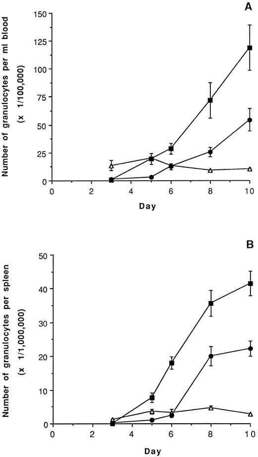
The marked and selective enhancement of G-CSF production by SCH 14988 led us to investigate whether this compound was capable of increasing neutrophil levels in vivo. To test this possibility, BALB/cJ mice were rendered neutropenic by treatment with a single sublethal dose of CY on day 0. The animals were then treated daily, starting on day 1, with 40 mg/kg SCH 14988 (a dose determined to be optimal from preliminary experiments) or CMC vehicle. Mice were sacrificed on days 3, 5, 6, 8, and 10, and leukocyte populations in peripheral blood and spleen were analyzed by flow cytometry. At all time points, tissues from SCH 14988- or vehicle-treated animals were compared with untreated, age-matched normal mice (that did not receive CY). As illustrated in Fig 4, SCH 14988 administration accelerated granulocyte recovery in blood and spleen with a return to normal numbers by day 5 in blood and day 4 in spleen. Cytological examination indicated that these granulocytes were exclusively segmented neutrophils, with no evidence for any increase in eosinophils or basophils (data not shown). Neutrophil recovery in CY-dosed animals given vehicle was consistently delayed by 1 to 2 days compared with SCH 14988-treated mice. SCH 14988 treatment also produced a greater degree of neutrophil granulocytosis in blood and spleen compared with administration of vehicle. This stimulatory effect of SCH 14988 on neutrophil levels was observed in 23 separate experiments. Doses of SCH 14988 greater than 40 mg/kg did not result in any further acceleration of recovery from neutropenia nor enhancement of peripheral neutrophil numbers. At no time did we observe any evidence of gross toxicity or abnormalities. A twofold to fivefold increase in the number of neutrophils present in the peritoneal cavity of SCH 14988-treated, compared with CY-dosed, vehicle-treated, animals was also observed (data not shown). In contrast to the pronounced effect of SCH 14988 on the neutrophil population, no effect on the number of Gr-1− Mac-1+ monocytes, CD4+ or CD8+ T cells, B220+ sIgM− pre-B cells, or B220+ sIgM+ B cells was noted in peripheral blood, spleen, or peritoneum (data not shown). Thus, the in vivo activity of SCH 14988 appears to be restricted to accelerating the recovery of the neutrophil compartment, consistent with the observed in vitro induction of G-CSF.
Acceleration of recovery from CY-induced neutropenia by SCH 14988. Mice were dosed as described in Materials and Methods and sacrificed on days 3, 5, 6, 8, or 10. Peripheral blood cells or spleen cells were collected, washed, and enumerated, and Gr-1 and Mac-1 staining were analyzed. Data is expressed as the mean ± standard error of mean (SEM) number of granulocytes (Gr-1+Mac-1+) per milliliter of blood (A) or per spleen (B). ▵ indicates normal age-matched control mice; • indicates CY-treated, vehicle-treated mice; ▪ indicates CY-treated, SCH 14988-treated mice. Differences between the vehicle-treated and SCH 14988-treated groups from days 5, 6, 8, and 10 are statistically significant at P < .05, as determined by the Students' t-test. Administration of the two likely metabolic products of SCH 14988 (see Results) or of analogs that did not induce G-CSF production in vitro had no effect on neutrophil recovery (data not shown).
Acceleration of recovery from CY-induced neutropenia by SCH 14988. Mice were dosed as described in Materials and Methods and sacrificed on days 3, 5, 6, 8, or 10. Peripheral blood cells or spleen cells were collected, washed, and enumerated, and Gr-1 and Mac-1 staining were analyzed. Data is expressed as the mean ± standard error of mean (SEM) number of granulocytes (Gr-1+Mac-1+) per milliliter of blood (A) or per spleen (B). ▵ indicates normal age-matched control mice; • indicates CY-treated, vehicle-treated mice; ▪ indicates CY-treated, SCH 14988-treated mice. Differences between the vehicle-treated and SCH 14988-treated groups from days 5, 6, 8, and 10 are statistically significant at P < .05, as determined by the Students' t-test. Administration of the two likely metabolic products of SCH 14988 (see Results) or of analogs that did not induce G-CSF production in vitro had no effect on neutrophil recovery (data not shown).
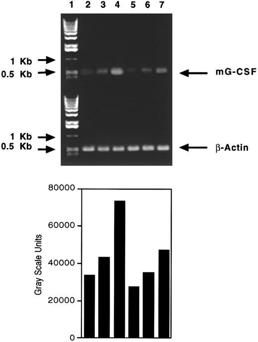
To confirm that SCH 14988 was acting in vivo by enhancing endogenous G-CSF production, we examined steady-state G-CSF mRNA levels in bone marrow, spleen, and peritoneal cells in mice depleted by CY and treated with SCH 14988. As depicted in Fig 5, SCH 14988 treatment reproducibly enhanced G-CSF mRNA levels in bone marrow by twofold and in peritoneal cells to a lesser extent. An increase in G-CSF mRNA levels was occasionally observed in spleen as well (data not shown). We also observed a twofold to threefold increase in serum G-CSF levels in SCH 14988-treated mice compared with vehicle-treated mice (Table 1). This augmentation of G-CSF mRNA and serum protein levels support and extend our previous results and suggest that the ability of SCH 14988 to enhance the recovery of CY-induced neutropenia is because of its G-CSF–stimulating activity.
Steady-state G-CSF mRNA levels in bone marrow and peritoneum after SCH 14988 treatment in neutropenic mice. Lane 1, 1-kb ladder; lane 2, bone marrow from normal, age-matched mice; lane 3, bone marrow from CY mice dosed with vehicle; lane 4, bone marrow from CY mice dosed with SCH 14988 at 40 mg/kg; lane 5, peritoneal cells from normal, age-matched animals; lane 6, peritoneal cells from CY mice dosed with vehicle; lane 7, peritoneal cells from CY mice treated with SCH 14988.
Steady-state G-CSF mRNA levels in bone marrow and peritoneum after SCH 14988 treatment in neutropenic mice. Lane 1, 1-kb ladder; lane 2, bone marrow from normal, age-matched mice; lane 3, bone marrow from CY mice dosed with vehicle; lane 4, bone marrow from CY mice dosed with SCH 14988 at 40 mg/kg; lane 5, peritoneal cells from normal, age-matched animals; lane 6, peritoneal cells from CY mice dosed with vehicle; lane 7, peritoneal cells from CY mice treated with SCH 14988.
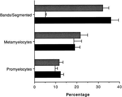
Acceleration of neutrophil recovery in SCH 14988-treated mice may occur by stimulating neutrophil granulopoiesis at the level of the bone marrow. To further examine the mechanism by which this compound enhances neutrophil recovery in CY-treated animals, we assessed bone marrow granulopoiesis by cytological examination. These studies showed significant increases in the proportion of segmented and banded neutrophils from SCH 14988-treated mice on day 6 with no change in the percentage of either promyelocytes or metamyelocytes (Fig 6). Thus, the ability of SCH 14988 to accelerate neutrophil recovery in the periphery correlated with an enhancement of granulopoiesis in the bone marrow, which was likely stimulated by the augmentation of G-CSF production. Attempts to accurately assess granulopoiesis at times earlier than day 6 by this method were unsuccessful because of the poor cell architecture of marrow cells still recovering from CY-induced effects.
Enhancement of banded/segmented neutrophils in bone marrow of SCH 14988-treated mice. BALB/cJ mice were treated with CY and SCH 14988 or vehicle as described in Fig 4 legend, and femoral bone marrow cells were prepared on day 6. Cells were placed on glass slides and stained with Wright-Giemsa before evaluation. Data are expressed as mean ± SEM and are representative of three separate experiments. ▪ indicates normal, age-matched control mice; □ indicates CY-treated, vehicle-treated mice; ▧ indicates CY-treated, SCH 14988-treated mice. The difference in the percentage of segmented neutrophils between the vehicle and SCH 14988 treatment groups is statistically significant at P < .001 as determined by the Students' t-test.
Enhancement of banded/segmented neutrophils in bone marrow of SCH 14988-treated mice. BALB/cJ mice were treated with CY and SCH 14988 or vehicle as described in Fig 4 legend, and femoral bone marrow cells were prepared on day 6. Cells were placed on glass slides and stained with Wright-Giemsa before evaluation. Data are expressed as mean ± SEM and are representative of three separate experiments. ▪ indicates normal, age-matched control mice; □ indicates CY-treated, vehicle-treated mice; ▧ indicates CY-treated, SCH 14988-treated mice. The difference in the percentage of segmented neutrophils between the vehicle and SCH 14988 treatment groups is statistically significant at P < .001 as determined by the Students' t-test.
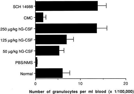
Consideration of alternative therapies for accelerating recovery from neutropenia in the clinic mandates that these alternatives show comparable or greater activity than rhG-CSF. We directly compared SCH 14988 with rhG-CSF in our animal model by administering SCH compound as above or rhG-CSF via subcutaneous dosing. As shown in Fig 7, the ability of SCH 14988 to enhance peripheral blood neutrophil numbers was comparable with that of 250 μg/kg rhG-CSF and was greater than that observed in animals treated with 50-125 μg/kg rhG-CSF. This dose response for rhG-CSF is consistent with that described previously.4-12 Thus, the in vivo activity of SCH 14988 appears to be similar to or greater than that of rhG-CSF in our in vivo model of neutropenia.
The effect of in vivo SCH 14988 treatment on blood neutrophil levels is similar to that of hG-CSF treatment in CY-dosed mice. BALBc/J were dosed with CY on day 0 (except for normal, age-matched) and daily administration with hG-CSF or SCH 14988 begun on day 1. Mice were sacrificed on day 6 and analyzed as described in Fig 4 legend. Data represent mean ± SEM and are representative of three separate experiments. The differences between the PBS/NMS and G-CSF treatment groups, and between the CMC and SCH 14988 treatment groups is statistically significant at P < .01 as determined by the Students' t-test. The differences between the SCH 14988 and the 50- and 125-μg/kg human G-CSF treatment groups are also statistically significant at P < .05. Cytological examination confirmed that granulocytes were exclusively segmented neutrophils.
The effect of in vivo SCH 14988 treatment on blood neutrophil levels is similar to that of hG-CSF treatment in CY-dosed mice. BALBc/J were dosed with CY on day 0 (except for normal, age-matched) and daily administration with hG-CSF or SCH 14988 begun on day 1. Mice were sacrificed on day 6 and analyzed as described in Fig 4 legend. Data represent mean ± SEM and are representative of three separate experiments. The differences between the PBS/NMS and G-CSF treatment groups, and between the CMC and SCH 14988 treatment groups is statistically significant at P < .01 as determined by the Students' t-test. The differences between the SCH 14988 and the 50- and 125-μg/kg human G-CSF treatment groups are also statistically significant at P < .05. Cytological examination confirmed that granulocytes were exclusively segmented neutrophils.
DISCUSSION
These studies have disclosed a novel and specific small molecular weight stimulator of G-CSF production that is capable of significantly accelerating recovery from chemotherapy-induced neutropenia in vivo. SCH 14988 specifically enhanced G-CSF production in cultures of PHA-stimulated human PBMNCs, as well as in cultures of LPS-stimulated human peripheral monocytes. No effect on the in vitro production of a variety of cytokines and CSFs that have shown granulopoietic or neutrophil enhancing activities, such as TNF, IL-1, IL-3, IL-6, IL-8, or GM-CSF was observed. This effect of SCH 14988 on G-CSF induction was observed at the level of steady-state mRNA, implying that a particular event(s) involved in G-CSF gene expression is modulated by SCH 14988 treatment. In contrast, we were unable to detect any changes to the steady-state mRNA levels of other selected cytokines. Administration of this molecule to mice rendered neutropenic by CY treatment resulted in the accelerated recovery of the neutrophil compartment in peripheral blood, spleen, and peritoneum and enhanced granulopoiesis in the bone marrow. These effects correlated with augmentation of G-CSF mRNA levels in the bone marrow as well as an increase in serum G-CSF protein levels. Moreover, the degree of stimulation of neutrophil recovery was similar to that observed after exogenous rhG-CSF treatment. No effect on the number of monocyte/macrophages, T cells, or B cells was seen in these tissues at any time. These results suggest that SCH 14988 may belong to a new class of small molecular weight molecules capable of selectively augmenting cytokine production, in this case G-CSF production, and indicate that it may be effective in the clinic for enhancing neutrophil recovery from radiation and chemotherapy.
In addition to the effects in neutropenic animals described here, we have also observed that SCH 14988 can enhance absolute peripheral neutrophil counts in untreated, normal mice by twofold to threefold (manuscripts in preparation). This activity is similar to that observed in normal animals treated with rhG-CSF, both in our laboratory and in the literature,5 but is less profound than we have observed in CY-depleted animals, in which we typically observe a threefold to eightfold enhancement of neutrophil levels (eg, Figs 4 and 7). These observations may relate to the fact that conditions in the hematopoietic microenvironment after chemotherapy or radiation treatment favor granulopoiesis and that steady-state neutrophil development in normal mice is not as amenable to modulation. Indeed, in contrast to the data shown in Fig 5, in which bone marrow G-CSF mRNA levels are enhanced in neutropenic mice after SCH 14988 treatment, elevations in marrow G-CSF mRNA levels are not consistently detected in normal SCH 14988-treated animals (Fine et al, manuscript in preparation). This suggests that the granulopoietic activity of SCH 14988 in vivo may require additional cofactors that are provided by CY treatment to enhance G-CSF production. That SCH 14988 was able to enhance in vitro G-CSF production by PHA-stimulated PBMNCs or by LPS-stimulated monocytes, but not in nonactivated cultures, may reflect the fact that these in vitro stimulation conditions may be necessary to approximate the altered in vivo microenvironment that favors enhanced G-CSF expression and subsequent granulopoiesis. Alternatively, signal transduction through the G-CSF receptor22-25 may be modulated in the presence of SCH 14988 to favor granulopoiesis. Our preliminary experiments indicate that SCH 14988 does not alter G-CSF receptor surface expression in vitro or in vivo, suggesting that the activity of SCH 14988 is not caused by augmented signalling via an increased number of G-CSF surface receptors.
Although absolute levels of G-CSF in the sera of LPS-treated animals can reach 200 ng/mL (Dr A. Wendel, personal communication), G-CSF levels in serum samples obtained from our neutrophil depleted, and SCH 14988-treated animals were only 100 to 200 pg/mL (Table 1). These relatively low (compared with LPS treatment) G-CSF serum levels may be caused by enhanced local consumption of this cytokine in hematopoietic tissues and, thus, may not accurately reflect endogenous production.26 These serum G-CSF levels in SCH 14988-treated neutropenic mice do stand in marked contrast to the lack of any detectable G-CSF in normal, untreated mice (Table 1).26,27 These observations, along with the fact that we only found increases in neutrophil numbers but did not observe any influx of monocyte/macrophages in any tissue, including peritoneal cavity, at any time, underscore the fact that SCH 14988 is not a potent proinflammatory agent or immunomodulator, but rather is a selective inducer of G-CSF as indicated by our in vitro studies. The lack of proinflammatory activity of SCH 14988 is further emphasized by its inability to significantly exacerbate responses in an in vivo murine model of antigen-specific delayed-type hypersensitivity (M. Grace and T. Kung, unpublished observations) or in a mouse graft-versus-host model (M. Grace et al). In combination with the enhancement of G-CSF mRNA levels in the bone marrow of SCH 14988-treated mice, these data imply that sustained low-level production of G-CSF, in the absence of any pronounced inflammatory events, is sufficient to stimulate granulopoiesis to the degree necessary to reconstitute the peripheral neutrophil compartment. Whether this low level G-CSF production is sufficient to mediate an expansion of the mitotic compartment1-3 remains unclear.
Staging of marrow granulopoiesis on day 6 post-CY by Wright-Giemsa staining indicated that in vivo SCH 14988 administration resulted in an increase in later stage bands and segmented neutrophils, but not in promyelocytic or metamyelocytic precursor cells (Fig 6). This stands in contrast to previous reports in which G-CSF was administered, which found an enhancement of precursor cells and a rapid shift of more mature granulocytes to peripheral compartments.1-3 The reason for this discrepancy is presently unclear. It is possible that at earlier times a more pronounced increase in the proportion of promyelocytes and metamyelocytes occurred; however, our attempts to accurately assess granulopoietic populations in marrow on days 2 to 4 post-CY were hindered by the poor architecture of the bone marrow cells we were able to obtain. Alternatively, this discrepancy may be caused by qualitative and/or quantitative differences between high-dose exogenous G-CSF administration and the lower level G-CSF production in SCH 14988-treated animals.
The mechanism by which SCH 14988 enhances G-CSF expression is currently under investigation. Although a role for post-transcriptional mechanisms has been shown in the regulation of G-CSF gene expression,1 at least three distinct cis regulatory elements have been shown to be required for LPS-mediated induction of G-CSF in murine monocyte lines or for constitutive expression of G-CSF in carcinoma lines.1 28-31 The fact that we have observed an increase in G-CSF production in different cell types in the absence of an increase in other cytokines or CSFs, such as GM-CSF, suggests that G-CSF expression is regulated independently of these other proteins in these cells. We expect that SCH 14988 will be a useful tool with which to probe these phenomena. Our preliminary data suggest that the augmentation of G-CSF expression by SCH 14988 is partially mediated by post-transcriptional mechanisms and that cycloheximide treatment of human monocytes incompletely prevents G-CSF induction by SCH 14988 (data not shown), implying that new protein synthesis may be partially involved in the activity of SCH 14988. Future studies will explore these areas in more depth.
In addition to the effects described above, we also have obtained preliminary evidence that in vivo administration of SCH 14988 enhances human granulopoiesis in severe combined immunodeficiency mice transplanted with human bone (data not shown). We also have observed a threefold to 10-fold increase in ex vivo phagocytic activity of peritoneal cells from SCH 14988-treated animals as well as a threefold to fourfold enhancement in the phagocytic and killing activity of human peripheral blood neutrophils cultured with SCH 14988 (manuscript in preparation). Thus, besides stimulating both murine and human granulopoiesis, SCH 14988 appears to be a significant inducer of phagocytic cell effector function, suggestive of a potential role of this molecule as a general anti-infective agent. These in vivo and in vitro properties of this compound show that it is possible to identify small molecular weight molecules that possess similar biological activity to recombinant CSFs and cytokines. Recently, we have identified additional structures with similar activity to SCH 14988. Future studies will further address the efficacy and mechanism of action of these compounds in preclinical and clinical models of immune modulation as well as mechanisms of G-CSF gene regulation.
ACKNOWLEDGMENT
We are grateful to Dr A. Wendel for determining serum G-CSF levels in one of our three experiments and to Dr S. Nagata for providing the mG-CSF cDNA clone. We also thank Drs P. Zavodny and L. Sullivan for reviewing the manuscript and Dr D. Solomon for SCH 14988 synthesis and for helpful discussions.
Address reprint requests to Jay S. Fine, PhD, Schering-Plough Research Institute, Department of Immunology, #K-15-2/2700, 2015 Galloping Hill Road, Kenilworth, NJ 07033.

![Fig. 1. Chemical structure of SCH 14988, N-[4-[[[6-(3-methyl-2-pyridinyl)- 1,7-naphthyridin-8-yl]amino]sulfonyl]phenyl]acetamide.](https://ash.silverchair-cdn.com/ash/content_public/journal/blood/90/2/10.1182_blood.v90.2.795/4/m_bl_0017f1.jpeg?Expires=1769261375&Signature=U9hI1vY0u18JX9IfFXA8qXTA7K9i4dryfm8sBsXHPXUO-xysrHVYM2fyS-9buOgJhXu2QtiYPlzLB9P1rBAkFgORBr7mFbyaKfRfvw6mNeMBmd4yLxCH3q4fNvZnghJYKUDt9GYtJ9fkKUlCjMsz1B1PUxrTkp1fQmwkWjh9Cp3Swc9OA4IldF0XC~x5nwhIGCqkkxbkvFDWtR8IDLxJ3sj-ziv7riRirmRNlY9Mp6Wnuih9I6oXvhdkkdHg9IoFILHhJFvgWdvB6MinwxKTWXIpf78h8Q2al4fZfPyWKWbSYPnT5bJYpRffvpBWSKzYx-I-GGAsVtzyytHUSRFocw__&Key-Pair-Id=APKAIE5G5CRDK6RD3PGA)
