Abstract
The idiotype (Id) of the Ig expressed on the surface of non-Hodgkin's lymphoma cells is a suitable target for immunotherapy. Indeed, treatment with monoclonal anti-Id antibodies (Abs) can induce long-lasting clinical remissions. However, some of the treated patients relapse with a tumor expressing Ig with point mutations in the idiotope recognized by the particular monoclonal antibody (MoAb). The alternative approach of active immunization with tumor Id can cure the disease in mice with established tumors and is now being studied in clinical trials. Here, we tested the hypothesis that active immunization with the idiotype would evoke a polyclonal immune response that would cover mutated tumor variants. As a test system, we chose the tumor from a patient who had achieved a complete remission after therapy with anti-Id Ab but subsequently relapsed with a mutated tumor variant no longer binding the treatment Ab. Mice were immunized with proteins and genetic vaccines derived from the original tumor, including (1) Id-keyhole limpet hemocyanin protein, (2) Id single-chain variable fragment (scFv) granulocyte-macrophage colony-stimulating factor (GM-CSF) protein, (3) DNA encoding the Id, and (4) an adenovirus encoding the Id. All immunized mice developed a specific immune response detecting tumor-derived Id proteins from the original tumor and from all tumor variants. We conclude that active immunization with tumor Id can induce a polyclonal immune response and therefore may prevent the escape of mutated tumor variants.
IMMUNOTHERAPY FOR tumors depends on the existence of tumor-specific target antigens.1 The idiotype (Id) of the Ig expressed on the surface of non-Hodgkin's lymphoma (NHL) cells is a unique tumor marker. Both variable regions of heavy and light chains (VH and VL ) contribute to the Id and result from unique gene rearrangements.2,3 Apparently, malignant transformation to lymphoma occurs in a cell that has already undergone Ig gene rearrangements, since all lymphoma cells of an individual patient express a unique Id. Previous investigation from our group has shown that passive therapy with a monoclonal antibody (MoAb) binding to the Id can induce long-lasting remissions in patients with NHL.4-7 Importantly, some patients who initially responded to anti-Id MoAb eventually relapsed with a tumor that did not bind the MoAb used for the treatment.8 Molecular analysis of the relapse biopsies revealed the existence of tumor cells bearing mutations in the expressed Ig genes.9 10
One such patient (C.J.) with a follicular NHL has been well-described previously.8-10 He had achieved remission after treatment with a murine MoAb (7D11) in 1983. Within a few months, he relapsed with a lymphoma of identical histology. Relapsed tumor cells still expressed a surface Ig; however, they no longer bound the treatment MoAb 7D11. Sequence analysis of the genes coding for the variable regions of the tumor Ig proved the clonal origin of all of the tumor cells but revealed extensive point mutations. Further investigation showed that these mutations had been present in the original tumor specimen before any immunotherapy. Not all of the mutations led to amino acid changes, and not all of the changes in the protein sequence resulted in a loss of binding by the MoAb used for treatment. However, in the tumor cells leading to the relapse, a change of one or two amino acids in the second complementarity-determining region (CDR2) of the heavy chain seemed to be responsible for the loss of binding to the treatment MoAb. Thus, a particular epitope within the Id, in this case the 7D11-idiotope, was no longer expressed in the relapsed tumor cells.
These findings suggested a change in strategy, namely the investigation of an active vaccination approach.11-14 We hypothesized that active immunization with the tumor Id would lead to a polyclonal immune response capable of recognizing mutated tumor variants. Using murine B-cell lymphoma models, our group and others have shown that Id chemically coupled to keyhole limpet hemocyanin (KLH) elicited an immune response and protected mice from a subsequent tumor challenge.15,16 Moreover, this vaccine in combination with chemotherapy was able to cure the disease in mice already bearing the lymphoma.17 Later, we demonstrated that it is possible to substitute for the carrier protein and for an adjuvant by genetically fusing granulocyte-macrophage colony-stimulating factor (GM-CSF) to the Id protein.18,19 Most recently, naked plasmid DNA encoding Id was used to vaccinate mice to elicit a protective immune response.20 21 Ongoing experiments are evaluating the use of adenovirus (Adv) encoding the Id as a vaccine in the mouse lymphoma model (Caspar C, Hakim I, Syrengelas A, Levy R, submitted).
However, the Ig of the mouse B-cell lymphoma model does not undergo somatic mutation and therefore does not allow a test of whether a polyclonal response can cover the somatic mutants that actually occur in a clinical setting. We thus returned to the mutating tumor of patient C.J. We vaccinated mice with his original Id and then tested the reactivity of the immune mouse sera with Id variants that had occurred in this patient. Several different forms of C.J. Id vaccine were evaluated: Id protein chemically coupled to KLH, Id protein fused to murine GM-CSF, naked plasmid DNA encoding Id, and AdV encoding Id. Each of these types of vaccine was able to induce antibodies that recognized all of the mutated variants contained within the C.J. tumor population.
MATERIALS AND METHODS
Tumor
As previously reported, a patient with low-grade NHL was treated in 1983 with a murine MoAb (7D11) raised against the Id expressed by his lymphoma cells.7,8 In the initial tumor biopsy, all B cells expressed a surface IgMκ that was recognized by the treatment antibody. The patient achieved remission after a course of treatment with the MoAb, but relapsed within a few months with tumor cells that expressed the same level of IgMκ but were no longer recognized by the MoAb. Historically, the mutated cells were called Id-negative.8 However, the Id consists of several epitopes or idiotopes to which MoAbs may bind. Therefore, the mutated Ids not bound by the treatment MoAb 7D11 are referred to here as 7D11-idiotope–negative. Heterohybridomas were made from the original tumor biopsy and from the tumor that relapsed after antibody therapy (Table 1). Sequences of three 7D11-idiotope–positive clones (2A12, 1H1, and 2C12) and one 7D11-idiotope–negative variant clone (1B11) have been previously described.9,10 The analysis showed multiple differences between the DNA sequences of the variable regions of all of these clones resulting from somatic point mutations. Identity in CDR3 proved their common clonal origin. In our previous analysis, a change in one or two amino acids in the VH CDR2 in relapsing tumor cells seemed critical for the loss of binding to the treatment MoAb and the resulting tumor escape.10
Tumor Proteins
Id proteins of individual tumor cells were produced by heterohybridomas secreting human IgMκ (1H1, 2C12, and 1B11; Table 1). One of the original heterohybridomas (2A12) had lost protein production. This protein was therefore reexpressed in a baculovirus/insect cell expression system both as a human IgGκ and as a single chain variable fragment (scFv) GM-CSF fusion protein as described later herein. To obtain additional Id proteins for this study from a posttreatment lymph node biopsy, the rearranged VH gene was amplified by polymerase chain reaction (PCR) from the cDNA. The product was cloned into an expression vector that contained the human IgGκ constant regions and the tumor-derived VL region. Single clones of transfected bacteria were screened by PCR using a CDR3-specific 3′ primer and the VH3 family–specific leader 5′ primer. The tumor-derived VH gene segments were sequenced and compared against those previously derived from this tumor. These newly isolated VH genes from clones a to h were transiently transfected22 (Lipid DMRI/DOPE; Vical, San Diego, CA) into the human melanoma cell line UM449 (gift from Dr Mark Cameron, University of Michigan, Ann Arbor, MI). After 3 days of incubation, the supernatants were harvested and assayed by enzyme-linked immunosorbent assay (ELISA) for IgGκ expression (Table 1).
Vaccines
Vaccine formulations (Fig 1) were evaluated by immunizing 7- to 10-week-old female C3H/Hen mice (Charles River Laboratory, Wilmington, MA). Groups of five mice were immunized according to schedules indicated later herein. Sera were obtained by tail vein bleeding and analyzed.
Id constructs used for the vaccination: (a) Id protein coupled to KLH 50 μg injected subcutaneously twice 2 weeks apart, (b) scFv-muGM-CSF protein 50 μg injected intraperitoneally 3 times 2 weeks apart, (c) Id plasmid DNA (with human IgGκ constant regions) 100 μg injected IM 3 times 1 week apart, and (d) AdV containing the sequence encoding the scFv injected once IM at a dose of 3 × 10⁷ pfu.
Id constructs used for the vaccination: (a) Id protein coupled to KLH 50 μg injected subcutaneously twice 2 weeks apart, (b) scFv-muGM-CSF protein 50 μg injected intraperitoneally 3 times 2 weeks apart, (c) Id plasmid DNA (with human IgGκ constant regions) 100 μg injected IM 3 times 1 week apart, and (d) AdV containing the sequence encoding the scFv injected once IM at a dose of 3 × 10⁷ pfu.
Protein Vaccines
Id-KLH protein.The Ig produced by the heterohybridoma clone 1H1 (Table 1) was purified by affinity chromatography from tissue culture supernatant and chemically coupled to KLH by glutaraldehyde as described previously.14 23 Fifty micrograms of Id-KLH protein was injected subcutaneously twice 2 weeks apart. No adjuvant was used for these immunizations. Blood was drawn 10 days after the second injection.
scFv-GM-CSF fusion protein.Ig VH and VL were cloned from the heterohybridoma 2A12 and linked together by a sequence coding for a flexible linker (Gly3Ser1 )4 . The gene coding for murine GM-CSF was inserted downstream of the VL sequence. This construct was cloned into the pAcGP67 transfer vector, cotransfected with BaculoGold viral DNA into Sf9 cells (Pharmingen, San Diego, CA), and expressed in infected insect cells (High Five; Invitrogen, San Diego, CA) adapted to growth in suspension in serum-free media (Ex-Cell 401; JRH Biosciences, Lenexa, KS). The protein was purified by immunoaffinity chromatography using an anti-Id column (7D11 coupled to cyanogen bromide–activated Sepharose; Pharmacia, Piscataway, NJ) (Table 1). Five mice were immunized three times with purified scFv-muGM-CSF protein 50 μg intraperitoneally 2 weeks apart. Blood was drawn 10 days after the third immunization.
Naked DNA Vaccine
The variable regions from the C.J. tumor (expressed in heterohybridoma 2A12) were cloned into a plasmid encoding human IgG1 and κ constant regions under the control of two independent cytomegalovirus (CMV) promoters.24 Plasmid DNA was purified from transfected bacterial culture with Wizard Megaprep (Promega, Madison, WI) columns. One hundred micrograms of DNA in 100 μL normal saline was split and injected intramuscularly (IM) in the hindlegs three times 1 week apart. Blood was drawn 3 weeks after the last injection.
Adv Vaccine
The gene coding for the scFv (2A12) was cloned into a transfer vector, pAdXCJL1/CMV (originally from Dr Frank Graham; a gift from Drs Yifan Dai and Inder Verma, Salk Institute, La Jolla, CA).25 It was integrated in this vector into the site of the previous E1 region and expressed under the control of a CMV promoter. The circular AdV DNA pJM1726 with deleted E1 regions was used with the transfer vector to cotransfect 293 cells.27 The oversized pJM17 cannot be packaged correctly and does not produce infectious virus.28 Recombination with the transfer vector results in a DNA size suitable for packaging into an AdV envelope. The resulting AdV type 5 is replication-defective due to a deletion in early region 1 (E1). It is replicated only in 293 cells that contain the E1 genes. The virus was plaque-purified and amplified in 293 cells. The infected cells were harvested and lysed by two cycles of freezing and thawing. The virus was then purified by two cycles of cesium chloride centrifugation and dialyzed against phosphate-buffered saline (PBS), pH 7.4. The final viral titer was determined by plaque assay. Mice were immunized with a single dose of 3 × 10⁷ plaque-forming units (pfu) IM divided between the hindlegs. Blood was drawn 3 weeks after immunization.
Antibody Response
The immune response of the vaccinated mice was measured by ELISA. To exclude nonspecific binding to human constant or framework regions, the sera were diluted 1:20 in 2% bovine serum albumin (BSA) in PBS and absorbed onto polyclonal human IgG covalently bound to Sepharose (Sigma, St Louis, MO) and a mixture of five unrelated class-matched monoclonal IgM proteins (different from the ones used for later testing) covalently bound to Sepharose. To remove any possible antiallotypic reactivity, the mouse sera used for specificity testing were subsequently passed over a Sepharose column containing covalently bound normal Ig from patient C.J. (isolated from banked serum by protein A chromatography).
Ig proteins used for targets in the ELISA were captured on microtiter plates (Nunc, Naperville, IL) coated with goat anti-human IgG or anti-human IgM Ab (Caltag, South San Francisco, CA) as appropriate at 2.5 μg/mL. The absorbed mouse antisera were then serially diluted over eight wells. Mouse anti-human κ or λ constant region Abs served as standards. The treatment anti-Id MoAb (7D11) controlled for the presence of the 7D11-idiotope. Preimmune sera, as well as sera from mice immunized with control vaccines, were used as negative controls as indicated in the figures. Bound murine Abs were detected with horseradish peroxidase (HRPO)-conjugated goat anti-mouse IgG Abs (human-absorbed; Southern Biotechnology, Birmingham, AL) used at a 1:5,000 dilution. ABTS in citrate buffer with hydrogen peroxide was used as the substrate. The plates were read on an ELISA reader by measuring OD at 405 nm. The level of anti-Id Ab response is given in micrograms per milliliter as calculated from the standards.
IgG Subclasses
The sera of five mice from each group were pooled and serially diluted on plates coated with anti-human IgG and Id proteins as described earlier. Bound Abs were detected with HRPO-conjugated goat Abs specific for the murine IgG subclasses (IgG1 , IgG2a , IgG2b , and IgG3 ) (Southern Biotechnology). Ab levels are expressed as relative amounts within each of the subclasses.
RESULTS
Molecular Rescue of Id Proteins From the Relapsed Tumor Biopsy
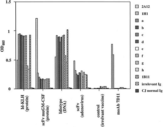
The aim of this study was to test the hypothesis that active immunization with Id induces a polyclonal immune response that would cover mutated Id variants that did not react with the anti-Id MoAb (7D11) used for therapy. Somatic mutations within the tumor cells gave rise to 7D11-idiotope–negative variants that were selectively spared in the patient. Such variants led to tumor relapse after initially successful anti-Id treatment. A single 7D11-idiotope–negative heterohybridoma clone (1B11) was previously produced from this patient (Table 1). We knew from previous investigation10 that the 7D11 Ab reacts with the isolated heavy chain. To obtain more test proteins derived from the relapse tumor biopsy, we cloned the VH gene from the relapse tumor and produced eight additional Id proteins by transient expression in mammalian cells. None of these Id proteins reacted with the treatment MoAb 7D11 (Table 1 and Fig 2). A detailed analysis of these proteins will be presented later herein.*
Reactivity of tumor-derived Id proteins with the treatment MoAb 7D11 and with sera from immunized mice. Sera from individual mice were absorbed against polyclonal human Ig and on normal Ig from patient C.J. The absorbed sera were then reacted at a final dilution of 1:50 in 2% BSA/PBS with the 7D11-idiotope–positive proteins (2A12 and 1H1), with mutated 7D11-idiotope–negative Id variants (1B11 and a to h), and with class-matched irrelevant human Ig or with C.J. normal Ig. Bound murine Ig was detected with HRPO-conjugated goat anti-murine IgG Abs (human-absorbed). The mean OD at 405 nm per vaccine group of 5 mice is given for each Id protein. Reactivity with the treatment MoAb 7D11 controlled for the presence of the specific idiotope in Id protein 2A12 and 1H1 and its absence in the Id variants a to h, 1B11, and the human control Igs. Sera from mice immunized with irrelevant Igs were negative in this assay.
Reactivity of tumor-derived Id proteins with the treatment MoAb 7D11 and with sera from immunized mice. Sera from individual mice were absorbed against polyclonal human Ig and on normal Ig from patient C.J. The absorbed sera were then reacted at a final dilution of 1:50 in 2% BSA/PBS with the 7D11-idiotope–positive proteins (2A12 and 1H1), with mutated 7D11-idiotope–negative Id variants (1B11 and a to h), and with class-matched irrelevant human Ig or with C.J. normal Ig. Bound murine Ig was detected with HRPO-conjugated goat anti-murine IgG Abs (human-absorbed). The mean OD at 405 nm per vaccine group of 5 mice is given for each Id protein. Reactivity with the treatment MoAb 7D11 controlled for the presence of the specific idiotope in Id protein 2A12 and 1H1 and its absence in the Id variants a to h, 1B11, and the human control Igs. Sera from mice immunized with irrelevant Igs were negative in this assay.
Mutated Id Proteins From the Relapsed Tumor React Specifically With the Polyclonal Abs Induced by Id Vaccine
Mice were immunized with four different formulations of tumor Id vaccines: Id protein coupled to KLH, scFv-muGM-CSF fusion protein, plasmid DNA coding for the Id protein, and AdV containing the gene encoding the scFv (Fig 1). Specific anti-Id Ab levels in the sera of immunized mice were measured by ELISA. To exclude nonspecific binding to human constant or framework regions and to allotypic epitopes, the immune sera were preabsorbed on irrelevant human Igs and on normal Ig obtained from the banked serum of patient C.J. In addition, three class-matched irrelevant human Igs (different from the ones used for preadsorption) and C.J. normal Ig were used as negative controls in the ELISA. All mice developed a specific anti-Id immune response after vaccination (Fig 2). These hyperimmune sera reacted specifically with all of the Id proteins, whereas reactivity was not seen with VH family–matched human Ig proteins or with C.J. normal Ig. This result was independent of the Id vaccine formulation. Most remarkably, all nine Id proteins that failed to react with the treatment MoAb 7D11 were detected by the polyclonal Abs in the sera of the individual immunized mice. Serum from a mouse immunized with an irrelevant Ig did not react with any of the tumor-derived proteins.
Quantification of the Anti-Id Immune Response
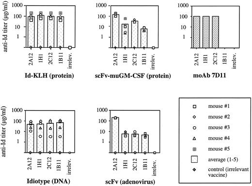
Reactivity of mouse sera after adsorption on irrelevant human Ig was quantified by testing with four different Id proteins of this patient: three 7D11-idiotope–positive (2A12, 1H1, and 2C12) and one 7D11-idiotope–negative (1B11). All immunized mice developed a significant and specific anti-Id immune response (Fig 3). Titers obtained by the different vaccination strategies are shown for the five individual mice, with the bar indicating the mean anti-Id titer. Serum from a mouse immunized with an irrelevant protein was used as a negative control. As an additional specificity control, the hyperimmune sera were tested against three irrelevant class-matched monoclonal human Igs. These human Igs were tested individually but are shown here as a single bar (irrelev. Ab) since they all were equally nonreactive. Sera with the highest and most consistent antibody titers were obtained from mice immunized with Id protein coupled to KLH. Injections with Id plasmid DNA resulted in slightly lower titers. The weakest humoral immune responses were obtained using either the scFv AdV construct or the scFv-muGM-CSF fusion protein. In contrast, immunization with plasmid DNA encoding Id without constant regions failed to evoke a significant anti-Id immune response (data not shown).
Anti-Id titers in vaccinated and control mice measured by ELISA. Sera were diluted 1:20 in BSA 2% in PBS, preabsorbed on polyclonal human Ig, and then serially diluted on plates with 1 of 4 Id proteins (7D11-idiotope–positive: 2A12, 1H1, and 2C12; 7D11-idiotope–negative: 1B11) or with 1 of 3 class-matched irrelevant human Igs (same VH family as the Id). The irrelevant Igs were tested separately but are shown as a single bar since they were all equally negative. Reactivity of the target proteins with treatment MoAb 7D11 is shown.
Anti-Id titers in vaccinated and control mice measured by ELISA. Sera were diluted 1:20 in BSA 2% in PBS, preabsorbed on polyclonal human Ig, and then serially diluted on plates with 1 of 4 Id proteins (7D11-idiotope–positive: 2A12, 1H1, and 2C12; 7D11-idiotope–negative: 1B11) or with 1 of 3 class-matched irrelevant human Igs (same VH family as the Id). The irrelevant Igs were tested separately but are shown as a single bar since they were all equally negative. Reactivity of the target proteins with treatment MoAb 7D11 is shown.
Isotypes
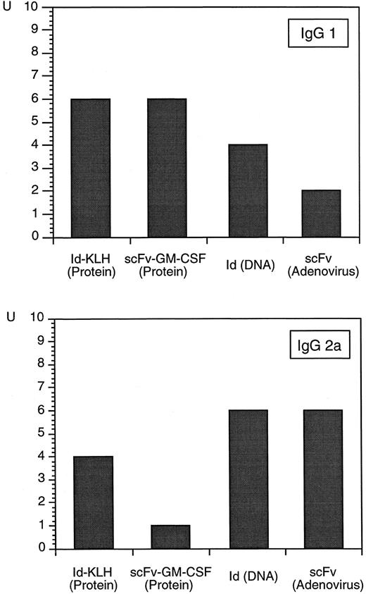
The relative amount of the different murine Ig isotypes elicited by the different vaccine strategies is shown in Fig 4. A high proportion of IgG2a was seen in the groups immunized with scFv AdV and with Id DNA IM, whereas the two groups immunized with scFv-GM-CSF protein and Id-KLH protein had a stronger IgG1 immune response. Interestingly, intradermal DNA immunization induced IgG1 rather than IgG2a antibodies with overall lower titers (results not shown). No significant differences between the groups were found for IgG2b , and only minimal IgG3 could be detected (data not shown).
Relative contribution of IgG isotypes to the anti-Id immune response measured by ELISA comparing the different vaccine formulations. Id protein was indirectly captured on the plates, and the sera of 5 mice per group were pooled and serially diluted. Bound murine IgG was detected with subclass-specific HRPO-conjugated Abs. Results are presented as relative units.
Relative contribution of IgG isotypes to the anti-Id immune response measured by ELISA comparing the different vaccine formulations. Id protein was indirectly captured on the plates, and the sera of 5 mice per group were pooled and serially diluted. Bound murine IgG was detected with subclass-specific HRPO-conjugated Abs. Results are presented as relative units.
Sequence Analysis of the Id Proteins
Previous study from our group has shown that somatic point mutations in the CDR2 of the VH gene of this patient's tumor were responsible for the loss of binding of the tumor Id to the treatment MoAb 7D11.10 In the current study, we rescued eight additional Id proteins from the relapsed tumor specimen and determined their VH gene sequences. All VH genes derived from this patient's tumor differed from each other by point mutations clustered in the CDR. Amino acid changes were most abundant in CDR2, the presumed binding site of the MoAb used in the initial treatment of the patient. All 7D11-idiotope–negative clones had at least one change in amino acids 51 to 61 that was not observed in the clones reactive with the treatment MoAb 7D11 (Fig 5). Interestingly, changes also occurred in the adjacent framework region 3 (FR3). This may indicate that FRs contribute to the epitope recognized by the treatment MoAb. The predicted three-dimensional structure of the VH shows that CDR2 mutational hot spots are in the immediate vicinity of FR3. The fact that none of the Id proteins derived from the relapsed tumor biopsy reacted with the treatment MoAb supports the conclusion that this antibody was extremely effective in eliminating 7D11-idiotope–positive cells.
Protein sequence of the VH of Id proteins derived from the original and the relapsed tumor. Amino acids were deduced from DNA sequences of the respective clones and compared against the previously published consensus sequence.10 Dashes in the sequence indicate identity with the consensus sequence, and differences are indicated by single letters. X indicates undetermined amino acids. Positions of the CDR are marked. Changes in CDR2 thought to be critical for the loss of binding to treatment antibody 7D11 are underlined twice.
Protein sequence of the VH of Id proteins derived from the original and the relapsed tumor. Amino acids were deduced from DNA sequences of the respective clones and compared against the previously published consensus sequence.10 Dashes in the sequence indicate identity with the consensus sequence, and differences are indicated by single letters. X indicates undetermined amino acids. Positions of the CDR are marked. Changes in CDR2 thought to be critical for the loss of binding to treatment antibody 7D11 are underlined twice.
DISCUSSION
Our study addresses the question of whether active immunotherapy using the Id of a lymphoma can be expected to induce an immune response capable of binding to tumor cells expressing mutated Id proteins. It is logical to assume that the polyclonal immune response contains sufficient diversity to cover all somatic mutations that would occur in a given Ig expressed by a monoclonal lymphoma. To prove that this is the case, we chose a tumor in which we had documented escape from monoclonal anti-Id therapy due to point mutations in the expressed Id variable regions. We immunized mice with a single Id sequence and analyzed the immune response against a panel of mutated Ids expressed by the patient's tumor cells. We decided to use this xenogeneic test system because murine lymphomas do not show spontaneous somatic mutations. Furthermore, the mutations we survey here actually occurred in a clinical situation, and it is relevant to ask whether they could ever be recognized by the immune system after immunization with a single Id variant. The best test of the hypothesis would have been to immunize the human subject. However, this was not possible. The limitation of a nonsyngeneic model is that xenogeneic or allogeneic epitopes in the variable regions may contribute to the generation of an anti-Id immune response or interfere with the analysis of anti-Id responses. However, the variable regions alone proved to be poorly immunogenic in our test system. Vaccination with plasmid DNA encoding Id without human constant regions failed to evoke a significant anti-Id immune response. To remove any possible antihuman and antiallotypic reactivity, we absorbed the immune sera against polyclonal human Ig and against normal Ig of patient C.J.
We tested several Id-vaccine formulations (Fig 1): Id protein coupled to KLH, scFv-muGM-CSF fusion protein, naked plasmid DNA encoding the gene segments of the tumor variable regions together with human IgG κ constant regions, and AdV containing the gene segments coding for the scFv. The vaccines that contained only scFv from the patient have no human Ig constant regions and focus the test of immunogenicity on the Ig V regions. All immunized mice developed a significant and specific immune response against the Id (Figs 2 and 3). Most importantly, this included the nine Id proteins derived from biopsy of the relapsed tumor, none of which bound the treatment MoAb 7D11 (Fig 2). All Id proteins tested differed in terms of amino acid sequence of the VH . Specifically, all individual 7D11-idiotope–negative proteins had different amino acid substitutions in CDR2, which was critical for the binding of the treatment MoAb (Fig 5). Interestingly, we found a mutational hot spot in the adjacent FR3, indicating a possible contribution of this region to the binding of the treatment MoAb.
Although it was not the purpose of this study to reach a conclusion about the ultimate efficacy of the different types of vaccines used, some general points of comparison can be stated. In the present study, the highest titers of anti-Id Abs were obtained in the group immunized with Id-KLH protein, followed by the group immunized with DNA IM. Titers observed after vaccination with AdV and scFv-GM-CSF protein were slightly lower (Fig 3). The Ig subtypes contributing to this immune response were analyzed. A predominance of IgG2a over IgG1 indicates a bias of T-helper cell 1 (Th1) over a Th2 response.29 In general, Th1-directed immune responses have been associated with favorable antitumor effects.30 The high titer of anti-Id IgG2a measured in mice immunized with DNA and with AdV is especially promising despite lower titers of total antibody (Fig 4). In fact, vaccination by AdV has been found to induce strong cellular immune responses31 and is protective in a mouse lymphoma model (Caspar C, Hakim I, Syrengelas A, Levy R, submitted). Similarly, the route of DNA immunization may influence the Th type of the immune response.21 In the current study, immunization with intradermal DNA resulted in overall lower anti-Id responses compared with the IM route. Moreover, this route induced a higher IgG1 /IgG2a ratio, indicating a Th2-like rather than a Th1-like response.
Since the Id vaccine has to be custom-made, the time and effort required for its production are critical and potentially rate-limiting in the wide application of this approach. Sufficient amounts of Id protein can be produced by rescue hybridoma technology in approximately 3 to 6 months. Production of the scFv-GM-CSF fusion protein in eukaryotic cells takes about the same amount of time, but yields tend to be lower than those achievable by hybridomas. Bacterial scFv proteins can be made faster. However, in our experience, such proteins are often insoluble within the bacteria (inclusion bodies), making purification difficult. The use of baculovirus in an insect cell system has been shown to be suitable for antibody production,32 and an adaptation has resulted in sufficient quantities of soluble Id protein for vaccine use. Naked DNA vaccination is clearly the most streamlined approach. The gene segments coding for VH and VL can be identified and amplified by PCR. They can then be cloned into an expression plasmid and used directly for vaccination within weeks of obtaining the tumor sample. Production of an AdV as a carrier of the vaccine involves the same initial molecular cloning steps. In addition, transfection, plaque purification, and safety-testing are needed, requiring an additional 6 to 8 weeks. The viral vaccine requires only a single injection to evoke a strong immune response, which may be an advantage over naked DNA.
We conclude that the polyclonal immune response evoked by active vaccination with tumor Id can cover bona fide tumor variants that escaped treatment in a patient during therapy with an anti-Id MoAb. The clinical relevance of this approach is being tested in ongoing studies of active vaccination with tumor Id protein in patients with B-cell lymphoma.13 33 We consider the use of naked DNA to be a streamlined and promising approach solving major problems in the production of a customized vaccine. However, the most effective method of immunization of patients with lymphoma remains to be established.
Supported by Grants No. CA33399 and AI31219 from the National Institutes of Health, and in part by a Cure for Lymphoma Foundation Fellowship, a grant from the Kurt and Senta Hermann Foundation, a grant from the Swiss Institute for Applied Cancer Research/Farmitalia (all to C.B.C.), and an American Cancer Society Clinical Research Professorship (R.L.).
Address reprint requests to Ronald Levy, MD, Stanford University School of Medicine, Division of Oncology M207, Stanford, CA 94305-5306.


